Thorax
Cards 3-1 to 3-26
Bones and Joints
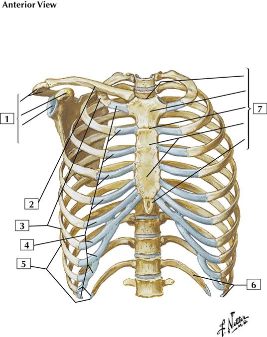
3-1 Bony Framework of Thorax
Comment:
The thoracic cage is part of the axial skeleton, which also includes the skull and vertebral column. Bones of the thorax include the sternum, the 12 pairs of ribs, and the respective articulations of the ribs. The clavicle and scapula are part of the pectoral girdle, which is associated with the upper limb.
The articulations of the thorax include the sternoclavicular joint (which is a saddle-type synovial joint with an articular disc), the sternocostal joints (which are synchondroses), and the costochondral joints (which are primarily cartilaginous joints).
The opening at the top of the thoracic cage is the superior thoracic aperture, and the opening at the bottom of the cage is the inferior thoracic aperture, which is closed by the abdominal diaphragm.
Atlas Plate 183
See also Plate 243
3-2 Costovertebral Joints
Comment:
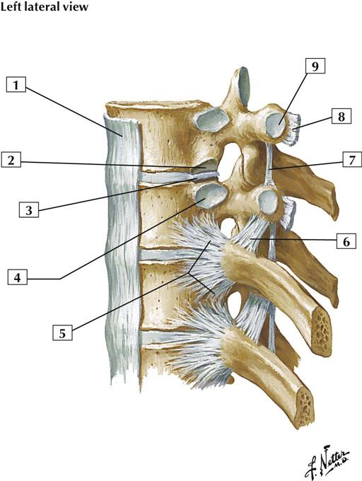
The superior and inferior articular processes (facets) articulate and form plane synovial joints (zygapophysial joints). Each articulation is surrounded by a thin capsule. Accessory ligaments unite the laminae, transverse processes, and spinous processes. These articulations permit some gliding movements between adjacent vertebrae during flexion, extension, and limited lateral bending.
Costovertebral joints are plane synovial joints that occur between the head of a rib and the costal facets of a vertebra. Costotransverse plane synovial joints (ribs 1-10) occur between a tubercle of a rib and a transverse process of a vertebra. Gliding movements occur at these joints.
Atlas Plate 184
See also Plate 154
Muscles
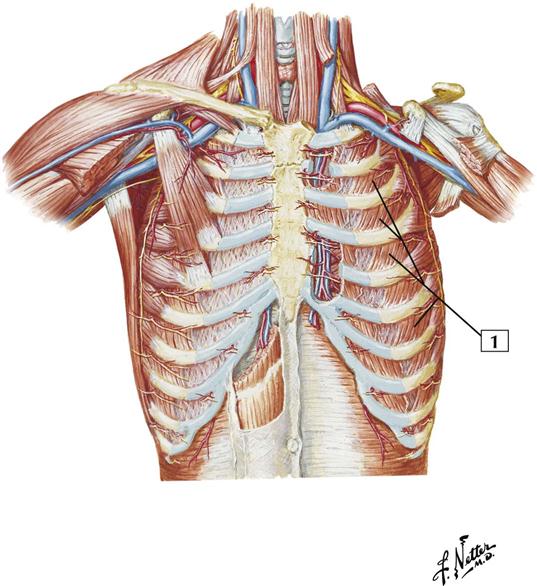
3-3 Anterior Thoracic Wall
Origin (superior attachment):
Arises from the lower border of a rib.
Insertion (inferior attachment):
Attaches to the upper border of the rib below its origin.
Action:
It is generally accepted that the external intercostal muscles are active during inspiration and that they elevate the ribs.
Innervation:
These muscles are innervated by intercostal nerves, which are numbered sequentially according to the intercostal interspace. The 4th intercostal nerve supplies muscles that occupy the 4th intercostal space, between the 4th and 5th ribs.
Comment:
Because these muscles fill the intercostal spaces, there are 11 external intercostal muscles on each side of the thorax.
All of the intercostal muscles keep the intercostal spaces rigid, preventing them from bulging out during expiration and being drawn in during inspiration.
Atlas Plate 186
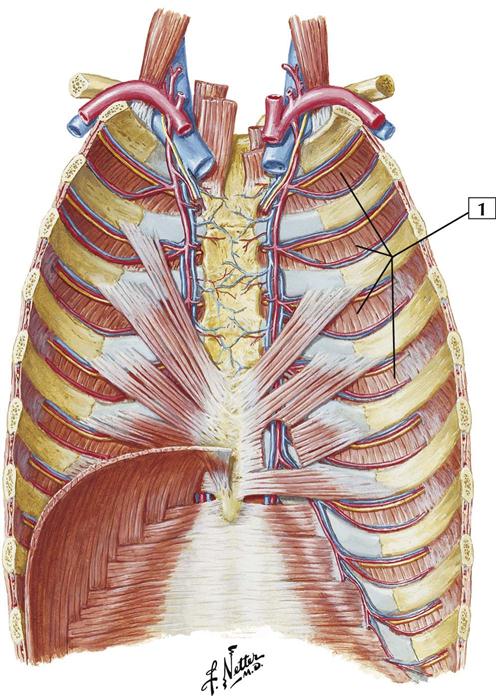
3-4 Anterior Thoracic Wall: Internal View
Origin (superior attachment):
These muscles arise from a ridge on the inner surface of the inferior aspect of each rib and from the corresponding costal cartilage.
Insertion (inferior attachment):
Each muscle attaches to the upper border of the rib below its origin.
Action:
The portions of the upper 4 or 5 internal intercostal muscles that interconnect with the costal cartilages elevate the ribs. The more lateral and posterior portions of the muscles, where the fibers run more obliquely, depress the ribs and are active during expiration.
Innervation:
Intercostal nerves.
Comment:
In general, the fibers of the internal intercostals are roughly perpendicular to those of the external intercostal muscles.
All of the intercostal muscles keep the intercostal spaces rigid, preventing them from bulging out during expiration and being drawn in during inspiration.
Atlas Plate 187
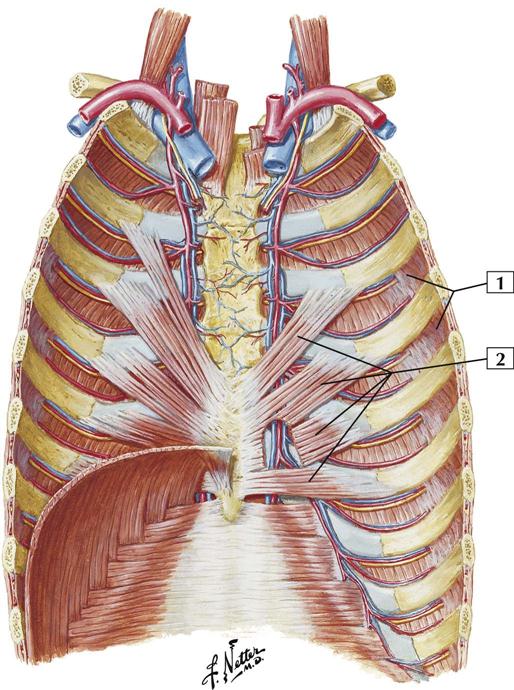
3-5 Anterior Thoracic Wall: Internal View
Origin:
Each innermost intercostal arises from the lower border of a rib. The transversus thoracis arises from the posterior surface of the lower portion of the body of the sternum and the xiphoid process.
Insertion:
Each innermost intercostal attaches to the upper border of the rib below its origin. The transversus thoracis attaches to the inner surfaces of costal cartilages 2-6.
Action:
The action of the innermost intercostals is controversial, but these muscles are thought to elevate the ribs. The transversus thoracis muscle depresses the ribs.
Innervation:
Intercostal nerves.
Comment:
The innermost intercostal muscles frequently are poorly developed and may be fused to the overlying internal intercostals.
The transversus thoracis muscle is variable in its attachments.
All of the intercostal muscles keep the intercostal spaces rigid, preventing them from bulging out during expiration and being drawn in during inspiration.
Atlas Plate 187
3-6 Posterior and Lateral Thoracic Walls
Origin:
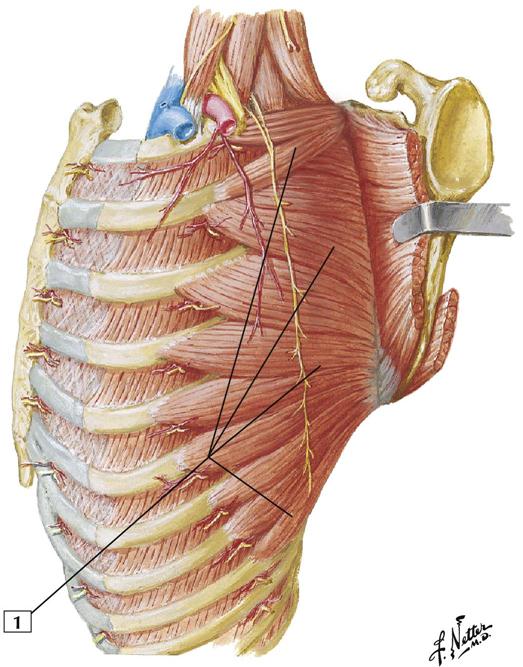
Arises by fleshy digitations from the outer surfaces and superior borders of the first 8 to 9 ribs.
Insertion:
The muscle fibers pass backward, closely apply themselves to the chest wall, and insert on the ventral aspect of the vertebral border of the scapula.
Action:
This muscle pulls the medial border of the scapula anteriorly toward the thoracic wall, preventing the bone from protruding (winging). Its fibers also rotate the scapula upward by laterally rotating the inferior angle. This action helps abduct the arm at the shoulder. Abduction above 90° (above the horizontal) can be accomplished only by lateral rotation of the inferior angle of the scapula.
Innervation:
Long thoracic nerve (C5, C6, and C7).
Comment:
The serratus anterior is particularly important in abduction of the arm above 90°.
Atlas Plate 414
3-7 Musculature of Esophagus
Comment:
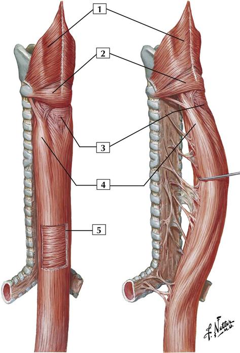
The esophagus is a muscular canal that extends from the pharynx to the stomach. Its muscular coat is organized into 2 planes: an external plane of longitudinal fibers and an internal plane of circular fibers. The esophageal muscle transitions from skeletal to smooth muscle as it descends from the pharynx to the stomach.
Atlas Plate 231
Nerves
3-8 Typical Thoracic Spinal Nerve
Comment:
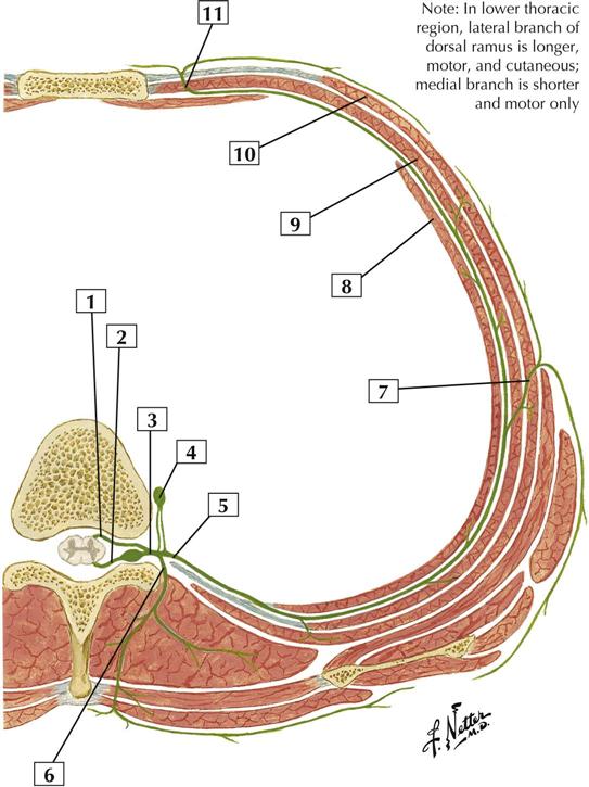
This thoracic nerve is a typical example of a spinal nerve. Dorsal and ventral roots combine to form the spinal nerve, which divides into a small dorsal ramus that supplies the intrinsic muscles of the back and a larger ventral ramus (intercostal nerve) that innervates all the muscles lining the trunk. The ventral ramus divides into a lateral cutaneous branch at the midaxillary line; anteriorly, and laterally to the sternum, it gives rise to an anterior cutaneous branch. The intercostal nerves course between the internal intercostal and innermost intercostal muscles.
Stay updated, free articles. Join our Telegram channel

Full access? Get Clinical Tree


