The Yeasts


The Yeasts


General Characteristics


Yeasts are eukaryotic, unicellular organisms that are round to oval and range in size from 2 to 60 µm. The microscopic morphologic features have limited usefulness in helping to differentiate or identify these organisms. The microscopic morphology on cornmeal agar is most useful when considered in conjunction with the biophysical profile (i.e., a combination of the biochemical and physical characteristics used in the identification of a microorganism) obtained using a commercial system. Differentiation of yeasts in direct microscopic and histopathologic examination of clinical specimens is often impossible, but sometimes particular characteristics are seen that suggest the identification or are pathognomonic (i.e., unique) for a particular organism. Important morphologic characteristics that are useful in differentiating yeasts include the size of the yeasts, the presence or absence of a capsule, and broad-based or narrow-necked budding. For example, variability in size with evidence of a capsule and narrow-necked budding are features that can be helpful for distinguishing Cryptococcus spp. from Candida spp. The medically important yeasts and yeastlike organisms belong to different taxonomic groups, including the Ascomycota, Basidiomycota, and Deuteromycota.


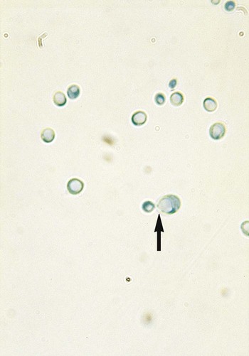
In general, the yeasts reproduce asexually by blastoconidia formation (budding) (Figure 63-1) and sexually by the production of ascospores or basidiospores. The process of budding begins with a weakening and subsequent outpouching of the yeast cell wall. This process continues until the bud, or daughter cell, is completely formed. The cytoplasm of the bud is contiguous with the cytoplasm for the original cell. Finally, a cell wall septum is created between mother and daughter yeast cells. The daughter cell often eventually detaches from the mother cell, and a residual defect occurs at the budding site (i.e., a bud scar).


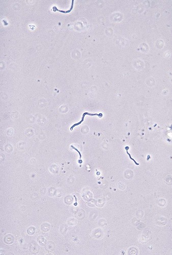
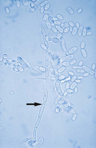
With certain environmental stimuli, yeast can produce different morphologies. An outpouching of the cell wall that becomes tubular and does not have a constriction at its base is called a germ tube; it represents the initial stage of true hyphae formation (Figure 63-2). Alternatively, if buds elongate, fail to dissociate, and form subsequent buds, pseudohyphae are formed; to some, these resemble links of sausage (Figure 63-3). Pseudohyphae have cell wall constrictions rather than true intracellular septations delineating the fungal cell borders.


The number of fungal infections caused by yeasts and yeastlike fungi has increased significantly during recent years. Most of these infections have been caused by various Candida spp. However, other yeasts also cause significant disease, particularly in immunocompromised hosts, as yeasts are the cause of many opportunistic infections. In addition to causing disease in immunocompromised patients, infections also are common in postsurgical patients, trauma patients, and patients with long-term indwelling venous catheters. Some of these yeasts are resistant to commonly used antifungal agents, which emphasizes the need for prompt, appropriate identification and, in some cases, antifungal susceptibility testing.


The extent to which the laboratory should identify all yeast species is the subject of debate. Each laboratory director must decide how much time, effort, and expense is to be spent on the identification of yeasts in the laboratory.


The development of commercially available yeast identification systems has provided laboratories of all sizes with accurate, standardized methods. However, these methods should be used in conjunction with cornmeal agar morphology to prevent misidentifications. Some commercial systems have extensive computer databases that include biochemical profiles of a large number of yeasts. Variations in the reactions of carbohydrates and other substrates utilized are considered in the identification of yeasts provided by these systems.


Commercially available systems are recommended for all laboratories. They may be used in conjunction with some less expensive and rapid screening tests that provide presumptive identification of C. neoformans and definitive identification of Candida albicans.


More recent diagnostic tools that have been introduced for quicker characterization of yeasts include CHROMagar Candida (BD Diagnostics, Sparks, Maryland) and C. albicans PNA FISH (AdvanDX, Woburn, Massachusetts). Because some laboratories might prefer to use conventional systems, the information presented in this section discusses rapid screening methods for presumptive identification of yeasts, commercially available systems, and a conventional schema for identifying commonly encountered yeast species.


Epidemiology


Candida spp.


Candida spp. are responsible for the most frequently encountered opportunistic fungal infections. Currently Candida spp. are the fourth most common cause of hospital-acquired bloodstream infections (BSIs) in the United States, and a mortality rate as high as 49% has been seen with these infections. Candida infections are caused by a variety of species. C. albicans is the most commonly isolated yeast, but other, emerging species include Candida glabrata, Candida parapsilosis, Candida tropicalis, Candida dubliniensis, and Candida krusei. The frequency with which these organisms are isolated varies by institution. Until recently C. albicans was the most common yeast isolated from infections, accounting for at least 60% to 70% of yeast infections. Epidemiologic data for the past decade reveal a paradigm shift in candidal infections. Studies from intensive care units (ICUs) confirm an increase in C. glabrata and C. krusei (isolated in approximately 25% of infections) and C. albicans (about 50% of infections). C. albicans and other Candida spp. are part of the human body’s microbiota (i.e., normal flora), but they also have become endemic in most hospitals. Infections may be caused by endogenous yeasts or may be acquired in the hospital. Differentiating among the Candida spp. in the clinical laboratory is very important because of the differences in the virulence of the species and in their susceptibility to antifungal drugs.


Cryptococcus spp.


Cryptococcus neoformans-Cryptococcus gatti complex has been divided into the two species and five serotypes. Serotype A has been referred to as C. neoformans var. grubii with some groups recognizing the variant as a distinct species. Serotype D is now classified as C. neoformans var. neoformans. Both C. neoformans serotypes produce the teleomorphic state Flobasidiella neoformans. A C. grubii/C. neoformans hybrid exists that is a variant of serotypes A and D. Serotypes B and C are recognized as the independent species, C. gattii. Both serotypes B and C produce the teleomorph referred to as Filobasidiella bacillispora. Standard laboratory tests do not differentiate among the serotypes of C. neoformans and C. gattii.Cryptococcus exists as a saprobe in nature. It is most associated with avian excreta, particularly pigeons. C. neoformans is thought to be widely distributed in nature, and aerosolization is a prerequisite to most infections.


Pathogenesis and Spectrum of Disease


Candida Albicans


Candidiasis is an infection caused by a Candida spp. It may include oroesophageal candidiasis, intertriginous candidiasis (in which skin folds are involved), paronychia, onychomycosis, perlèche respiratory infections, vulvovaginitis, thrush, pulmonary infection, eye infection, endocarditis, meningitis, fungemia or candidemia, or disseminated infection. Paronychia is an infection of the tissues surrounding the nails, and onychomycosis is an infection of the nail and nail bed. Thrush, an infection of the mucous membranes in the mouth, is considered a localized infection. Thrush can be seen in newborns, patients with human immunodeficiency virus (HIV) infection, individuals with diabetes, and patients undergoing chemotherapy. Creamy patches or colonies appear on the tongue and mucous membranes. Candida organisms may be recovered from the oropharynx, gastrointestinal (GI) tract, genitourinary tract, and skin.


The clinical significance of candidal organisms recovered from respiratory tract secretions is difficult to determine, because Candida spp. are considered part of the normal oropharyngeal flora of humans. A study at the Mayo Clinic evaluated the clinical significance of yeasts other than C. neoformans that are recovered from respiratory secretions. These researchers concluded that such yeasts are part of the normal flora and do not need to be routinely identified. Similarly, Barenfanger et al. demonstrated that routine identification of yeasts from respiratory specimens results in unnecessary antifungal therapy, an extended hospital stay, and increased health care costs without demonstrable benefit. Simultaneous recovery of the same species of yeast from several body sites, including urine, is a good indicator of disseminated infection and fungemia.


The pathogenesis of candidal infections is extremely complex and probably varies with each species. Adhesion of Candida organisms to the epithelium of the gastrointestinal or urinary tract is a crucial factor. Candida spp. commonly colonize mucosal surfaces. Their ability to invade and cause infection depends on adherence to the surface before infection. Three distinct aspartyl proteases have been described in C. albicans, and strains with high levels of proteases have been shown to have an increased ability to cause disease in experimental animal models. Hydrophobic molecules on the surface of Candida spp. also appear to be important in pathogenesis, and a strong correlation exists between adhesion and surface hydrophobicity. In addition, high levels of phospholipase, found in strains of C. albicans, have correlated with a higher mortality rate in experimental animals compared with experimental infections caused by strains that produce a lower level of phospholipase. Phenotypic switching (i.e., the ability to produce pseudohyphae and hyphae), seen in C. albicans, also may play a role in pathogenesis.


Non-Albicans Candida


The other Candida spp. (also called non-albicans Candida), once thought not to cause disease, are emerging as agents of infection in certain patient populations. The incidence of infection with C. glabrata is higher in older adults than in young adults and children. Recent studies have demonstrated the ability of C. glabrata to become resistant to common antifungal drugs. C. tropicalis has been shown to be prevalent in patients with hematologic malignancies, especially those who are neutropenic. Mouse models of infection and human studies have shown C. tropicalis in the tissues surrounded by necrotic tissues, which may indicate that the organism can invade the GI tract efficiently, particularly in oncology patients. This phenomenon most likely is due to the expression and secretion of aspartyl proteases and tropiase (acid proteinase), a virulence factor found in Candida organisms. Because C. krusei is inherently resistant to the azole class of antifungal drugs, identification of this species is essential to proper clinical management of the patient. C. parapsilosis is the primary cause of fungemia in the neonatal intensive care unit (NICU). C. parapsilosis is also the second most frequently isolated Candida spp. in positive blood cultures; this could be due partly to its known selective growth in hyperalimentation solutions and also to its ability to grow on intravascular catheters. Historically C. parapsilosis was categorized into three groups (I to III); however, it now has been typed by molecular methods into three different Candida species: C. parapsilosis, C. orthopsilosis, and C. metapsilosis.


Cryptococcus Neoformans


Genus Cryptococcus


Cryptocococcosis is an acute, subacute, or chronic fungal infection that has several manifestations. Crytococcus neoformans var. grubii, C. neoformans var. neofromans, and C. gatti are considered the major human pathogens. Differences in the infections by Cryptococcus neoformans appear to be dependent on the host immune status and not the variant. C. neoformans infections can present initially as a chronic or subacute pulmonary infection. C. neoformans eventually makes its way to the central nervous system, where the yeast can cause cryptococcal meningitis. Disseminated disease with meningitis is commonly seen in immunocompromised patients. Patients with a moderately compromised immune system or who are early in the disease process of cryptococcal fungemia may present without concomitant meningitis. Disseminated cryptococcosis and cryptococcal meningitis became well recognized in patients with acquired immunodeficiency syndrome (AIDS), and they remain an important cause of morbidity and mortality in these patients in resource-poor countries that do not have access to highly active antiretroviral therapy.


Patients with disseminated infection may have painless papular skin lesions that may ulcerate. Other, less common manifestations of cryptococcosis include endocarditis, hepatitis, renal infection, and pleural effusion. Interestingly, a review of patient records at the Mayo Clinic revealed that more than 100 immunocompetent patients with C. neoformans colonization of the respiratory tract did not develop subsequent infection. Follow-up on these patients was as long as 6 years, and none in this group were considered to be immunocompromised. This makes the clinical significance of C. neoformans somewhat difficult to assess. However, given the severity of disease it can cause, its presence in clinical specimens should be considered significant. In many instances, the clinical symptoms are suppressed by corticosteroid therapy, which is a risk factor for disease, and culture or serologic evidence (detection of cryptococcal antigens) provides the earliest proof of infection. Cryptococcal infection is strongly associated with such debilitating diseases as leukemia and lymphoma and the immunosuppressive therapy that may be required for these and other underlying diseases. In some cases the presence of C. neoformans in clinical specimens precedes the symptoms of an underlying disease.


C. neoformans can exhibit a very characteristic polysaccharide capsule. The capsule collapses and protects the yeast from desiccation under drying conditions. The capsule of C. neoformans is thought to help the organism survive through the pigeon gut before it is excreted. The reduction in the yeast’s cell size caused by capsular collapse places the organism in the ideal size range for alveolar deposition in the human host. In addition, a virulent property of the polysaccharide capsule is that it contains compounds that phagocytes do not recognize. The so-called acapsular strains of C. neoformans, which actually just have a very reduced capsule, are more easily phagocytosed. In some instances, C. neoformans elicits minimal tissue response in infected individuals, particularly severely immunocompromised patients.


Phenoloxidase, an enzyme found in C. neoformans, is responsible for melanin production. Some have speculated that melanin might act as a virulence factor by making the organism resistant to leukocyte attack. Evidence also has been presented that increased melanin production can decrease immune system functions, such as lymphocyte proliferation and tumor necrosis factor production. Whether phenoloxidase is truly a virulence factor has yet to be determined (Li SS, et al, 2010. (Cryptococcus. 2010 Proc Am Thorac Soc Vol 7 pp 186-196, 2010). An interesting question is whether the interactions of substances in the brain that are known to react with phenoloxidase may play a role in the affinity of C. neoformans and certain neurotropic dematiaceous fungi for invading the central nervous system (Li et al, 2010).


Trichosporon spp.


Trichosporonosis is caused by a variety of Trichosporon spp., which have undergone changes in nomenclature based on DNA sequence comparisons. The yeastlike fungus causes disease almost exclusively in immunocompromised patients, particularly those with leukemia. Disseminated trichosporonosis is the most common clinical manifestation. Skin lesions accompanied by fungemia are frequently seen. Endocarditis, endophthalmitis, and brain abscess have been reported. Trichosporon organisms occasionally are recovered from respiratory tract secretions, skin, the oropharynx, and the stool of patients with no evidence of infection and may represent transient fungal colonization of those individuals.


White piedra, an uncommon fungal infection of immunocompetent patients, is found in both tropical and temperate regions of the world. It is characterized by the development of soft, yellow or pale brown aggregations around hair shafts in the axillary, facial, genital, and scalp regions of the body. The Trichosporon spp. that cause this disease frequently invade the cortex of the hair, causing damage.


Laboratory Diagnosis


Specimen Collection, Transport, and Processing


See Chapter 59.


Stains


Candida spp.

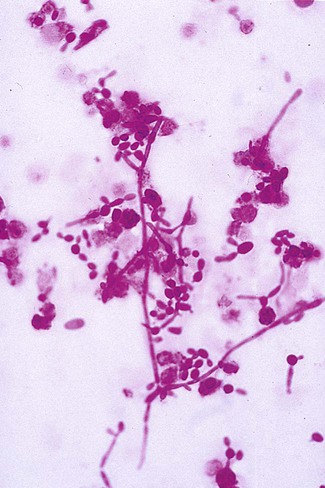
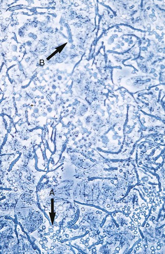
Direct microscopic examination of clinical specimens containing Candida organisms reveals budding yeast cells (blastoconidia) 2 to 4 μm in diameter and/or pseudohyphae (Figure 63-4) showing regular points of constriction, resembling links of sausage. True septate hyphae (filamentation) may also be produced by C. albicans and C. dubliniensis. The blastoconidia, hyphae, and pseudohyphae are strongly gram positive. The approximate number of such forms should be reported, because the presence of large numbers in a fresh clinical specimen may have diagnostic significance. Microscopically C. glabrata blastoconidia are notably smaller (at 1 to 4 µm) than those of other medically significant Candida spp.


Cryptococcus spp.

Traditionally, the India ink preparation has been the most widely used method for the rapid detection of C. neoformans in clinical specimens. This method is still used as a rapid and inexpensive assessment tool in many institutions and has considerable diagnostic value in resource-poor settings. This method delineates the large capsule of C. neoformans, because the ink particles cannot penetrate the capsular polysaccharide material. Although this test is useful, many laboratories have replaced it with the more sensitive cryptococcal latex agglutination test that detects cryptococcal antigen. (The cryptococcal antigen detection [CAD)]test is described later in the chapter.) The India ink preparation is commonly positive in specimens from patients with AIDS and has been shown to have a sensitivity of 50% in patients who do not have HIV or AIDS. Laboratories that examine many specimens from these patients may want to continue using this procedure in combination with the CAD test and culturing.


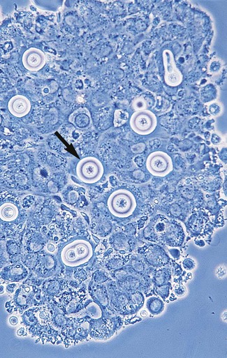
Microscopic examination of other clinical specimens, including respiratory secretions, can be valuable for making a diagnosis of cryptococcosis. C. neoformans appears as a spherical, single or multiple budding, thick-walled yeast 2 to 15 μm in diameter. It usually is surrounded by a wide, refractile polysaccharide capsule (Figure 63-5). Perhaps the most important characteristic of C. neoformans is the extreme variation in the size of the yeast cells; this is unrelated to the amount of polysaccharide capsule present. It is important to remember that not all isolates of C. neoformans exhibit a discernible capsule.


Trichosporon spp.

Microscopic examination of clinical specimens that contain Trichosporon spp. reveals hyaline hyphae, numerous round to rectangular arthroconidia, and occasionally a few blastoconidia. Usually hyphae and arthroconidia predominate. In white piedra, white nodules are removed and observed using the potassium hydroxide (KOH) preparation after light pressure is applied to the coverslip to crush the nodule. Hyaline hyphae 2 to 4 μm wide and arthroconidia are found in the preparation of the cementlike material that binds the hyphae together. The organism may be identified in culture by the presence of true hyphae, blastoconidia, and arthroconidia in conjunction with a positive urease (see Figure 60-40). Although Trichosporon asahii may be distinguished from other Trichosporon species by its biophysical profile (carbohydrate and substrate utilization), these organisms are likely best distinguished at the species level with molecular tools, such as DNA sequencing.


Molecular Assays


Nucleic acid amplification tests (NAATs) have been developed for a variety of yeast species. However, these are usually performed in research settings. Most are labeled as “laboratory-developed tests” or “home-brewed tests,” and few have been approved by the U.S. Food and Drug Administration (FDA). Real-time polymerase chain reaction (PCR) methods are now commercially available in the TaqMan system (Applied Biosystems, USA) and LightCycler (Roche Molecular Systems, Indianapolis, Indiana). These methods are very expensive but have proven to be much more specific than conventional yeast identification methods. Ligouri et al. recently demonstrated the Multiplex PCR method for identifying Candida spp. that has high concordance with commercially available phenotypic identification systems, such as the API 20C AUX (bioMérieux, Durham, North Carolina) and Vitek 2 YST card (bioMérieux, Durham, North Carolina). The Multiplex PCR method is much faster and more sensitive than the current phenotypic tests. A newer molecular test, the PNA FISH kit, uses in situ hybridization to detect Candida organisms in blood culture bottles by targeting specific rRNA sequences. Subculturing should follow this method, because whether more than one species is present cannot be determined. Colonies may also display protrusions from the colony, resembling a star or foot-like projections on blood agar.


Cultivation


Stay updated, free articles. Join our Telegram channel

Aug 25, 2016 | Posted by in MICROBIOLOGY | Comments Off on The Yeasts

Full access? Get Clinical Tree

Get Clinical Tree app for offline access