The Respiratory Tract


The Respiratory Tract


Louis P. Dehner, M.D.

J. Thomas Stocker, M.D.

Haresh Mani, M.D.

D. Ashley Hill, M.D.

Aliya N. Husain, M.D.


DEVELOPMENT OF THE LUNG

The lung begins as a pouch or groove originating from the primitive foregut in week 3 of embryologic development, when the embryo is 3 mm long. As the groove enlarges caudally, a tubular lung bud is formed; the upper portion develops into the epithelium of the larynx and the caudal portion into the epithelium of the tracheobronchial tree (1).

The embryonic period of lung development (Table 12-1) begins in week 4 of gestation as the single lung bud from the foregut divides into two primary bronchial buds, the forerunners of the right and left lungs (Figure 12-1A to C). During week 5 of gestation, the primary bronchi divide. Each forms three lobar buds that, by the end of week 6 of gestation, divide again to form 10 segmental bronchi on the right and 8 to 9 on the left. These potential airways consist of a central core of epithelial cells surrounded by loose primitive mesenchyme that contains widely separated capillaries. The primitive pulmonary arteries begin to form from the sixth aortic arch, near the end of the embryonic period. The pulmonary veins begin as evaginations of the left atrium during week 4 of gestation and coalesce with the mesenchymal capillary plexus early in week 5. These early events with the branching morphogenesis and alveolarization are accompanied by microvascularization of the small airspaces for postnatal gas exchange (2). There are several important signaling pathways involved in early lung development (3,4,5). Lung has been discussed by Correia-Pinto and associates (6). MicroRNAs (miRNA) are important in the regulation of lung development at the levels of proliferation, apoptosis, differentiation, and morphogenesis (7). DICER1 is an RNase III enzyme that performs the final step in generating immature miRNAs (5). The loss of DICER1 in lung epithelium early in development results in loss of branching and the formation of lung cysts (8).

The pseudoglandular period (weeks 6 to 16 of gestation) begins with the completion of the proximal airways and encompasses the development of the conducting airway system to the level of the terminal bronchioles (Figure 12-2A, B). The pseudostratified columnar epithelium of the proximal airway displays cilia at week 10 of gestation. The appearance of cilia extends to the epithelial cells of the peripheral airways by week 13. Goblet cells appear in the bronchial epithelium at weeks 13 to 14 of gestation, and submucosal glands begin as solid buds originating from the basal layers of the epithelium by weeks 15 to 16. Smooth muscle cells develop around airways by the end of gestational week 7 and organize to form an identifiable wall to the larger bronchi by week 12. Lymphatics appear first in the hilar region of the lung in gestational week 8 and in the lung itself by week 10. Cartilage is first seen in week 4 of gestation and forms distinct rings along the trachea and main bronchi by the end of week 10.


TABLE 12-1 PHASES OF INTRAUTERINE LUNG DEVELOPMENT


Phase


Gestation Period


Major Event


Embryonic


26 days to 6 weeks


Development of major airways


Pseudoglandular


6-16 weeks


Development of airways to terminal bronchioles


Acinar or canalicular


16-28 weeks


Development of acinus and its vascularization


Saccular


28-34 weeks


Subdivision of saccules by secondary crests to term


Alveolar


34 weeks (and beyond)


Alveolar acquisition


Adapted from Langston C. Prenatal lung growth and pulmonary hypoplasia. In: Stocker JT, ed. Pediatric Pulmonary Disease. Washington, DC: Hemisphere, 1989:2, with permission.


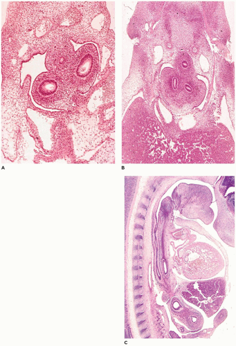
FIGURE 12-1 • Embryonic periods in respiratory tract development. A: At 29 to 31 days gestation (stage 14), the primary bronchial buds are surrounded by primitive mesenchyme. Note the small esophagus above and between the bronchi. B: By 35 to 37 days (stage 16), the primary bronchi have divided into secondary and early tertiary buds. Note the centrally located esophagus and the large amount of hepatic parenchyma (lower half). C: In a sagittal plane of a 37- to 40-day (stage 17) embryo, the relationship between the esophagus (nearest vertebral column) and trachea (between esophagus and heart) can be seen. The heart and liver are ventral to the foregut structures.

The acinar or canalicular period extends from weeks 17 to 28 of gestation and is characterized by the development of the basic structure of the gas-exchanging portion of the lung (Figure 12-3A, B). Smooth-walled respiratory bronchioles, lined by cuboidal epithelium, subdivide into multiple, irregular alveolar ducts. By week 20 of gestation, the cells lining the ducts develop into type II pneumocytes with lamellar and multivesicular bodies associated with surfactant synthesis.
Type I pneumocytes then differentiate from type II cells to form the thin air-blood interface required for gas exchange. As the interstitium thins in the latter portion of the acinar period, the capillaries of the interstitium proliferate and come to lie beneath the type I cells. Submucosal glands in the trachea and bronchi progress from tubules to mucuscontaining acini. By week 24, the cartilage has extended to the most distal bronchi.


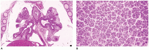
FIGURE 12-2 • Pseudoglandular period. A: At 9 weeks of gestation, the proximal airways are present throughout the right and left lobes. B: By 13 weeks, bronchiolar development is well under way, and early division into lobules and clusters of acini is apparent.

The saccular period begins at week 28 of gestation with the development of secondary crests, which are formed as distal airspaces divide into smaller units (Figure 12-4A, B). With an accompanying marked decrease in the interstitial tissue and further increase in the capillary bed, a complex, interwoven capillary network develops in the wall of the saccules. This provides for effective gas exchange as alveoli begin to develop at the end of the period (32 to 36 weeks of gestation).


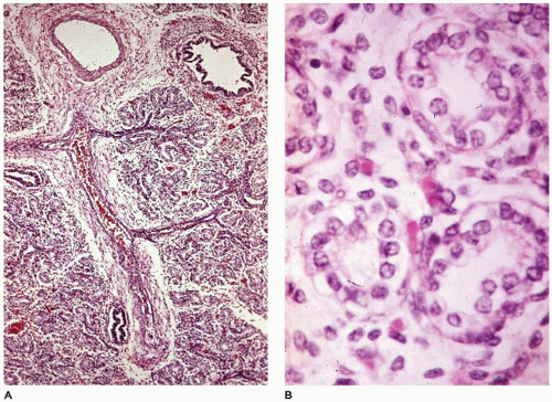
FIGURE 12-3 • Acinar period. A: In a 370-g fetus, acinar development is characterized by pulmonary arteries and proximal bronchioles surrounded by alveolar ducts still widely separated by mesenchymal tissue. B: The alveolar duct structures are lined by cuboidal epithelium (early type II cells), and blood-filled capillaries are present just beneath the cells.


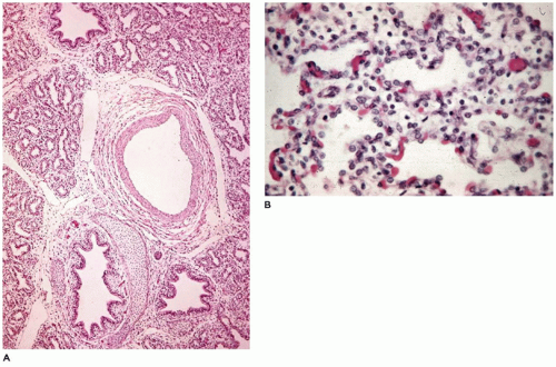
FIGURE 12-4 • Saccular period. A: In a 650-g fetus, discrete acini are identifiable within a lobule. B: Secondary crests are covered by thinning type I cells, which expose capillary beds immediately beneath the cells.

The final period of development, the alveolar period, begins in utero at 32 to 36 weeks of gestation and extends until 18 to 24 months after birth. Alveoli develop as flask-shaped structures with thin walls whose double capillary network meshes to appear as a single capillary bed (Figure 12-5). At term, type I pneumocytes are extremely thin, resulting in an air-blood barrier of only 0.2 mm including the type I cell, the underlying basement membrane, and the cytoplasm of the capillary endothelial cell. Lymphatic channels are distributed around pulmonary arteries, bronchi, and bronchioles and extend along interlobular septa to anastomose with a plexus beneath the pleura. Lymphatic spaces do not exist between alveoli.


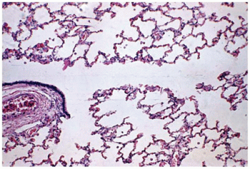
FIGURE 12-5 • Alveolar period. At 2 months of age, a respiratory bronchiole (left) gives rise to alveolar ducts, alveolar saccules, and thin-walled alveoli.

The vascular supply of the lungs changes appreciably in late gestation and infancy. The bronchial arterial circulation, originating from the aortic arch, supplies the bronchi, bronchioles, and interlobular septa in older children and adults; however, the bronchial artery contributes substantially to the circulation of the alveolar ducts and alveoli in the central portions of the lungs through bronchopulmonary artery anastomoses in utero and in early infancy.

At birth, the surface area of the lung is about 4 m2, with the number of alveoli ranging from 10 to 150 million (mean of 53 million). Alveoli increase in number after birth, reaching the adult range of 300 to 600 million alveoli by 2 years of age. Thereafter, lung growth occurs in terms of volume and alveolar size, with no further increase in the number of small air spaces (alveoli).


NASOPHARYNX


Choanal Atresia

Choanal atresia occurs in about 1 in 5-8000 live births and consists of unilateral or bilateral occlusion of the airway between the posterior nasal passage and the nasopharynx (9). The entity has been seen in monozygotic twins and has also been noted following radiotherapy for nasopharyngeal carcinoma. The septum blocking the airway is usually composed of bone or cartilage, but in as many as 20% to 50% of cases, it may be composed of a mucous membrane alone (10). Choanal atresia may exist as an isolated sporadic lesion, in an autosomal dominant form, or possibly in an autosomal
recessive form. It has been associated with palatal defects, tracheoesophageal fistula (TEF), congenital heart malformations, trisomy 6, Pfeiffer syndrome, Treacher Collins syndrome, the fetal carbimazole syndrome, and the CHARGE (Coloboma, Heart defect, choanal Atresia, Retardation, Genital, Ear anomaly) association (CHD7 mutation on 8q12.2) of which it is a major component.


Cleft Lip and/or Palate

Cleft lip, with or without unilateral and bilateral involvement of the hard palate, the soft palate, or both, is the most common malformation of the respiratory tract. It occurs once in 750 live births as an isolated anomaly or as part of a wide variety of chromosomal, inherited, and noninherited syndromes, of which over 250 have been described (11). Cleft palate is associated with other anomalies in 47% of cases, cleft lip and palate in 37%, and cleft lip alone in 14%. Anomalies most frequently seen are those in the central nervous system (CNS) and skeletal system, followed by urogenital and cardiovascular anomalies. Maternal cigarette smoking and alcohol use are associated with a 1.6- to 2.0-fold increase in isolated cleft lip, cleft palate, or both (12). The incidence of cleft lip and palate is dose related, increasing with increased cigarette smoking.


Laryngocele

A laryngocele is an abnormal dilation of the laryngeal saccule that extends upward within the false vocal fold and is in communication with the laryngeal lumen. It is filled with air, although mucoid or purulent contents may be seen following obstruction and infection, respectively. Laryngoceles occur rarely in childhood but may present as airway obstruction or as a neck mass in a neonate or an older child (13,14). The lesion, seen predominantly in boys and containing air or fluid or both, may be within the larynx behind the thyroid cartilage (33%), external to the larynx (25%), or involving both locations (15). Currently, laryngoceles are classified as either internal (confined medial to the thyrohyoid membrane) or combined (when it extends further laterally), since the existence of a purely external laryngocele has been refuted (16). Etiologic factors include congenital increased laryngeal pressure, or mechanical obstruction (such as by tumors including laryngeal papillomas). Since they are outpouchings of the laryngeal wall, they are lined by either respiratory or squamous epithelium. Infection of the lesion may occur (pyolaryngocele), leading to acute respiratory distress.


Laryngomalacia

Stridor and feeding difficulties in the newborn may be caused by laryngomalacia due to flaccidity of a long epiglottis, short arytenoepiglottic folds, or bulky arytenoid swellings, resulting in partial obstruction of the larynx. Kay and Goldsmith (17) have developed a classification based on the underlying pathophysiologic processes, with type 1 characterized by a foreshortened or tight aryepiglottic fold, type 2 defined by the presence of redundant soft tissue in the supraglottis, and type 3 applying to cases caused by other etiologies. Potentially serious complications of the obstruction include pulmonary hypertension and cor pulmonale, sudden death during respiratory tract infections, failure to thrive, and possible impaired intellectual development secondary to episodes of hypoxia and hypercapnia. Twenty percent of these infants have severe neurologic compromise or multiple congenital anomalies. Surgical procedures including supraglottoplasty have been used in severe cases (about 10% to 15% of cases) and have been successful in relieving respiratory symptoms in 80% of those cases (18). Laryngomalacia-induced stridor has been reported in patients with Pierre Robin, acrocallosal, Marshall-Smith, cri du chat, fetal warfarin, Down, Freeman-Sheldon, and Mohr syndromes. A familial form of laryngomalacia has also been described.


Laryngeal Stenosis and Atresia

Supraglottic, glottic, and subglottic developmental webs may produce varying degrees of laryngeal stenosis and have been described in families with an autosomal dominant inheritance pattern. Fraser syndrome is associated with congenital stenosis or atresia in 30% of cases, along with other anomalies (19).

Subglottic stenosis, as an acquired lesion, has been seen secondary to short-term and long-term intubation in the neonatal intensive care unit (NICU) with increased incidence when the infant is intubated for longer periods (20). With acquired stenosis, dense submucosal fibrous connective tissue is present circumferentially in the subglottic area and may narrow the lumen significantly. Submucosal glands are usually absent, and the cricoid cartilage may display evidence of erosion.

Congenital laryngeal atresia occurs in three patterns (21):


  • Type 1, atresia of both supraglottic and infraglottic portions of the larynx


  • Type 2, atresia of the infraglottic region (Figure 12-6A, B)


  • Type 3, glottic atresia

Associated conditions include esophageal atresia (EA), TEF, “total sequestration” of the lungs in the absence of TEF, anal anomalies, urinary tract malformations, skeletal anomalies, and heart malformations. Many of the conditions are part of the vertebral, anal atresia, cardiac, TEF, renal, and limb (VATER or VACTERL) association. Other associations include partial diaphragmatic obliteration, Fraser syndrome, DiGeorge developmental field defect, and partial trisomy 9. Pulmonary hyperplasia has been noted in infants who have laryngeal atresia, with lung weights ranging from 150% to 300% of normal.


Laryngotracheoesophageal Cleft

Failure in formation of the tracheoesophageal septum, normally complete by day 35 of gestation, leads to the development of one of four forms of laryngotracheoesophageal cleft (22) (Figure 12-7A to D):


  • Supraglottic interarytenoid cleft (50% of cases)


  • Partial cricoid cleft


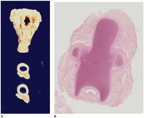
    FIGURE 12-6 • Laryngeal atresia. A: The larynx reveals a patent upper opening (upper piece) and a patent trachea (lower two cross sections). B: A histologic section from the area in the region of the cricoid cartilage reveals only a pinpoint lumen (bottom center).


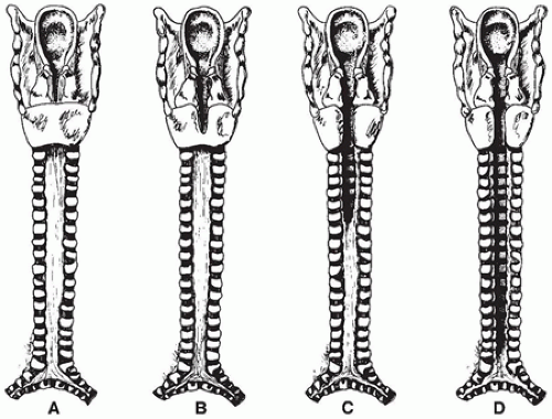
    FIGURE 12-7 • Types of laryngotracheoesophageal cleft. A: Supraglottic interarytenoid cleft. B: Partial cricoid cleft. C: Total cricoid cleft. D: Complete cleft to level of carina.


  • Total cricoid cleft


  • Complete cleft of the trachea to the level of the carina

Maternal polyhydramnios is seen in many cases, and a familial occurrence has been reported with relative frequency. Associated conditions include TEF and other elements of the VATER association, pulmonary hypoplasia, exstrophy of the bladder, polysplenia, double outlet right ventricle, and the G syndrome.


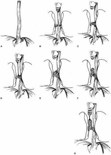
FIGURE 12-8 • Types of tracheal agenesis. A: Total pulmonary agenesis (8% of cases). B: Tracheal agenesis with main bronchi arising directly from the esophagus (10%). C: Tracheal agenesis with fused main bronchi and bronchoesophageal fistula (56%). D: Tracheal agenesis with the larynx joined by atretic strand to distal trachea, which has a fistulous connection with the esophagus (10%). E: Upper tracheal agenesis with large direct tracheoesophageal communication (5%). F: Tracheal agenesis with no communication with the esophagus (5%). G: Short-segment tracheal agenesis (5%).


TRACHEA


Tracheal Agenesis

Tracheal agenesis is a rarely occurring, uniformly fatal malformation that is usually associated with tracheoesophageal or bronchoesophageal fistula. Various classifications divide the entity into three to seven types (Figure 12-8A to G);
however, nearly 70% of cases consist of agenesis of the entire trachea with a small fistulous connection between the esophagus and the carina (Figure 12-8C to E) (23,24). The lungs may be normally developed or totally absent (pulmonary agenesis). In the rare cases of tracheal agenesis with no fistulous connection to the esophagus (i.e., total sequestration of the lungs), the lungs are uniformly distended, histologically resembling extralobar sequestration (25). There is a male predominance of approximately 2:1 and an association with maternal polyhydramnios in tracheal agenesis. In addition to the anomalies of the VATER association, tracheal agenesis has been seen in association with duodenal atresia, annular pancreas, syndactyly, and CNS malformations. Evans et al. (24) describe four groups based on the type of anomalies associated with the tracheal agenesis: group 1, anomalies restricted to the trachea, larynx, and cardiovascular system; group 2, severe cardiovascular anomalies and abnormal lung lobulation; group 3, a caudal component in addition to thoracic abnormalities, with anal and renal anomalies being common; and group 4, multisystem involvement with a high incidence of aberrant vessels, complex cardiac malformations, lung lobation defects, and anomalies of other foregut derivatives.


Tracheal Bronchus

This rare anomaly, also known as bronchus suis, is defined by a bronchus to the right upper lobe arising at the junction of the mid and distal one-third of the right lateral trachea (26).


Tracheal Stenosis

Laryngeal or tracheal stenosis is usually seen as an acquired lesion related to intubation or to the presence of a foreign body; congenital stenosis of the trachea is rare (27). Congenital stenosis may be diffuse, funnel-like, or segmental. Diffuse, generalized hypoplasia accounts for about 30% of cases, funnel-shaped or “carrot-shaped” stenosis for 20%, and segmental stenosis for the remaining 50%. Segmental stenosis may be due to complete tracheal cartilage rings, “napkin-ring” stenosis, or too small but normally shaped rings with a narrow pars membranosa (Figure 12-9) (28). Associated anomalies include anomalous bronchi, TEF, unilateral pulmonary agenesis, Crouzon syndrome, Larsen syndrome, Down syndrome, Alagille syndrome, and ventricular septal defect.

Tracheal stenosis, or narrowing, may also be produced by extrinsic pressure, most commonly by abnormally placed or abnormally large blood vessels including


Vascular ring due to double aortic arch

Vascular ring due to right aortic arch

Aberrant right subclavian artery

Anomalous innominate artery

Anomalous left carotid artery—aneurysmal left and right pulmonary arteries

“Sling” retrotracheal left pulmonary artery


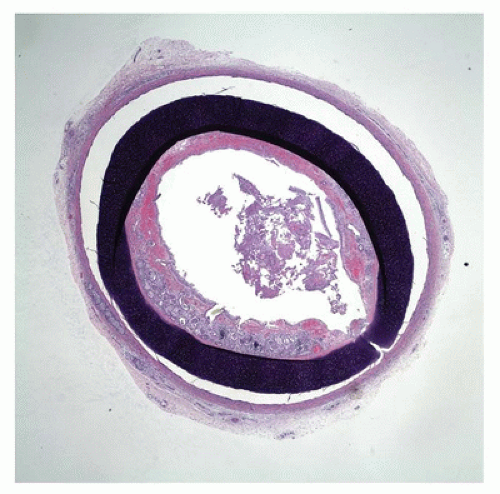
FIGURE 12-9 • Tracheal stenosis. A cross section from the mid trachea shows a complete cartilage ring beneath the mucosa, significantly narrowing the tracheal lumen.

Advances in surgical management of congenital tracheal stenosis have improved survival, especially since the advent of extracorporeal membrane oxygenation (ECMO) (29).


Tracheomalacia

Congenital tracheomalacia (i.e., soft or collapsing trachea) is exceedingly rare and overlaps with tracheal stenosis secondary to cartilage plate deficiency (30). Isolated cases have, however, been reported in association with Down syndrome, EA, CHARGE association, Larsen syndrome, pulmonary vascular sling, polychondritis, and various chondrodystrophies including Ellis-van Creveld syndrome, Langer-type mesomelic dwarfism, and diastrophic dysplasia. Aortopexy has been successfully employed in the treatment of tracheomalacia in infants. Acquired tracheomalacia may be seen in infants and young children who have been intubated for prolonged periods or as a result of trauma, radiation, or a neoplasm (31).


Tracheobronchiomegaly

Tracheobronchiomegaly, or the Mounier-Kuhn syndrome, usually involves men 20 to 40 years of age but has been reported in children of both sexes and has a familial occurrence, suggesting an autosomal recessive type of inheritance (32). The tracheal diameter exceeds the normal by three standard deviations. Saccular bulging of the intercartilaginous membranes is frequent. The disorder has been noted in a child with cutis laxa and in adults with Ehlers-Danlos syndrome.


Tracheoesophageal Fistula and Esophageal Atresia

EA, with or without TEF, occurs sporadically with an incidence of 1 in 3500 live births (33). Maternal polyhydramnios
is present in more than 30% of cases, and nearly 35% of the infants are premature. The anomaly can be divided into five (or more) types (Figure 12-10A to E). More than 95% of the patients have EA with the clinical findings of excessive oral and pharyngeal secretions or choking, cyanosis, or coughing during first attempts at feeding. While 98% are sporadic, the remaining have separate genetic factors (34).


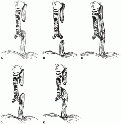
FIGURE 12-10 • Types of tracheoesophageal fistula (TEF) and esophageal atresia (EA). A: EA with TEF to the distal esophageal segment (>85% of cases in various series). B: EA without TEF (8%). C: TEF without EA (4%). D: EA with TEF to the proximal esophageal segment (1%). E: EA with TEF to both proximal and distal esophageal segments (1%).

TEF and EA can be most easily demonstrated at autopsy by removing the esophagus and trachea en bloc (see Chapter 1A, Chapter 1B, Chapter 1C, Chapter 1D) and then opening the esophagus lengthwise along its posterior or dorsal margin. EA is readily apparent as a blind pouch (Figure 12-11A, B), but a small fistula between the anterior or ventral portion of the esophagus and the trachea can also be visualized, as can the rare esophagobronchial fistula. Histologically, squamous metaplasia of the trachea and bronchi may be seen in 80% of patients, primarily along the posterior wall of the trachea but frequently extending around the entire internal surface of the trachea and into the bronchi. The segment of esophagus may show tracheobronchial remnants in the form of abnormal mucous glands and ducts, abnormal mucin secretion, the presence of cartilage, and a disorganized muscle coat (31). Aspiration of gastric contents may be present in the lung, producing pneumonia with foreign body giant cell reaction.

Associated anomalies are seen in 49% to 72% of infants with EA and TEF, with multiple anomalies frequently present (Table 12-2) (35). A nonrandom association of TEF with other malformations has been recognized in about 45% of cases and given the acronyms of VATER, VACTER, or VACTERL (vertebral, anal, cardiac, tracheoesophageal, renal, or radial and limb anomalies) (33). Other less frequently associated anomalies include congenital pulmonary airway malformation (CPAM), diaphragmatic hernia, duodenal atresia, biliary atresia, sirenomelia, trisomy 18,
and intracardiac epithelial cyst. TEFs may develop in burn patients, with foreign body impaction such as a disc battery (36), and following radiation and chemotherapy for mediastinal malignancies, including lymphoma (37).


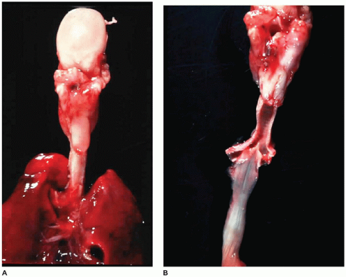
FIGURE 12-11 • TEF and EA. A: In a posterior view of the tongue (top), trachea, and lung, the esophagus is seen to end in a blind pouch (center). B: With the trachea and esophagus open posteriorly, a fistula can be seen connecting the carina with the distal end of the esophagus.

Postsurgical survival of patients with EA and TEF has increased steadily over the last 50 years, presently ranging from 75% to over 90% (38). The highest mortality rate occurs in infants with low birth weight or with coexisting cardiac malformations. TEF may recur after surgical repair in nearly 10% of cases (39). Tracheal narrowing may persist for years in nearly one-third of the patients, along with respiratory infections and gastroesophageal reflux. Histologically, esophageal inflammation may be seen in 51% of cases, Barrett esophagus in 6%, and Helicobacter pylori infection in 21% of cases (40). An increased incidence of esophageal adenocarcinoma in adulthood in patients with TEF has been suggested (41).


TABLE 12-2 ANOMALIES ASSOCIATED WITH ESOPHAGEAL ATRESIA AND TRACHEOESOPHAGEAL FISTULA


Organ System


Incidence (%)


Most Frequent Examples


Musculoskeletal


15-24


Vertebral defects, rib defects, radial amelia, caudal dysgenesis


Cardiovascular


11-49


Ventricular septal defect, patent ductus arteriosus, right aortic arch


Gastrointestinal


20


Imperforate anus, malrotation, duodenal atresia


Genitourinary agenesis


12-50


Renal malposition, renal cysts or hypospadias, horseshoe kidney


Craniofacial


10


Choanal stenosis, ear malformations, micrognathia


Central nervous system


7


Hydrocephalus


Pulmonary


2


CPAM, pulmonary hypoplasia


Adapted from Stocker JT. Congenital and developmental diseases. In: Dail DH, Hammer SP, eds. Pulmonary Pathology. 2nd ed. Heidelberg, Germany: Springer-Verlag, 1994:163, with permission.


BRONCHUS


Bronchial Atresia

Bronchial atresia is an entity seen almost exclusively in infants and is thought by some to be the underlying cause in the entire spectrum of parenchymal malformations from infantile (congenital) lobar emphysema (ILE), CPAMs to sequestration in almost 80% of cases (38). Cases of bronchial atresia with mild emphysema, however, have been reported in children from 1 day to 13 years of age (median, 4 years) with symptoms of chronic cough and fever in nearly all of the cases, often related to the recurrent pneumonia noted in more than 90% of cases (42). The atretic bronchus is connected to the right lower lobe, left upper lobe, and right upper lobe in decreasing order of frequency. Histologically, the affected bronchus may be obstructed by circumferential or eccentric luminal fibrosis with or without abnormalities of the cartilage plates and filled with mucin to create a “mucocele.” The fibrosis may be the result of in utero inflammation in the neonate or possibly postpartum inflammation in the case of children and adults.

Bronchial stenosis may also be associated with ILO. The lumen of the bronchus may be intrinsically narrowed by postinflammatory fibrosis or by an intraluminal mass such as aspirated meconium or other foreign material, bronchial adenoma, ectopic thyroid tissue, or bronchial mucosal web. Extrinsic causes of bronchial stenosis include parabronchial masses such as teratoma and bronchogenic cyst, enlarged or abnormally located pulmonary arteries, and cardiac or left atrial enlargement. Bronchial stenosis has also been associated with EA and TEF. More recently, studies have also suggested that bronchial stenosis and/or atresia are common features of CPAM, extralobar sequestration, intralobar sequestration (ILS), EA, and TEF (38,43). It is often difficult to demonstrate bronchial atresia and/or stenosis unless a dissecting microscope is used and the lesion is looked for carefully. The presence of an intrabronchial mucocele is helpful.


Bronchomalacia

Bronchomalacia and tracheobronchomalacia are seen most frequently in premature infants treated for prolonged periods with mechanical ventilation (39). Congenital bronchomalacia is rare and occurs when there is abnormal development of bronchial cartilage, leading to collapse of the lumen and possible development of secondary pneumonia. Bronchomalacia has also been suggested as a cause of sudden death in infancy and early childhood. Deficiency of subsegmental bronchial cartilage with bronchial collapse is also a feature of Williams-Campbell syndrome and has been noted in children with Larsen syndrome. Children with Down syndrome have a high incidence (up to 50%) of laryngomalacia, tracheomalacia, and bronchomalacia (44).

Histologically, the affected bronchus is decreased in size, with the usual cartilage plates replaced by scattered small islands of immature-appearing cartilage. The lung distal to the collapsed bronchus may show pneumonia or is distended in a pattern typical of ILO. Bronchial stents are used in the treatment of this abnormality but have been associated with complications including an aortobronchial fistula (39).


Bronchial Isomerism Syndromes

Bronchial isomerism results in “mirror-image” lungs (i.e., bilateral right or left lung) and is associated with five types of “polysplenia/asplenia” or heterotaxy syndromes (36).

Type 1, Ivemark asplenia syndrome, is a nonfamilial malformation complex involving bilateral right-sidedness, including absence of the spleen, intestinal malrotation, symmetric liver, and bilateral three-lobed “right” lungs with bronchi for both lungs. A variety of cardiac malformations are also associated with this type, including right aortic arch, symmetric venae cavae, transposition of the great vessels, and total anomalous pulmonary venous return.

Type 2, M-anisosplenia, involves boys who have one or more larger and one or more smaller spleens, along with congenital heart malformations, bilateral three-lobed “right” lungs, and relatively normal visceral situs.

Type 3, the polysplenia syndrome, is characterized by a bilateral two-lobed “left” lung bronchial pattern with intestinal malrotation, symmetric liver, congenital heart malformations, and 4 to 14 uniform small spleens.

Type 4, F-anisosplenia, involves females who have bilateral two-lobed “left” lungs, congenital heart malformation (usually double outlet right ventricle), and anisosplenia.

Type 5, O-anisosplenia, is characterized by bilateral two-lobed “left” lungs, an approximately 50% incidence of intestinal malrotation, multiple spleens, an equal sex ratio, and congenital heart malformations, particularly double-outlet right ventricle, ostium atrioventriculare commune, or both (see Chapter 13).


Abnormal Bronchial Branching and Origin

Abnormal branching patterns, mostly minor anomalies such as double stem superior segments of lower lobe bronchi and trifurcation of the left upper lobe bronchus, are seen in nearly 10% of bronchograms (45). However, major anomalies are also seen, including double right lobe bronchus, accessory cardiac bronchus, tracheal origin of the right upper lobe bronchus (also called preeparterial bronchus), and bridging bronchus (46).

McLaughlin et al. (47) noted a tracheal origin of a bronchus in 2% of 412 symptomatic patients younger than 5 years of age who were undergoing bronchoscopy. The various forms of tracheal bronchus (Figure 12-12) may lead to recurrent episodes of pneumonia requiring resection of the bronchus and lobe. Other anomalies are noted in more than 75% of patients with tracheal bronchus. Wells et al. (48) suggest that in patients with sling left pulmonary artery, the tracheal bronchus often associated with the right upper lobe may represent the “normal” origin of the bronchus,
and the bronchi supplying the right middle and lower lobes are branches of the left main bronchus that are crossing or “bridging” the mediastinum (Figure 12-13). They note that the origin of the tracheal bronchus is at the normal level of tracheal bifurcation (T4-5), and the bifurcation of the bronchi supplying the left lung and right middle and lower lobes is at the T6-7 level.


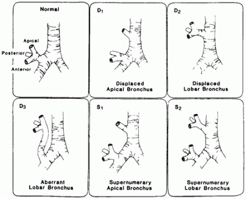
FIGURE 12-12 • Anatomic variations of right upper lobe bronchus. (From McLaughlin FJ, Strieder DJ, Harris GB, et al. Tracheal bronchus: association with respiratory morbidity in childhood. J Pediatr 1985;106:751, with permission.)


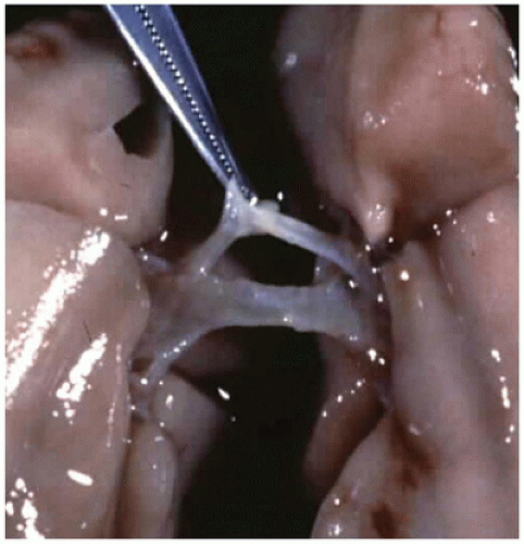
FIGURE 12-13 • A bronchus “bridges” the mediastinum in the case of tracheal agenesis.


Bronchobiliary and Bronchoesophageal Fistulae

Bronchoesophageal fistula probably represents a variation of TEF but may also be seen with infectious diseases such as tuberculosis and has been reported in association with Crohn disease.

Congenital bronchobiliary fistula rarely occurs; however, when it does, it is usually located between the right mainstem bronchus and the left hepatic duct (49). The bronchobiliary fistula is thought to represent a duplication of the upper gastrointestinal tract from its junction with the airway to the level of the ampulla of Vater. The fistula arises from the proximal portion of the right main bronchus, accompanies the esophagus through the diaphragm, and joins the biliary tree at the left hepatic duct. In its proximal portion, the tract resembles a bronchus with cartilage rings and respiratory epithelium, and in its distal portion, the tract resembles a bile duct or esophagus. Bronchobiliary fistulas may be seen in older children and adults and has been described secondary to biliary obstruction, liver infections (such as echinococcosis), and liver tumors (such as undifferentiated embryonal sarcoma).


Bronchogenic Cyst

The bronchogenic cyst is a discrete, extrapulmonary fluid-filled mass. It is most frequent in the hilar or middle-mediastinal area but may be present in a midline location from the subcutaneous region of the suprasternal area to beneath the diaphragm (1,50,51). They need to be differentiated from esophageal and enteric duplication cysts and pericardial cysts that may also be present in the mediastinal region. Bronchogenic cysts are rarely connected to the tracheobronchial tree or involve the pulmonary parenchyma. Case
reports of “intrapulmonary bronchogenic cysts” probably represent instances of type 1 CPAM (52).


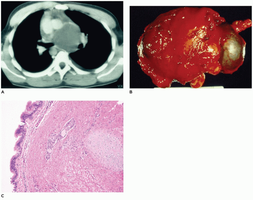
FIGURE 12-14 • Bronchogenic cyst. A: A CT of the chest displays a large mass in the middle mediastinum. B: A resected bronchogenic cyst, which was separate from the lung, is covered by connective tissue. C: Ciliated pseudostratified columnar epithelium overlies a wall composed of fibrous connective tissue, glands, and a cartilage plate in a bronchogenic cyst.

Bronchogenic cysts are seen most frequently in children and young adults as incidental findings on chest radiographs, at surgery, or at autopsy, but they may present with symptoms related to secondary infection of the cyst, including fever, hemorrhage, or perforation. In infants, bronchogenic cysts located near the trachea, especially the carina, may produce obstruction and respiratory distress.

In infants, the gross appearance of the cysts consists of a 1- to 4-cm, smooth-to-irregular, spheroid mass attached to, but not in communication with, the tracheobronchial tree (Figure 12-14A to C). The cysts may contain clear serous fluid, but if they are infected, the fluid may be turbid or hemorrhagic. In older patients, the cysts may reach a diameter of 8 to 10 cm and may be found throughout the mediastinum as well as in or beneath the diaphragm. Extrathoracic cysts are usually confined to the subcutaneous region in the suprasternal area (53).

Microscopically, the lining of the cyst is composed of ciliated cuboidal to pseudostratified columnar epithelium. Cartilage plates and seromucous glands are present in the wall, as is fibromuscular connective tissue (Figure 12-14C). The presence of striated muscle and stratified squamous or columnar epithelium suggests an esophageal cyst (Figure 12-15). Enteric cysts are lined by mucus-secreting columnar epithelium and contain gastric glands with parietal cells in the wall. All three types of cysts may display squamous metaplasia, mucosal ulceration, inflammation, extensive necrosis, or a combination of these, making an exact diagnosis difficult. Pericardial cysts are lined by mesothelium.

Bronchogenic cysts have been noted between the sequestration and the midline in association with extralobar sequestrations in older children. This suggests that the cysts have arisen from “rests” of bronchogenic cells along the abortive foregut tract that gave rise to the sequestration (54).


Plastic Bronchitis

Children with cardiac defects, especially in those requiring a Fontan procedure (1% to 2% of cases) or an underlying pulmonary disease (asthma or allergic disease), may develop obstructive bronchial cast (55,56). There are two types of casts: type I, cellular casts made up of inflammatory cells with fibrin, and type II, acellular casts composed mainly
of mucin. Other underlying causes include cystic fibrosis (CF), neoplasia, alpha-thalassemia, beta-thalassemia, and acute chest syndrome of sickle cell disease. Secretory hyperresponsiveness with excess mucin secretion is regarded as the basis for the disorder (57).


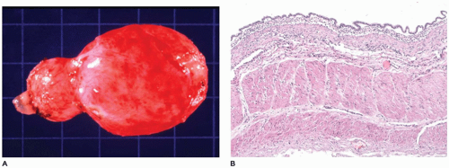
FIGURE 12-15 • Esophageal cyst. A: A cystic structure was resected from the middle mediastinum adjacent to the esophagus. B: Columnar epithelium overlies a wall composed of thick muscular bands in this esophageal cyst from the mediastinum.

Grossly, the cast is a partial or complete mold of the bronchial tree with either tube-like features or partially or completely solid cores. Those composed of acellular mucin may be partially clear to opaque. Microscopically, the cast has a mucofibrinous appearance with or without inflammatory cells and cellular debris. In the case of hypersensitivity airway disease or asthma, the cast contains eosinophils and Charcot-Leyden crystals.


LUNG


Pulmonary Agenesis

Complete absence of both lungs is extremely unusual and incompatible with life. However, unilateral agenesis, involving one or more lobes, has been seen in 1 in 10,000 to 20,000 autopsies and, in the absence of other severe anomalies, is compatible with long-term survival (58). There is a 1.3:1 female predominance with unilateral agenesis; the right and left lungs are absent with equal frequency. Associated anomalies are noted in about 75% of cases and include, in decreasing order of frequency, cardiovascular, gastrointestinal, skeletal, and urogenital systems (59). Cardiovascular malformations include dextrocardia, septal defects, patent ductus arteriosus, and total anomalous pulmonary venous return. Skeletal anomalies include hemivertebrae and a high frequency of thumb malformations, especially triphalangeal thumb (Mardini-Nyhan association) (60). Along with the radial and vertebral anomalies, imperforate anus and TEF have been described, suggesting an association of pulmonary agenesis with the VATER or VACTERL association (61). There are also some phenotypic similarities to the syndrome associated with inactivation of ERK/MAP kinase genes MEK1 and MEK2 (62).

The larynx and upper trachea are usually well-formed in unilateral pulmonary agenesis, although with bilateral agenesis the total trachea may be absent. The lower trachea in unilateral agenesis may continue directly into the existing lung as a tracheobronchus or bifurcate at the carina, giving rise to a rudimentary blind-ending bronchus on the side of the agenesis. The pulmonary artery and vein to the side of the agenesis are absent or hypoplastic and may have an unusual course to the lung, often forming a pulmonary sling (63). Shift of the mediastinum to the side of the agenesis is usually present, often giving the appearance of dextrocardia in right-sided agenesis. Studies in older infants have demonstrated an absolute increase in the number of alveoli in the existing lung despite a reduced number of bronchial generations and pulmonary artery branches.


Abnormal Lobation, Location, and Shape

Abnormalities of lobation of the lung are usually of little clinical significance unless they are associated with other anomalies such as the asplenia or polysplenia syndrome (discussed earlier in chapter). Lobes may be fused to give the appearance of a single lobe on the right or left, or pleural fissures may produce the appearance of multiple lobes. The appearance of multiple lobes may be seen in infants with long-standing healed bronchopulmonary dysplasia (64). Fusion of the lungs in the midline behind the heart produces a conjoined, or “horseshoe,” lung analogous to the horseshoe kidney. Additional anomalies are often present in association with horseshoe lung, including those of the VATER association and the PAGOD syndrome (pulmonary hypoplasia, agonadism, omphalocele, dextrocardia,
and diaphragmatic defect), among others (65). Bronchial supply to both lungs may be anatomically normal, but there is usually an anomalous pulmonary artery supply and venous drainage resembling that seen in scimitar syndrome.

Herniation of the lung across the mediastinum into the opposite hemithorax, associated with ILO, CPAM, extralobar sequestration, and other conditions, occurs relatively frequently (1). The lung can also herniate outside the thoracic cavity, usually into the neck. Cervical herniation or “protrusion” is most frequently reported as a “normal variant” in some infants and children. It may also be seen, however, as a result of trauma or surgery, and in association with iniencephalus, the Klippel-Feil syndrome, and the cri du chat syndrome. A familial occurrence has been noted, and the condition is thought to be an autosomal dominant hereditary disease. Herniation through the diaphragm and intercostal spaces may also occur.


Pulmonary Sequestration

Sequestration is defined by a segment of lung parenchyma with its own systemic arterial blood supply independent of the normal lung. There are two basic types of sequestrations: extralobar (ELS) and intralobar (ILS) (1).


Extralobar Sequestration

Extralobar sequestrations of the lung are distinctly different from ILSs in that they are discrete masses of pulmonary parenchyma outside the normal pleural investment of the lung and are not connected to the tracheobronchial tree (25). They apparently originate from an outpouching of the foregut, and separate from the normally developing lung (Figure 12-16A). This outpouching then loses its connection with the foregut, isolating the parenchyma from the tracheobronchial tree (54).

Extralobar sequestrations are diagnosed prenatally in about 25% of cases, and about 60% of these patients present by 3 months of age (67). Presenting symptoms, often noted on the first day of life, include cyanosis, dyspnea, and difficulty in feeding. Approximately 10% of patients are asymptomatic. Fetal nonimmune hydrops, anasarca, pleural effusion, or localized edema may be present along with maternal polyhydramnios. Extralobar sequestrations may be seen in older children, occasionally in association with a bronchogenic cyst, and they have been reported in adults as old as 81 years of age. There is a slight female predominance.


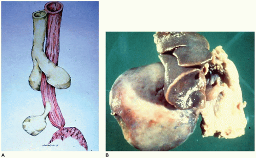
FIGURE 12-16 • Extralobar sequestration. A: The normal lung develops as an evagination from the foregut (top half). A second evagination (bottom) from the foregut gives rise to lung tissue not attached to the normally developing lung. B: A large right-sided thoracic mass is attached to the mediastinum by a thin vascular pedicle. Note the hypoplasia of the right lung.


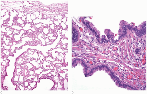
FIGURE 12-16 • (Continued) C: The pulmonary parenchyma is uniformly dilated from the bronchioles to the most distal alveoli. D: Back-to-back bronchiole-like structures typical of CPAM type 2 are seen in 50% of extralobar sequestrations. Note also the rhabdomyomatous dysplasia.

Associated anomalies are present in more than 65% of cases of extralobar sequestration, with 50% of lesions containing CPAM type 2 within the sequestration or, less frequently, in a lobe of the “normal” lung. The ELS/CPAM cases are seen more frequently in the first 3 months of life and on the left side (67). Other anomalies include bronchogenic cyst, cardiovascular malformations, bronchopulmonary foregut connection, pectus excavatum, absence of pericardium, and diaphragmatic hernia with concomitant pulmonary hypoplasia. High levels of CA19-19 have been reported in a few cases of extralobar sequestration.

Extralobar sequestration is usually a single round to ovoid lesion ranging from 0.5 to 15 cm in diameter (Figure 12-16B). In a report of 50 cases, 48% of the lesions were located in the left hemithorax, 20% in the right hemithorax, 8% in the anterior mediastinum, 6% in the posterior mediastinum, and 18% beneath the diaphragm (67). The blood supply to the extralobar sequestration is through a direct branch of the thoracic or abdominal aorta in over 75% of cases. The remaining receive their blood supply from smaller systemic arteries, the pulmonary artery, or rarely, from a systemic artery (25). Venous drainage is through the systemic circulation in over 80% of cases; the remaining 20% of cases are drained either partially or completely by the pulmonary veins, or, rarely, by the portal vein (68).

Grossly, the lesion is covered by a smooth to wrinkled pleura overlying a fine, reticular network of lymphatics. These lymphatics may be prominent in 30% or more of cases. Cut sections of the lesion display homogenous, pink-to-tan tissue resembling normal pulmonary parenchyma, or clusters of small cysts. Prominent subpleural lymphatics may also be seen.

Microscopically, extralobar sequestrations consist of uniformly dilated bronchioles, alveolar ducts, and alveoli in a normal acinar pattern (Figure 12-16C). Bronchioles are usually tortuous with undulating cuboidal to columnar epithelium. In 50% of cases, the lesion may consist partially or entirely of back-to-back, dilated, bronchiole-like structures typical of CPAM type 2 (Figure 12-16D). Lymphatics are unremarkable in the majority of cases but may be dilated and increased in number beneath the pleura and around bronchovascular bundles, occasionally resembling congenital pulmonary lymphangiectasia (CPL) (Figure 12-16C). Although they are rare, infarction, arteritis, and inflammation may be present in an extralobar sequestration. In the absence of severe anomalies, survival is good, although with large intrathoracic lesions, the associated pulmonary hypoplasia may be severe enough to cause death. Rhabdomyomatous dysplasia is seen in 25% to 30% of cases (Figure 12-16D).


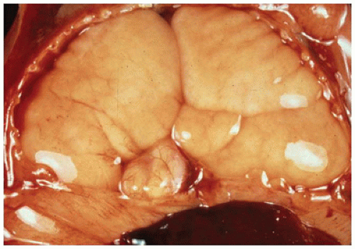
FIGURE 12-17 • Hyperplastic lungs in the case of laryngeal atresia are massively enlarged, displaying the markings of the ribs on their surface.


“Total” Sequestration with Pulmonary Hyperplasia

Infants with laryngeal or tracheal atresia without TEF have, in effect, “total” sequestration of the lungs and display a histologic appearance virtually identical with that seen in extralobar sequestration. The lungs are often two to four times the expected weight and crowd the chest cavity, flattening the diaphragm and leaving an impression of the ribs on the visceral pleural surface (Figure 12-17) (25). Similar pulmonary changes may be seen with in utero hyperextension of the neck that appears to compress and obstruct the larynx. Scurry et al. (69) have noted normal sized or hyperplastic lungs in infants with varying degrees of upper airway obstruction despite the presence of renal dysgenesis and oligohydramnios, conditions more frequently associated with pulmonary hypoplasia. Lymphatics are unremarkable. Atresia or obstruction of a single bronchus to a lobe may lead to hyperplasia of that lobe and the development of one form of ILE (see below) (66).


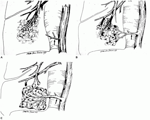
FIGURE 12-18 • Sequence of events in the formation of intralobar pulmonary sequestration. A: Occlusion of a bronchial branch by means such as aspirated material or inflammatory debris can lead to the development of pneumonia distal to the occlusion. B: As the pneumonia persists or progresses, the lung seeks oxygenated blood to aid in resolution and repair. If pulmonary artery flow is inadequate, systemic blood supplying pleural granulation tissue through the pulmonary ligament arteries may be “parasitized.” C: As the pneumonia resolves (or progresses or recurs), the major arterial supply to the sequestered portion of lung is derived from the hypertrophied pulmonary ligament artery (or arteries). (From Stocker JT, Malczak HT. A study of pulmonary ligament arteries: relationship to intralobar pulmonary sequestration. Chest 1984;86:611, with permission.)


Intralobar sequestration

Intralobar sequestration (ILS), by definition, consists of a portion of lung within the normal pleural investment that is isolated (sequestered) from the tracheobronchial tree and is supplied by a systemic artery (Figure 12-18) (25,70). Although a small percentage of ILSs are clearly congenital in origin and might more correctly be called arteriovenous malformations (71), the vast majority of ILSs are probably acquired lesions formed through
repeated episodes of pneumonia. During the course of these episodes, normal pulmonary ligament arteries become hypertrophic to provide the systemic artery supply (Figure 12-18A to C) (70,72). Some examples of ILS may develop within a previously existing malformation (e.g., CPAM). The following evidence suggests the acquired nature of ILS (66,73):


ILS is rarely seen in the newborn (<15 cases described in children younger than 5 years of age).

ILS is infrequently associated with other congenital malformations.

ILS is limited to the lower lobes in 98% of cases, allowing access to normally occurring pulmonary ligament arteries.

ILS-affected patients have a frequent history of repeated pulmonary infections.

ILS presents with a clinical picture of chronic or recurrent pneumonia (e.g., cough, sputum production) in over 85% of cases.

ILSs involve the lower lobe in 98% of cases with this probably reflecting the availability, within pleural granulation tissues, of branches of normally occurring pulmonary ligament arteries or arteries within the diaphragm that are parasitized for access to oxygen-rich systemic blood. The pulmonary ligament arteries originate from the thoracic aorta and extend through the pulmonary ligament between the mediastinum and the lower lobes of the lung (70). No comparable arteries except the bronchial arteries are present for potential use by the upper lobes in cases of chronic or recurrent pneumonia.

Radiographic findings include cystic areas, some with fluid levels, along with homogeneous and inhomogeneous shadows (74). Lack of communication with the tracheobronchial tree is demonstrable by bronchography in about 85% of cases; the other 15% of cases show some communication between the bronchial tree and the sequestration. Arteriography demonstrates single (84%) or multiple (10%) systemic arteries (Figure 12-19A, B). The majority of the arteries (73%) originate from the thoracic aorta, but about 21% originate from the abdominal aorta or celiac axis and another 4% from the intercostal arteries. In rare instances, arteries may originate from the coronary, subclavian, innominate, internal thoracic, or pericardiophrenic arteries. Venous drainage occurs through the pulmonary veins in 95% of cases, and the remaining 5% of cases drain into the systemic circulation. Increased serum levels of CA19-19 and CA125 have been noted in patients with ILS (75).

ILS is located on the left side in 55% of cases and on the right in 45% of cases; bilateral involvement is rare. Grossly, the sequestered segment of lung displays variable pleural thickening with adhesions between mediastinal structures, the diaphragm, and the parietal pleura. Variably sized (1 mm to 5.0 cm) cysts filled with thin to viscid fluid are noted amid a dense fibrous parenchyma on cut section (Figure 12-19).
Microscopically, the pulmonary parenchyma is distorted by chronic inflammation and fibrosis (Figure 12-19D). The cysts are lined with cuboidal or columnar epithelium and are filled with amorphous eosinophilic material, foamy macrophages, or both. Elastic and muscular arteries are present within the interstitium and may show medial hypertrophy, thrombosis, and arteritis (76).


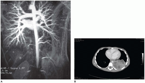
FIGURE 12-19 • Intralobar sequestration. A: An arteriogram demonstrates arteries arising from the descending aorta (mid right) supplying a portion of pulmonary parenchyma. B: A CT demonstrates a mass in the posterior area of the right hemithorax.


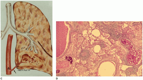
FIGURE 12-19 • (Continued) C: An artery arising from the descending aorta and passing through the pulmonary ligament supplies a cystic portion of the lung in the left lower lobe. D: Dense fibrous connective tissue containing lymphoid aggregates surrounds irregular cysts filled with debris and macrophages.


Pulmonary Hypoplasia

Pulmonary hypoplasia is the incomplete or defective development of the lung resulting in overall reduced size due to reduced numbers or size of acini (Figure 12-20A). Lung weight and lung weight-to-body weight ratio are the simplest means of determining whether hypoplasia exists. The normal lung weight-to-body weight ratio for term and near-term infants is 0.022 (range 0.012 to 0.025) (77). Emery and Mithal (78) describe a radial alveolar count using a line intersect method in which a line is drawn from a terminal bronchiole perpendicular to the nearest septal division or pleura surface (Figure 12-20B). The number of alveoli intersected by the line determines the count with the mean for term infants of 4.4 ± 0.9 (79). Alveolar counting and lung volume measurements may also be used to define hypoplasia (80). Two-dimensional or three-dimensional ultrasound and MRI have also been used in determining whether the lungs of an in utero fetus or newborn infant may be hypoplastic. However, secondary changes such as bronchopneumonia or pulmonary hemorrhage which increase the weight of the lungs create difficulties in the assessment for the presence of hypoplasia on basis of lung weight alone. One simple but less precise observation is the distance of a terminal bronchiole from the pleural surface.

Pulmonary hypoplasia is noted in more than 10% of neonatal autopsies and occurs in association with another malformation (or malformations) in more than 85% of cases (81). The most frequently occurring anomalies are diaphragmatic defects and renal malformations (Table 12-3), but a wide variety of anomalies have been described (1). The common feature of most of these anomalies is that they directly or indirectly compromise the thoracic space available for lung growth. The cause of the decreased thoracic space may be intrathoracic (e.g., abdominal contents herniated through a defect in the diaphragm) or extrathoracic (e.g., enlarged cystic kidneys). The thorax itself may be abnormal as in Jeune asphyxiating thoracic dystrophy, spondyloepiphyseal dysplasia congenita, and achondroplasia (82). In utero accumulation of fluid within the thorax as pleural effusion or chylothorax has also been implicated in the production of pulmonary hypoplasia.

Pulmonary hypoplasia may also occur in the absence of other anomalies or in cases of prolonged preterm premature rupture of amniotic membranes leading to oligohydramnios, loss of fetal cushioning, and lack of amniotic fluid pressure and growth factors which would normally be inhaled into

the lung (81). As with infants with hypoplasia secondary to other anomalies, these infants present with respiratory distress, are difficult to ventilate, and frequently have episodes of pneumothorax (PT) and interstitial pulmonary emphysema (IPE). Potter sequence with sloping forehead, flattened face and nose, receding chin, large ears, broad spade-like hands, and deformations of the limbs secondary to compression by the uterus in the absence of adequate amniotic fluid is a consistent finding in cases associated with oligohydramnios from any cause. Pulmonary hypoplasia has been noted in children with Down syndrome, but it is thought to result from failure of the lung to develop properly in the postnatal period (83).


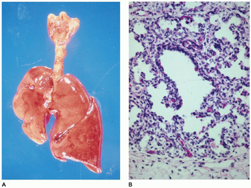
FIGURE 12-20 • Pulmonary hypoplasia. A: The right lung is markedly diminished in size, secondary to herniated abdominal organs through a right-sided diaphragmatic hernia. By weight, the left lung is also hypoplastic. B: At the periphery of an acinus in this hypoplastic lung, a radial alveolar count (RAC) is far below the normal of 4 to 6 for a term infant, confirming the diagnosis of hypoplasia.


TABLE 12-3 ANOMALIES ASSOCIATED WITH PULMONARY HYPOPLASIA


Common


Diaphragmatic hernia


Renal agenesis, bilateral


Renal dysgenesis, bilateral


Obstructive uropathy


Polycystic renal disease (autosomal recessive)


Large abdominal wall defects


Infrequent


Diaphragmatic hypoplasia or eventration


Anophthalmia/microphthalmia—usually in association with diaphragmatic hernia


Hemolytic disease of the newborn


Pleural effusion, as with nonimmune fetal hydrops


Musculoskeletal abnormalities, such as thoracic dystrophies


Anencephaly


Scimitar syndrome


Chromosomal anomalies, including trisomy 13, 18, and 21


Rare


Abdominal pregnancy


Ascites secondary to congenital cytomegalovirus infection


Cloacal dysgenesis


Congenital hydropericardium


Down syndrome (probably postnatal “hypoplasia”)


Eagle-Barrett syndrome


Giant cervical teratoma


Glutaric acidemia, type II


Homozygous β-thalassemia


Horseshoe lung


Hypoplasia of the arcuate nucleus


Laryngotracheoesophageal cleft


Neonatal hypophosphatasia


Pena-Shokeir syndrome, type I


Phrenic nerve agenesis


Right-sided cardiovascular malformation, as with hypoplastic right side of the heart and pulmonary valve or artery atresia


Rhabdomyoma in tuberous sclerosis


Thoracic neuroblastoma


Upper cervical spinal cord


Extralobar sequestration


At autopsy, the lungs may be either uniformly reduced in size or markedly asymmetric (e.g., with diaphragmatic hernia). In cases in which the pulmonary hypoplasia is the direct cause of death, the lung weight usually is less than 40% of expected and is often as low as 20% to 30%. Histologically, the acini are small for the infant’s gestational age, but alveolar and capillary development is usually consistent with the gestational age.


Infantile (Congenital) Lobar Emphysema (Overinflation)

ILO is the overinflation or hyperplasia of a pulmonary lobe as the result of a partial or complete obstruction of the bronchus to the lobe by intrinsic or extrinsic factors (66) (Table 12-4). More recently, it is being recognized that not all cases involve the whole lobe; rather a segment is involved, especially on the left side where the upper segments are involved, sparing the lingula. This is especially important to recognize, since these patients may be cured by segmentectomy (84). Boys are more frequently affected than girls (1.5:1). ILO presents in the first week of life in about 50% of cases (with about 40% presenting in the first day of life) and in the first 6 months of life in over 80%, but ILO can occasionally be seen in children and young adults from 7 months to 20 years of age. Symptoms are those of mild respiratory distress increasing over a period of hours to days to weeks; cyanosis, respiratory infections, vomiting, choking, and feeding difficulties may also be seen. Rarely, the lesion may present as a sudden PT (66). Imaging studies reveal, in the classic form (see below), a characteristic hyperlucent, overdistended lobe producing mediastinal shift and compression of the uninvolved lobes (85) (Figure 12-21A). In the polyalveolar lobe form (see below), imaging may display a lobe of normal lucency but one that occupies a disproportionate part of the hemithorax with mediastinal shift. Less frequently, retained lung fluid may be seen in the involved hyperexpanded lobe (usually the polyalveolar lobe type) on initial examination but which may clear over subsequent days. Associated anomalies are present in 5% to 40% of patients, and 70% of these anomalies are cardiovascular (86). The upper lobes are involved in over 95% of cases—the left slightly more often than the right. Multiple lobe involvement occurs in about 15% of cases, usually with at least one lobe being an upper lobe. Bilateral involvement has been reported in one case involving the left upper and right middle lobes. Lower lobe involvement is rarely seen except in the “acquired” form of ILO, as in premature infants receiving mechanical ventilation who develop granulation tissue obstruction of a lower lobe bronchus, probably as a result of endotracheal tube suctioning (87).


TABLE 12-4 CAUSES OF INFANTILE LOBAR OVEREXPANSION


Bronchial abnormality


Bronchial stenosis


Bronchial atresia


Abnormal origin of bronchus


Extrinsic obstruction of bronchus


Vascular anomaly


Pulmonary artery sling


Anomalous pulmonary venous return


Left-to-right shunting with dilated pulmonary arteries


External mass


Bronchogenic cyst


Intrinsic obstruction of bronchus


Aspirated meconium


Mucous plug


Granulation tissue


Bronchial mucosal folds


Torsion of bronchus


Foreign body


Adapted from Stocker JT. Congenital and developmental diseases. In: Dail DH, Hammer SP, eds. Pulmonary Pathology. Heidelberg, Germany: Springer-Verlag, 1989:55, with permission.


Grossly, the lobe in vivo and after resection is hyperexpanded with individual alveoli, which may be readily visualized (Figure 12-21B). Microscopically, two patterns (classic and polyalveolar) are identified. Nearly, 70% (the classic pattern) display a uniform overdistension of apparently normally developed acini with alveolar saccules and alveoli three to ten times the normal size but with radial alveolar counts (RAC) similar to those of age-matched controls (Figure 12-21C) (66). There may be focal disruption of alveolar walls. The other 30% (the polyalveolar pattern) show only little overdistension of what appear to be “complex” acini of the type seen in polyalveolar lobes and hyperplastic lungs (Figure 12-21D), and these have RACs that are two standard deviations beyond the mean of age-matched controls. Seventy-five percent of these infants with polyalveolar lobe present clinically within the first day or two of life and are likely to show radiologic features of retained lung fluid (88,89). Examination of the bronchus to the lobe may reveal stenosis, atresia, or intrinsic obstruction, or the bronchus may be unremarkable if extrinsic compression was present. Cartilage abnormalities of the bronchial wall have been described, but special techniques must be employed to demonstrate these changes convincingly.
Surgical resection of the involved lobe is curative, although nonsurgical management has been successful in unusual cases (85).


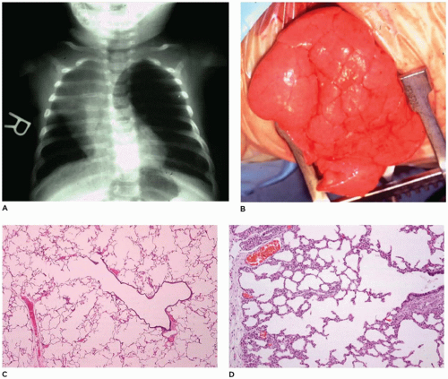
FIGURE 12-21 • Infantile lobar overinflation. A: A hyperinflated left lung shifts the mediastinum to the right. B: At surgery, the hyperinflated lung bulges from the opening in the thorax. C: “Classic” form of ILO. The alveolar duct and alveoli are dilated to 3 to 10 times the normal size but are otherwise unremarkable. D: “Hyperplastic” form of ILO. While not overinflated, this lung displays a complex acinar formation with a larger number of alveoli (and consequently a large radial alveolar count) than would be expected at this age.


Congenital Pulmonary Lymphangiectasis

CPL is a rare, usually fatal disorder that presents in the first hours to days of life (90); 5% to 10% of affected infants are stillborn. It is characterized by the presence of dilated thin-walled to thick-walled lymphatics within the interlobular septa and beneath the pleura of the lung. CPL may be seen as a primary disorder or as secondary to obstructive cardiovascular lesions, particularly total anomalous pulmonary venous return, but it may occur as part of a generalized lymphangiectasis or as an isolated pulmonary lesion (91). There is a distinct male predominance of over 2.5:1. Symptoms include cyanosis and acute respiratory distress. Fluid abnormalities including chylothorax, pleural effusion, fetal hydrops, and maternal polyhydramnios have been described in utero and postpartum (92). In addition to the 60% of cases with cardiovascular anomalies, CPL is associated with renal malformations, generalized lymphangiectasis, and other anomalies, in another 20% of cases (1). The diagnosis of CPL in the absence of cardiovascular or other anomalies, should be strongly suspect.

The lungs in CPL are bulky, firm, noncompressible, and covered by a milky network of dilated subpleural lymphatics (Figure 12-22A). Rarely, a single lobe is involved by this process (93). On cut section, the lymphatics are fluid-filled and extend from the interconnecting subpleural network into the interlobular septa and around the bronchovascular bundles (Figure 12-22B). Microscopically, the lymphatics are diffusely
and uniformly dilated and may appear to be increased in number. Identification of lymphatics can be aided by CD31 and D2-40 immunohistochemistry and can help differentiate the lesion from IPE. These small, irregular cysts are lined with a thin layer of endothelial cells and surrounded by a loose myxoid to occasionally dense connective tissue that often contains foci of extramedullary hematopoiesis. Clusters of lymphatics surround bronchovascular bundles within the interlobular septa and may separate acini beneath the pleura (Figure 12-22C, D). This is in contrast to the air-filled, larger, “unlined” cysts of IPE that are limited to the interlobular septa and do not extend laterally beneath the pleura.


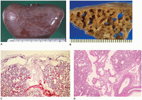
FIGURE 12-22 • Congenital pulmonary lymphangiectasis. A: A fine network of dilated lymphatics is present beneath the pleura, most notably where interlobular septa intersect the pleura. B: Cut section of the lung from an infant total anomalous pulmonary venous return reveals enormously dilated lymphatics. C: Dilated lymphatics extend laterally beneath the pleura (top) and centrally along an interlobular septum (center). Note the slight increase in connective tissue between the channels. D: Numerous dilated lymphatics extend along interlobular septa surrounding bronchovascular bundles.


Congenital Pulmonary Airway Malformation

CPAM is a developmental anomaly of the lung, with an incidence of about 1 in 5000 live births, that can be separated into four major types based on clinical and pathologic features (Figure 12-23) (94). The former designation “CCAM” was changed to “CPAM” to reflect the fact that the lesions as described below are “cystic” in only two of the four types and “adenomatoid” in only one type (type 3). CPAM more accurately encompasses three types in this classification. CPAMs are the most common surgically resected pulmonary malformation in children. CPAM is a unilateral lesion in about 95% of cases and involves a single lobe in 80% to 90% of cases. The right and left sides of the lung are nearly equally involved, with the lower lobes affected in about 60% of cases.

CPAM, type 0, also known as acinar dysplasia or agenesis, is a rarely occurring, infrequently described malformation that is largely incompatible with life (95). It may be regarded as the most extreme form of pulmonary hypoplasia. It is seen in term and premature infants who are cyanotic at birth and survive only a few hours and is associated with cardiovascular abnormalities and dermal hypoplasia. Grossly, the lungs are small and firm and have a diffusely granular
surface (Figure 12-24A). Microscopically, tissue consists of bronchus-like structures with muscle, glands, and numerous cartilage plates (Figure 12-24B). Prominent mesenchymal tissue separates these structures and contains extramedullary hematopoiesis, large thin-walled vascular channels, and collections of amorphic basophilic debris. Rarely, structures resembling proximal bronchioles are present, along with a few scattered acini at the periphery of the lesion.


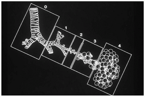
FIGURE 12-23 • Classification of CPAM. The classification is based on the similarity in appearance of the hamartomatous components of the lesion with the various areas of the normal tracheobronchial tree. Type 0, composed of bronchuslike structures, appears to be a malformation of the most proximal tracheobronchial tree. Type 1, containing bronchuslike and proximal bronchiole-like structures, mimics the distal bronchial tree and proximal acinus. Type 2, composed of bronchiole-like structures, resembles the bronchiolar segment of the acinus. Type 3, composed of bronchiole-like structures and alveolar ducts and saccules lined by cuboidal epithelium, resembles the midacinar region. Type 4, with thin-walled structures lined by type 1 alveolar lining cells, should be diagnosed correctly as PPB type 1. (From Stocker JT. Congenital and developmental diseases. In: Dail HD, Hammer SP, eds. Pulmonary pathology. 2nd ed. New York: Springer-Verlag, 1994:182, with permission.)


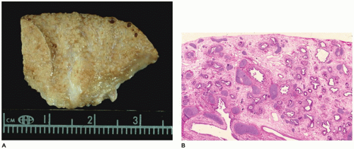
FIGURE 12-24 • CPAM, type 1 (congenital acinar dysplasia). A: A small nodular mass representing the right lung is largely devoid of air. A similar lung was present on the left side. B: Bronchial-like structures are surrounded by irregular cartilage plates and loose mesenchyme-containing thin-walled vascular structures.

CPAM, type 1, the large or predominant cyst type, presents primarily within the first week to month of life but can be seen in older children and even young adults (Figure 12-25A). It accounts for nearly 65% of cases and is usually readily amenable to surgery with a good prognosis. Grossly, the type 1 lesion is characterized by single or multiple large cysts (3 to 10 cm in diameter) surrounded by smaller cysts and compressed normal parenchyma (Figure 12-25B, C). Microscopically, the larger cysts are lined with ciliated, pseudostratified columnar epithelium and the smaller ones by cuboidal to columnar epithelium (Figure 12-25D, E). More than 45% of the cases display segments of mucus-producing cells among the epithelial lining of the larger cysts or in bronchioles and alveolar duct-like structures adjacent to the larger cysts (Figure 12-25F). These mucous cells are recognized to be precursors for adenocarcinomas (Figure 12-26) (see below—Tumors) that have been described in association with type 1 CPAM (96,97). The walls of the CPAM, type 1 cysts are composed of elastic tissue overlying fibromuscular connective tissue, and in 5% to 10% of cases, cartilage plates.

CPAM, type 2, the medium cyst type, accounts for 10% to 15% of cases, is seen exclusively within the first year of life and has a poorer outcome owing to its more frequent association with other anomalies, some of which are incompatible with life (e.g., renal agenesis). The type 2 lesion is composed of cysts 0.5 to 2.0 cm in diameter (rarely larger) that are evenly distributed and blend with the adjacent normal parenchyma (Figure 12-27A). The cysts occasionally surround normal appearing bronchi. The typical back-to-back bronchiole-like structures are lined by cuboidal to columnar epithelial cells with a thin underlying fibromuscular layer (Figure 12-27B).


Mucous cells and cartilage plates are absent except as components of “entrapped” normal bronchi. A variant or subgroup of the type 2 lesion, termed rhabdomyomatous dysplasia, contains ribbons of striated muscle fibers throughout the lesion, both in association with the cysts and between alveolar ducts and around blood vessels (Figure 12-27C). The cysts of this rhabdomyomatous variant may be less prominent than other type 2 lesions. Rhabdomyosarcoma has been reported to originate from CPAM, but this likely represents a primary pleuropulmonary blastoma (PPB) rather than being secondary to CPAM. CPAM, type 2-like features are present in 50% of extralobar sequestrations (67).


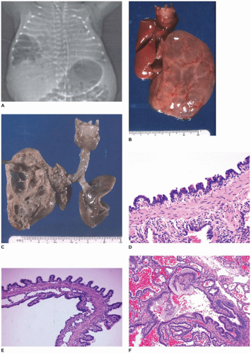
FIGURE 12-25 • CPAM, type 1. A: A cystic mass is present in the lower right hemithorax in a newborn with respiratory distress. B: Multiple large, fluid-filled cysts distend the left lobe from a fetus in the second trimester. C: When opened, the mass consists of intercommunication cysts. D: Cysts of type 1 CPAM are characteristically lined by ciliated intercommunicating in a sawtooth configuration with underlying fibromuscular connective tissue. E: A larger cyst wall (top) is covered by columnar epithelium in a papillary configuration. Note the columnar epithelial lining of the smaller cysts as well. F: Clusters of mucogenic cells are present along the cyst lining.


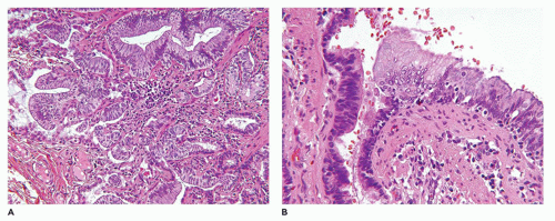
FIGURE 12-26 • CPAM type I. A: Mucinous metaplasia in the lung of an 18-year old who presented with a right middle lobe cyst. B: The mucinous epithelium has low-grade cytologic features, but it is thought that the changes are a precursor to mucinous adenocarcinoma.


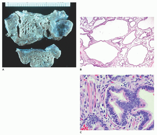
FIGURE 12-27 • CPAM, type 2. A: Small cysts (0.2 to 0.5 cm) are scattered throughout the lobe and blend with normal parenchyma. B: The back-to-back bronchiole-like structures are separated by structures resembling alveolar ducts. C: In a variant of type 2, striated muscle fibers are present in the connective tissue between and around cysts.

CPAM, type 3, the small cystic or solid type, occurs infrequently (5% of cases), is seen exclusively in the first days to month of life, has a notable male predominance, and owing to its large size and association with maternal polyhydramnios and fetal anasarca, has a high mortality rate. Increased maternal levels of serum alpha-fetoprotein have been anecdotally noted in the second trimester in association with CPAM type 3. CPAM, type 3, the original lesion described by Ch’in and Tang (98), consists of a large, bulky, parenchymal mass involving an entire lobe or even an entire lung (Figure 12-28A, B). The mass effect of the lesion consistently produces mediastinal shift and often results in hypoplasia of the uninvolved lung. Cysts are rarely larger than 0.2 cm in diameter, with the exception of scattered larger bronchiole-like structures. Microscopically, the lesion resembles an immature lung devoid of bronchi. Irregular, stellate-shaped, bronchiole-like structures lined with cuboidal epithelial cells are surrounded by alveolar ductules and saccules that are also lined by cuboidal cells, imparting the “adenomatoid” appearance for which this lesion was originally named (Figure 12-28C). Mucous cells, cartilage, and rhabdomyomatous cells are not present, and there is a paucity of vessels within the lesion.


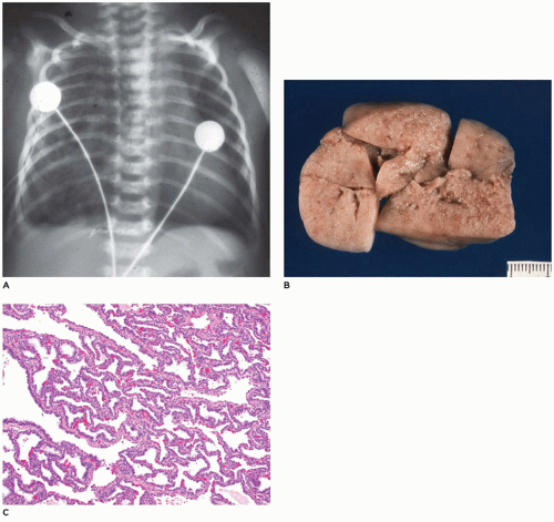
FIGURE 12-28 • CPAM, type 3. A: A large air-containing mass in the right hemithorax pushes the mediastinum to the left. B: The resected lesion is nearly solid with only a few slit-like openings. C: Randomly distributed irregular bronchiole-like structures are separated by dilated alveolus-like structures all of which are lined by cuboidal epithelial cells, imparting an adenomatoid (or gland-like) appearance.


CPAM, type 4, the peripheral acinar cyst type, is composed of large cysts lined by pneumocytes and it is very important to recognize it as the purely cystic pleuropulmonary blastoma (type 1 PPB). It is recommended that all children should be tested for DICER 1 mutation which is the first hit (of two-hits, see below) and is present in 70% of PPBs including those that are nonrecurring. Complete resection and close long-term follow-up are necessary (97). Once type 1 PPB has been excluded, CPAM type 4 is indeed rare if it exists at all. It is seen equally in boys and girls, with an age range of newborn to 4 years and accounts for 10% to 15% of cases. Before the recognition of type 1 PPB, most of these cases were included in the CPAM type 1 category. Clinically, the type 4 lesions may present with mild respiratory distress, sudden respiratory distress from tension PT, pneumonia, or on occasion, as an incidental finding with no symptoms (94). Radiographically, the lesion displays large air-filled cysts with mediastinal shift. The lesion involves a single lobe in about 80% of cases and rarely may be bilateral. Grossly, large thin-walled cysts are present at the “periphery” of the lobe and appear to be lined by a smooth membrane (Figure 12-29A). Microscopically, the cysts are lined by flattened
epithelial cells (type I and II pneumocytes), with occasional low cuboidal epithelium seen (Figure 12-29B to D). The wall of the cyst is composed of loose mesenchymal tissue with prominent arteries and arterioles. It must be emphasized that PPB type 1 and CPAM, type 4 are one in the same and should be viewed and managed as PPB.


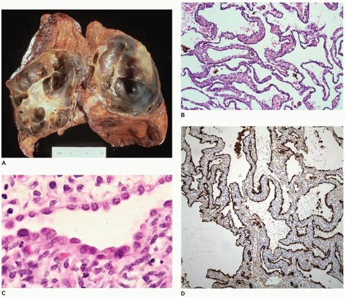
FIGURE 12-29 • PPB, type 1 (in the past diagnosed as CPAM type 4). A: The lung is distended by thin, almost translucent cyst walls. B: The walls of the cysts are composed of loose mesenchyme covered by an indistinct epithelial lining not apparent at this magnification (hematoxylin and eosin stain, original magnification ×25). C: The cyst walls are variously covered by an attenuated epithelium of alveolar lining cells (hematoxylin and eosin stain, original magnification ×150). D: The epithelium stains positively for cytokeratin (H&E, ×50). Note: Because of the similarities between PPB type 1 and CPAM type 4, it is best to consider these lesions as having the potential to progress to PPB types 2 and 3. All patients with such cystic lesions must have complete surgical excision with clear margins. If any doubt exists as to the correct diagnosis, the case should be referred to the International PPB Registry to ensure correct follow-up and management of the patient.

Ultrasonography has been demonstrated to be a highly useful modality in the in utero diagnosis of CPAM. In utero serial sonography has demonstrated the gradual reduction in the size of CPAM, type 1 and 2, with subsequent normal development of the uninvolved lung (99,100). Anomalies are noted in 15% to 20% of all cases of CPAM, particularly in association with the type 2 lesion (94,101) including bilateral renal agenesis/dysgenesis, extralobar pulmonary sequestration, cardiovascular malformation, diaphragmatic hernia, hydrocephalus and macrocephaly, myelomeningocele, jejunal atresia, prune belly syndrome, sirenomelia, bilateral nephromegaly, Pierre Robin syndrome, pulmonary hypoplasia, skeletal malformation, bile duct hypoplasia, left heart hypoplasia, polycytosis of a solitary medial kidney, and congenital nephrotic syndrome (diffuse mesangial sclerosis).


Congenital Alveolar Capillary Dysplasia

Congenital alveolar capillary dysplasia with or without misalignment of pulmonary veins (ACD/MPV) is a uniformly fatal rare entity that presents as respiratory distress, progressive hypoxemia, and pulmonary hypertension in the newborn (102). Familial occurrence has been noted (103); most cases are sporadic, with familial cases showing paternal imprinting (Table 12-5). Many cases have been reported to harbor microdeletions in the FOX gene cluster on 16q24.1 and mutations in the FOXF1 gene, a transcription factor that is involved in organogenesis (104). It is characterized by the failure of formation and ingrowth of alveolar capillaries. Broad alveolar septa with large alveolar capillaries within the septal wall are the hallmark of this disorder (Figure 12-30A to D) (105). Capillaries are centrally placed well beneath the basement membrane of the alveolar lining cells and surrounded by loose mesenchyme (Figure 12-30C). Ectatic veins are present within bronchovascular bundles, occasionally within the adventitia of the pulmonary arteries, and may form an intermittent ring around the bronchiole (Figure 12-30B). Small muscularized arteries are also present within the acini, extending to the precapillary area (Figure 12-30D). Other changes include paucity of alveolar capillaries and abnormal capillary size and location, seen as centrally located thin-walled dilated and congested alveolar wall vessels. Special stains including Movat pentachrome, trichrome, elastic, CK7 (for pulmonary epithelium), CD31 (for microvasculature), and SMA (for vascular smooth muscle) may be used to highlight the constellation of histologic changes. High capillary apposition and density may correlate with survival beyond the neonatal period. About a third of the patients with ACD/MPV also have pulmonary lymphangiectasis (106,107) and about 80% have anomalies of other organ systems (108). Treatment with inhaled nitric oxide and ECMO has prolonged life but has been uniformly unsuccessful in changing the fatal outcome of this disorder without lung transplantation.


TABLE 12-5 ANOMALIES ASSOCIATED WITH CONGENITAL CAPILLARY ALVEOLAR DYSPLASIA


Anophthalmia and distinct eyebrows


Familiary microphthalmia


Degeneration of the anterior segment of the eye


Atrioventricular septal defect and quadricuspid pulmonary valve


Down syndrome


Gastrointestinal


Duodenal atresia and anorectal anomaly


Intestinal malrotation


Total colonic Hirschsprung disease


Duodenal atresia


Left-right asymmetry


Arteriovenous malformation of the liver


Bilateral ureteropelvic junction obstruction


Atrioseptal defect


Abnormally lobulated lungs


Hydronephrosis


Urethral atresia


Atrioventricular canal


Peripheral Cysts of the Lung

Peripheral, air-containing cysts of the lung can be seen in neonates, infants, and young children; it occurs in association with Down syndrome, as a result of pulmonary infarction, or in association with idiopathic spontaneous PT (109). Occlusion of the pulmonary artery in infants can result in peripheral infarction of the lung, which, with necrosis and organization, can produce subpleural cysts of varying size. Gonzalez et al. (110) reported peripheral cysts in 18 of 98 patients with Down syndrome and suggested that the cysts are an intrinsic feature of the disease that may result from reduced postnatal production of peripheral small air passages and alveoli. The 0.2- to 1.0-cm air-filled cysts are located beneath the pleura and are formed of vascular fibrous connective tissue walls lined by alveolar lining cells (Figure 12-31A, B). The cysts communicate with more centrally located bronchioles and alveolar ducts. The cysts resemble those seen in the upper lobes of adult males with idiopathic spontaneous PT and have also been noted in a case of ILE (66).


Acute Lung Injury and Respiratory Distress Syndrome

Acute lung injury is a pattern of diffuse alveolar damage (DAD) seen in association with clinical acute respiratory distress syndrome (RDS). Previously referred to as hyaline membrane disease (HMD), acute lung injury is characterized by firm, atelectatic lungs with an uneven air expansion pattern, focal hemorrhage, edema fluid in alveoli, and hyaline

membranes along terminal and respiratory bronchioles and alveolar ducts. In the pediatric age group, DAD has been typically described in neonates with surfactant deficiency, although other causes include infections, inhalational injury, sepsis, and systemic shock. Breathing difficulty is common in the early neonatal period, occurring in up to 7% of newborn infants, and is not synonymous with RDS. Other common causes of breathing difficulty in term newborn infants include transient tachypnea of the newborn, pneumonia, meconium aspiration syndrome (MAS), persistent pulmonary hypertension of the neonate, and PT (111).


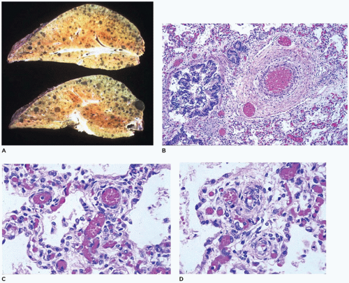
FIGURE 12-30 • Congenital alveolar capillary dysplasia. A: Bulky stiff lungs display focal hemorrhage and prominent interlobular septa. B: Dilated veins are present adjacent to and within the adventitia of a pulmonary artery (center-right). C: Broad alveolar septa contain many centrally located capillaries with only a few of them approaching the alveolar epithelium. D: Muscularized arteries are present within alveolar septa well away from bronchioles.


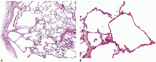
FIGURE 12-31 • Peripheral lung cysts in a 4-year-old male with trisomy 21. A: This lung biopsy demonstrates the presence of multiple pulmonary cysts residing beneath the pleural surface. B: These cysts represent a growth abnormality in the formation of small airspaces.


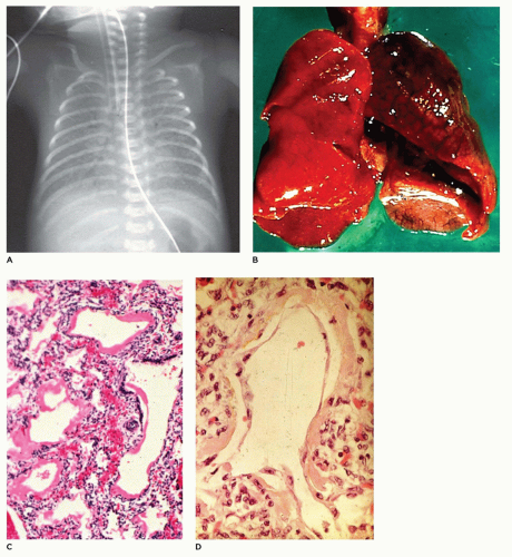
FIGURE 12-32 • Acute lung disease— HMD of the newborn. A: In this 24-hour-old, 1,050-g infant with respiratory distress, the lungs display a classic “ground-glass” opacity. B: The lungs in HMD are often atelectatic and display focal hemorrhage. C: Dilated bronchioles and alveolar ducts are lined by thick hyaline membranes. D: At 72 hours of age, the membranes are being covered by regenerating alveolar lining cells.

Infants present with tachypnea, intercostal retractions, and hypoxemia and display a typical x-ray image of ground-glass alterations of the lungs with an air bronchogram and diffusely scattered reticulogranular opacities (Figure 12-32A) (1). Grossly, the lungs are firm and resemble the liver more than the lung. Microscopically, there is an uneven air expansion pattern with atelectatic acini and
dilated bronchioles and alveolar ducts (Figure 12-32B). Scattered foci of alveolar hemorrhage and edema are present, but most striking is the presence of smooth, homogeneous, pink membranes lining terminal and respiratory bronchioles and alveolar ducts, particularly at points of division or branching (Figure 12-32C). These hyaline membranes are composed of necrotic alveolar lining cells, plasma transudate, inhaled amniotic fluid including squames and fibrin. Hemorrhage is often present. Hyaline membranes may be seen in infants who die as early as 3 to 4 hours after birth and are uniformly present as well-formed structures by 12 to 24 hours in infants with RDS. In the absence of severe disease requiring high oxygen tensions and ventilatory pressures, at 36 to 48 hours, the membranes begin to organize and separate from the underlying wall to be replaced by alveolar lining cells or bronchiolar cuboidal or columnar epithelium (Figure 12-32D). Bacteria may alter the appearance of the membranes by producing fragmented, faintly basophilic structures, with organisms often readily demonstrable by Gram stain on or within the membranes. Conditions associated with hyperbilirubinemia (e.g., kernicterus, intraventricular hemorrhage, intrahepatic bile stasis, disseminated intravascular coagulation) may produce, in infants surviving 3 or more days, yellow hyaline membranes as a result of the presence of unconjugated bilirubin.

Surfactant replacement therapy, although radically decreasing the incidence of acute lung injury in premature infants and its morbidity and mortality in these infants, does not appear to alter the pathologic features of DAD in infants dying of RDS. However, treated children may show a slightly higher incidence of pulmonary hemorrhage and a lower incidence of IPE and PT (112). Surfactant therapy appears to accelerate the rate of epithelial cell regeneration (113).

The pathophysiology of acute RDS in the neonate has been divided into eight categories: alveolar fluid transport, surfactant, innate immunity, apoptosis, coagulation, direct alveolar epithelial injury by bacterial products, ventilator-associated lung injury, and repair. The biological baseline network of genes and protein expression is reportedly different in children with RDS, as compared to adults (114). Beyond the neonatal period, causes of RDS are similar to those in adults. In a prospective, multicenter, observational study covering 21 pediatric intensive care units, pneumonia and sepsis were the most common causes of acute RDS; the authors found a lower acute RDS incidence and mortality in children than those reported for adults (115). In a masked, randomized, placebo-controlled trial in 24 children’s hospitals in six different countries, surfactant did not improve outcomes relative to placebo in children with direct lung injury/acute RDS (116).


Bronchopulmonary Dysplasia (Chronic Lung Disease of Prematurity)

Bronchopulmonary dysplasia was first described in 1967 by Northway et al. (117). In a retrospective study, they described the clinical and pathologic features of 19 infants dying following mechanical ventilation with high concentrations of oxygen for severe HMD (RDS). The pathology was correlated with clinical and radiographic findings and included a 2- to 3-day period of acute RDS, followed by a weeklong period of “regeneration,” another 10-day period of transition to chronic disease, and a final period of chronic disease extending beyond 1 month of life. The pathologic features in the first stage included the typical findings of HMD (e.g., atelectasis, uneven air expansion pattern, hemorrhage, and hyaline membranes). During the second stage, there was necrosis of bronchiolar and alveolar epithelium with persistence of hyaline membranes (Figure 12-32A to C). In the transition to chronic disease, injury to alveolar epithelium continued, along with widespread bronchial and bronchiolar mucosal metaplasia and marked mucus secretion. Clusters of hyperexpanded alveoli alternated with areas of atelectasis.

In the chronic stage, bronchioles displayed marked peribronchiolar smooth muscle hypertrophy associated with clusters of “emphysematous alveoli.” The birth weights of the 19 infants dying of bronchopulmonary dysplasia varied from 900 to 2466 g, with two-thirds of them weighing more than 1300 g. Bonikos et al. (118) suggested that bronchopulmonary dysplasia was due to the toxic effects of oxygen, poor bronchial drainage, and the effects of mechanical ventilation. In the 10 years following that initial brief description of the pathology of BPD, a number of other studies described in more detail the pathologic features including the changes noted in alveoli, airways, lymphatics, vessels, and connective tissue as criteria for the staging of bronchopulmonary dysplasia. In 1976, Bonikos et al. (119) described a severe necrotizing bronchiolitis in the acute stages of BPD and implicated prolonged exposure to high levels of oxygen as a major feature in the cause of bronchiolitis. Since that time, a number of additional factors, including infection, inflammation, poor nutrition, dehydration, and others, have been implicated in the pathogenesis of BPD (120). In addition to the bronchiolitis, there is a prominent alveolar septal fibrosis in the healed stages along with an increased incidence of cardiac hypertrophy.

The sequelae of this necrotizing bronchiolitis were described by Stocker in 1986 in a series of 28 patients with long-standing “healed” bronchopulmonary dysplasia, who died at 3 to 40 months of age (64). Noting the presence of deep pleural fissures and acini with varying degrees of alveolar septal fibrosis (Figure 12-33), it was suggested that the necrotizing bronchiolitis seen in the acute phases, while prohibiting adequate ventilation, often served to “protect” acini from damage by mechanical ventilation or high levels of oxygen (Figure 12-34A to C). Stocker also suggested that the alveolar fissures might represent areas of complete loss of acini corresponding to the marked decrease in internal surface area and number of alveoli noted by Sobonya et al. (121). The 6- to 10-fold reduction in number of alveoli suggested not only an absolute loss of some acini but a generalized reduction in lung growth. In 1991, Margraf et al. (122) confirmed the reduction in lung volume and small airway density noted by Sobonya.


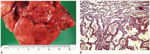
FIGURE 12-33 • Long-standing healed bronchopulmonary dysplasia. A: Irregular clefting and fissuring of pulmonary lobes probably represent the loss of acini during the acute phases of BPD. B: The acinus at top represents the one “protected” by occluded bronchioles from the damage of barotrauma and high oxygen pressures. At the bottom, this acinus displays the diffuse alveolar septal fibrosis caused by previous exposure to barotrauma and high oxygen pressures (Masson trichrome, ×25).

In recent years, with the advent of surfactant replacement therapy and increased sophistication in the use of mechanical ventilation (including high frequency jet ventilation) and oxygen supplementation, another stage in the evolution of the pathology of bronchopulmonary dysplasia has been seen. Although the occasional case of “classic” acute bronchopulmonary dysplasia with necrotizing bronchiolitis, alveolar cell hyperplasia, and peribronchiolar and alveolar septal fibroplasia is still seen along with focal alveolar septal fibrosis in the older patient (Figure 12-35A to D), the few infants who now die from bronchopulmonary dysplasia display what might best be described as “acinar simplification.” These simplified acini are characterized by uniformly dilated alveoli whose walls consist of thin alveolar septa with little or no interstitial fibrosis (123).


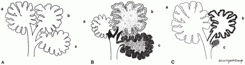
FIGURE 12-34 • Bronchopulmonary dysplasia (BPD) before the advent of surfactant replacement therapy. A: Schematic representation of three uniformly distended acini (a to c) with associated bronchiole, alveolar ducts, and alveoli. B: In the early stages of BPD, hyaline membranes or necrotic debris may totally occlude a bronchiole (a) protecting the distal acinus. Bronchioles that remain partially or completely open (b, c) allow the distal acinus to be exposed to varying degrees of injury from barotrauma and high oxygen tension. C: In the healed stages of BPD with resolution of the bronchiolar obstruction in (A), the “protected” distal acinus expands and continues to develop new alveoli. Depending on the degree of injury, acini may atrophy and disappear (c), producing pleural fissures (see Figure 12-33A), or display varying degrees of alveolar septal fibrosis (b) and be inhibited from further alveolar development. (From Stocker JT. Pathologic features of long-standing “healed” bronchopulmonary dysplasia: a study of 28 3- to 40-month-old infants. Hum Pathol 1986;17:943, with permission.)

The bronchioles are similarly unremarkable, with only an occasional mild increase in peribronchiolar musculature. These changes seem to represent an “arrest” of development of the acini, with a resulting markedly decreased number of alveoli within each acinus (Figure 12-36A to C). As a result, the surface area of the lung is significantly decreased even in the absence of significant pathology (e.g., alveolar septal fibrosis).

Although high concentrations of oxygen over prolonged periods of time are known to cause alveolar cell hyperplasia and necrotizing bronchiolitis with resulting alveolar septal fibrosis,

it is possible that low levels of oxygen (25% to 35%), while not producing significant alteration in the epithelial lining of the lung or not causing damage sufficient to cause septal fibrosis, may, in very immature infants, inhibit growth of the lung, that is, the development of new alveolar ducts and alveoli. Although the lung appears to “mature” and alveolar septa appear to thin and expand to resemble the septa of term infants, there is no accompanying significant increase in the surface area of the lung through an increase in number of alveoli. Thus, although recent advances in mechanical ventilation have limited the amount of injury to the bronchiole (i.e., no necrotizing bronchiolitis), the continued patency of all bronchioles throughout the course of therapy allows equal injury or inhibition of growth to all acini from even low levels of oxygen therapy.


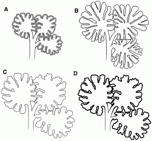
FIGURE 12-35 • Chronic lung disease of the premature (CLDP) since the advent of surfactant replacement therapy. A: Schematic representation of three normally expanded and aerated pulmonary acini in an immature infant. Note the appropriately thick septa of the developing lung. B: With normal lung growth and development, the acini not only increase in size [relative to (A)] but also in complexity with the appearance of “new” alveolar saccules and alveoli. C and D: In infants receiving surfactant replacement therapy who develop moderate-to-severe CLDP, the acini increase in size [relative to (A)] but show little, if any, increase in the number of alveolar saccules or alveoli. The alveolar septa in (C) appear normal in thickness compared with the less-injured or uninjured lung (B), or they may display a uniform mild alveolar septal fibrosis as in (D).


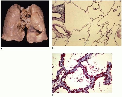
FIGURE 12-36 • Chronic lung disease of the premature. A: The lung appears largely unremarkable with an evenly aerated parenchyma. B: In this section of lung from a 4-month-old infant born at 26 weeks gestation who developed moderate respiratory distress and clinical BPD, the acini are simplified with dilated alveolar ducts and saccules and with very few alveoli arising from them. C: In this section of lung from a 2-month-old infant born at 28 weeks gestation who developed severe prolonged respiratory distress and clinical BPD, the alveolar septa of all acini show mild though uniform interstitial fibrous thickening.

Only gold members can continue reading. Log In or Register to continue

Stay updated, free articles. Join our Telegram channel

Sep 23, 2016 | Posted by in PATHOLOGY & LABORATORY MEDICINE | Comments Off on The Respiratory Tract

Full access? Get Clinical Tree

Get Clinical Tree app for offline access