The Cephalic, Oral, and Esophageal Phases of the Integrated Response to a Meal

CHAPTER 27 The Cephalic, Oral, and Esophageal Phases of the Integrated Response to a Meal


In this chapter we will look at the processes that occur in the gastrointestinal (GI) tract in the early stages of the integrated response to a meal. Even before food is ingested there are changes in the physiology of the GI tract, and in this so-called cephalic phase, as well as in the oral phase (when ingested food is in the mouth), the responses of the GI tract to the presence of food are mainly associated with preparing the GI tract for digestion and absorption. We will also look at the transfer of food from the mouth to the esophagus, the esophageal phase of the meal.


CEPHALIC AND ORAL PHASES


The main feature of the cephalic phase is activation of the GI tract in readiness for the meal. The stimuli involved are cognitive and include anticipation or thinking about the consumption of food, olfactory input, visual input (seeing or smelling appetizing food when hungry), and auditory input. The latter may be an unexpected link but was clearly demonstrated in the classic conditioning experiments of Pavlov, in which he paired an auditory stimulus to the presentation of food to dogs; eventually, the auditory stimulus alone could stimulate secretion. A real-life analogy is presumably being told that dinner is ready. All these stimuli result in an increase in excitatory parasympathetic neural outflow to the gut. Sensory input, such as smell, stimulates sensory nerves that activate parasympathetic outflow from the brainstem. Higher brain sites are also involved (such as the limbic system, hypothalamus, and cortex) in the cognitive components of this response. The response can be both positive and negative; thus, anticipation of food and a person’s psychological status, such as anxiety, can alter the cognitive response to a meal. However, the final common pathway is activation of the dorsal motor nucleus in the brainstem, the region where the cell bodies of the vagal preganglionic neurons arise; activation of the nucleus leads to increased activity in efferent fibers passing to the GI tract in the vagus nerve. In turn, the efferent fibers activate the postganglionic motor neurons (referred to as motor because their activation results in change of function of an effector cell). Increased parasympathetic outflow enhances salivary secretion, gastric acid secretion, pancreatic enzyme secretion, gallbladder contraction, and relaxation of the sphincter of Oddi (the sphincter between the common bile duct and the duodenum). All these responses enhance the ability of the GI tract to receive and digest the incoming food. The salivary response is mediated via the ninth cranial nerve; the remaining responses are mediated via the vagus nerve.


Many of the features of the oral phase are indistinguishable from the cephalic phase. The only difference is that food is in contact with the surface of the GI tract. Thus, there are additional stimuli generated from the mouth, both mechanical and chemical (taste). However, many of the responses that are initiated by the presence of food in the oral cavity are identical to those initiated in the cephalic phase because the efferent pathway is the same. Here we will discuss the responses specifically initiated in the mouth, which consist mainly of the stimulation of salivary secretion.


The mouth is important for the mechanical disruption of food and for the initiation of digestion. Chewing subdivides and mixes the food with the enzymes salivary amylase and lingual lipase and with the glycoprotein mucin, which lubricates the food for chewing and swallowing. Minimal absorption occurs in the mouth, although alcohol and some drugs are absorbed from the oral cavity and this can be clinically important. However, as with the cephalic phase, it is important to realize that stimulation of the oral cavity initiates responses in the more distal GI tract, including increased gastric acid secretion, increased pancreatic enzyme secretion, gallbladder contraction, and relaxation of the sphincter of Oddi, mediated via the efferent vagal pathway.


Properties of Secretion


General Considerations


Secretions in the GI tract come from glands associated with the tract (the salivary glands, pancreas, and liver), from glands formed by the gut wall itself (e.g., Brunner’s glands in the duodenum), and from the intestinal mucosa itself. The exact nature of the secretory products can vary tremendously, depending on the function of that region of the GI tract. However, these secretions have several characteristics in common. Secretions from the GI tract and associated glands include water, electrolytes, protein, and humoral agents. Water is essential for generating an aqueous environment for the efficient action of enzymes. Secretion of electrolytes is important for the generation of osmotic gradients to drive the movement of water. Digestive enzymes in secreted fluid catalyze the breakdown of macronutrients in ingested food. Moreover, many additional proteins secreted along the GI tract have specialized functions, some of which are fairly well understood, such as those of mucin and immunoglobulins, and others that are only just beginning to be understood, such as those of trefoil peptides.


Secretion is initiated by multiple signals associated with the meal, including chemical, osmotic, and mechanical components. Secretion is elicited by the action of specific effector substances, called secretagogues, acting on secretory cells. Secretagogues work in one of the three ways that have already been described in the previous chapter—endocrine, paracrine, and neurocrine.


Salivary Secretion


During the cephalic and the oral phase of the meal, considerable stimulation of salivary secretion takes place. Saliva has a variety of functions, including those important for the integrative responses to a meal and for other physiological processes (Table 27-1). The main functions of saliva in digestion include lubrication and moistening of food for swallowing, solubilization of material for taste, initiation of carbohydrate digestion, and clearance and neutralization of refluxed gastric secretions in the esophagus. Saliva also has antibacterial actions that are important for overall health of the oral cavity and teeth.


Table 27-1 Functions of Saliva and Chewing


Functional Anatomy of the Salivary Glands


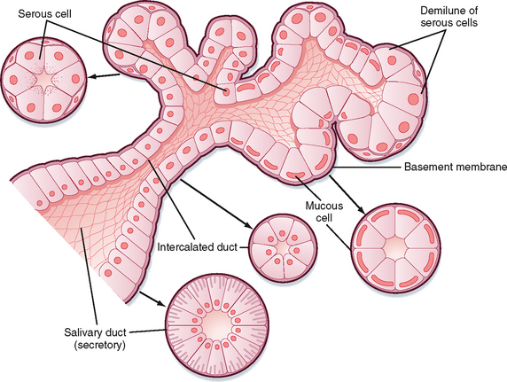
There are three pairs of major salivary glands: parotid, submandibular, and sublingual. In addition, many smaller glands are found on the tongue, lips, and palate. These glands are the typical tubuloalveolar structures of glands located in the GI tract (Fig. 27-1). The acinar portion of the gland is classified according to its major secretion: serous (“watery”), mucous, or mixed. The parotid gland produces mainly serous secretion, the sublingual gland secretes mainly mucus, and the submandibular gland produces a mixed secretion.


Cells in the secretory end pieces, or acini, are called acinar cells and are characterized by basally located nuclei, abundant rough endoplasmic reticulum, and apically located secretory granules that contain the enzyme amylase and other secreted proteins. There are also mucous cells in the acinus; the granules in these cells are larger and contain the specialized glycoprotein mucin. There are three kinds of ducts in the gland that transport secretions from the acinus to the opening in the mouth and also modify the secretion: intercalated ducts drain acinar fluid into larger ducts, the striated ducts, which then empty into even larger excretory ducts. A single large duct from each gland drains saliva to the mouth. The ductal cells lining the striated ducts, in particular, modify the ionic composition and osmolarity of saliva.


Composition of Saliva


The important properties of saliva are a large flow rate relative to the mass of gland, low osmolarity, high K+ concentration, and organic constituents, including enzymes (amylase, lipase), mucin, and growth factors. The later are not important in the integrated response to a meal but are essential for long-term maintenance of the lining of the GI tract.


The inorganic composition is entirely dependent on the stimulus and the rate of salivary flow. In humans, salivary secretion is always hypotonic. The major components are Na+, K+, HCO3, Ca++, Mg++, and Cl. Fluoride can be secreted in saliva, and fluoride secretion forms the basis of oral fluoride treatment for the prevention of dental caries. The concentration of ions varies with the rate of secretion, which is stimulated during the postprandial period.


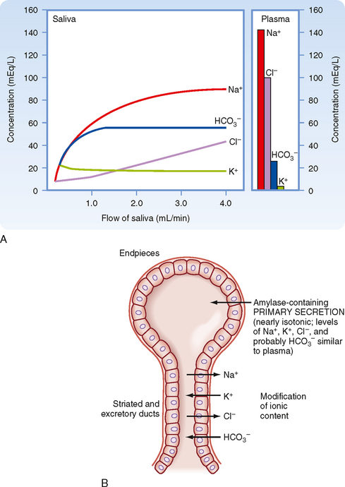
The primary secretion is produced by acinar cells in the secretory end pieces (acini) and is modified by duct cells as saliva passes through the ducts. The primary secretion is isotonic, and the concentration of the major ions is similar to that in plasma. Secretion is driven predominantly by Ca++-dependent signaling, which opens apical Cl channels in the acinar cells. Cl therefore flows out into the duct lumen and establishes an osmotic and electrical gradient. Because the epithelium of the acinus is relatively leaky, Na+ and water then follow across the epithelium via the tight junctions (i.e., via paracellular transport). Transcellular water movement may also occur, mediated by aquaporin 5 water channels. The amylase content and rate of fluid secretion vary with the type and level of stimulus. The excretory duct cells and the striated duct cells modify the primary secretion to produce the secondary secretion. The duct cells reabsorb Na+ and Cl and secrete K+ and HCO3 into the lumen. At rest, the final salivary secretion is hypotonic and slightly alkaline. Na+ is exchanged for protons, but some of the secreted protons are then reabsorbed in exchange for K+. HCO3, on the other hand, is secreted only in exchange for Cl, thereby providing excess base equivalents. The alkalinity of saliva is probably important in restricting microbial growth in the mouth, as well as in neutralizing refluxed gastric acid once the saliva is swallowed. When salivary secretion is stimulated, moreover, there is a decrease in K+ (but it always remains above plasma concentrations), Na+ increases toward plasma levels, Cl and HCO3 increase, and thus the secretion becomes even more alkaline. Note that HCO3 secretion can be directly stimulated by the action of secretagogues on duct cells. The duct epithelium is relatively tight and lacks expression of aquaporin, and therefore water cannot follow the ions rapidly enough to maintain isotonicity at moderate or high flow rates during stimulated salivary secretion. Thus, with an increase in the secretion rate, there is less time for modification by the ducts, and the resulting saliva more closely resembles the primary secretion and therefore plasma. However, [HCO3] remains high because it is secreted by duct and possibly acinar cells by the action of secretagogues (Fig. 27-2).


< div class='tao-gold-member'>

Stay updated, free articles. Join our Telegram channel

Jul 4, 2016 | Posted by in PHYSIOLOGY | Comments Off on The Cephalic, Oral, and Esophageal Phases of the Integrated Response to a Meal

Full access? Get Clinical Tree

Get Clinical Tree app for offline access