The Cardiovascular System


The Cardiovascular System


Ivan P. Moskowitz, M.D., Ph.D.

Selene Koo, M.D., Ph.D.

Aliya N. Husain, M.D.

Kathleen Patterson, M.D.


CONGENITAL MALFORMATIONS OF THE CARDIOVASCULAR SYSTEM


Incidence

The heart, first recognizable at 15 days of gestation, develops from a single tube into a four-chambered structure via an extraordinary series of loopings and septations (1). Given the complexity of cardiac development, it is not surprising that congenital cardiac defects account for the vast majority of cases of cardiac disease in childhood. The reported incidence of congenital heart disease (CHD) varies widely, ranging from 2.4/1000 to 8.8/1000 live births (2). The incidence statistics vary depending on the age of the patient population, the criteria used for diagnosis and inclusion in a study, and the length of study group follow-up. However, inclusion of defects that present later in life but that originate during fetal heart development, and are therefore congenital, such as bicuspid aortic valve, increases the incidence to 3% of live births (3).


Etiology

The etiology of congenital heart malformations has been historically thought to be multifactorial, with both genetic and environmental factors playing a role (4). However, no published epidemiologic work rules out a genetic etiology for the majority of CHD. Recent genetic advances suggest that the genetic basis of CHD is complex, potentially involving interactions between many genes (3). From 15% to 45% of patients with CHD have additional developmental anomalies, including chromosomal and nonchromosomal syndromes, malformation associations or sequences, and teratogen-associated defects (5). Genetic factors have long been recognized as a major player; historically, 10% of patients with CHD exhibit a trisomy, monosomy, duplication, or deletion on routine cytogenetic study, with trisomy 21 being the most common, whereas improved genetic techniques have allowed microdeletions and mutations of single genes to be identified in many of the developmental syndromes that include CHD as a major factor, with the 22q11 deletion, characteristic of the DiGeorge/velocardiofacial syndrome, being the most prevalent (4,6,7). The ever-increasing number of identified chromosomal abnormalities linked with heart malformations unfortunately does not mean that genetic screening can predict a specific form of CHD. Instead, it has become clear that mutations at multiple genetic loci can cause the same cardiac malformation and that mutations at a single locus can cause multiple different malformations. Meanwhile, molecular and biochemical analysis of normal gene products from many of these CHD-associated genes is leading to increased understanding of the developmental mechanisms in the heart (1). The advent of novel genetic techniques such as next-generation sequencing portends a rapid increase in our understanding of the genetic basis of CHD. Rather than providing a list of genes currently associated with CHD that will be quickly out of date, the reader is referred to recent reviews on the subject (e.g., 3).

CHD is fundamentally a defect of cardiovascular development, and increased understanding of cardiovascular development will help explain the etiology of the defects observed in human CHD. Cardiovascular development proceeds rapidly during early embryonic life through a series of well-defined steps (1). These include the generation of bilaterally paired groups of cardiac progenitor cells from lateral plate mesoderm, coalescing into the cardiac crescent, the migration of cardiac progenitors to the midline and the generation of the linear heart tube, the rapid growth and stereotyped rightward (D-looped) looping of the heart tube, the rapid growth of the cardiac chambers, and septation of the atria, ventricles, and outflow tract to form the final four-chambered heart with the capacity for distinct pulmonary and systemic blood flow. Cardiac development relies on both extrinsic developmental processes, such as left-right determination, and intrinsic developmental processes, such as cardiac septation. In the former, the proper establishment of the left-right axis of the body plan during gastrulation allows correct D-looping of the heart tube and appropriate development of each of the chambers with appropriate situs, or right- versus left-sided characteristics. Correct D-looping of the heart tube in turn establishes a necessary scaffold for
successful completion of the following steps in cardiovascular development. Appropriate pathologic evaluation of CHD specimens will independently evaluate the situs and other structural components of the specimen (see Classification section below).


Classification

An accurate classification of congenital heart malformations requires knowledge of the normal anatomy of the heart and a careful systematic approach to the examination. Normal values for heart weight relative to age and body size are readily available; normal values for the ventricular wall thickness and valve sizes have been reported for fetuses and newborns by Oyer et al. (8) and for infants and children by Rowlatt et al. (9) and Scholz et al. (10).


Cardiac Situs

Following a careful external examination of the shape and the position of the heart, the situs of the chambers of the heart is evaluated. With the relationships of the great vessels and venous drainage pattern, a sequential evaluation of the three segments of the heart (atria, ventricles, and great vessels) is undertaken. The connections of the segments, the relationship between the chambers within a segment, and the morphology of the segments are all assessed (11,12,13) (Table 13-2). When this segmental approach is used for classification, normal connections, relationships, and morphology are not incorporated into the diagnosis. In the majority of cases of CHD, a single defect is present (e.g., a ventricular septal defect [VSD]), and the heart is classified on the basis of that solitary anomaly. In more complex cases, situs relationships lead the diagnosis, and are followed by morphologic defects, in order of severity (13).


TABLE 13-2 SEQUENTIAL EXAMINATION OF HEART SEGMENTS


Atrial situs: determined by position of morphologic


RA


Situs solitus: RA on right; LA on left


Situs inversus: RA on left; LA on right


Situs ambiguus: indeterminate atrial situs


Bilateral right-sided atria


Bilateral left-sided atria


Atrioventricular connections


Atrioventricular concordance: RA→RV; LA→LV


Atrioventricular discordance: RA→LV; LA→RV


Ambiguous (indeterminate) connection


Double-inlet ventricle: RA + LA→one ventricle


Absence of one atrioventricular connection


Ventricular organization


Normal or D-looped: morphologic RV to the right of the morphologic LV


Inverted or L-looped: morphologic RV to the left of the morphologic LV


Ventricular morphology


Three normal components: inlet, trabecular, and outlet


Trabecular component determines morphologic right and left ventricles.


Rudimentary chamber: absent inlet portion


Ventriculoarterial connections


Ventriculoarterial concordance: RV→ PA; LV→Ao


Ventriculoarterial discordance: RV→Ao; LV→PA


Double-outlet ventricle: one ventricle→PA + Ao


Single outlet of heart: includes truncus, pulmonary atresia, aortic atresia


RA, right atrium; RV, right ventricle; LA, left atrium; LV, left ventricle; PA, pulmonary artery; Ao, aorta.


Situs refers to the position of a body part relative to other body parts. Because the human body has left-right differences, which develop either normally or abnormally, normal or abnormal situs can be determined. The morphologic features of the right (right atrium, liver) can be distinguished from those of the left (left atrium, stomach, spleen). There are three types of situs: (a) situs solitus, or normal situs; (b) situs inversus, the mirror image of situs solitus; and (c) situs ambiguus. Situs ambiguus is a general term for uncertain situs, characteristic of heterotaxy syndrome. This can include cases in which both sides of the body develop with “right” characteristics (asplenia syndrome) or “left” characteristics (polysplenia syndrome). The situs patterns of the atria and visceral organs are almost always the same. Normal situs allows normal cardiac/ventricular looping (D-loop). Inverted situs results in mirror-image looping (L-loop). Ambiguous situs will often cause abnormal looping, which in turn may cause structural abnormalities such as septal or conotruncal abnormalities (13).


Dextrocardia

Dextrocardia, in which the heart is located in the right side of the chest with a right-sided apex, occurs with situs inversus, situs ambiguus, and as an isolated finding (14). Except when occurring with situs inversus, the incidence of associated intracardiac and extracardiac anomalies is high (2). Dextrocardia should be distinguished from dextroposition, in which the heart is displaced to the right side of the chest with a left-sided apex (14).


Situs Ambiguus

Situs ambiguus (heterotaxia) occurs when the usual markers of situs are disorganized or missing as a result of disruption of the left-right axis determination early in development (15,16,17). The two best-described forms of situs ambiguus are asplenia (bilateral right sidedness) and polysplenia (bilateral left sidedness). The “sidedness” of the heart is determined by the atrial appendage morphology (16). The heterotaxic syndromes are frequently associated with complex congenital heart and venous malformations and a variety of extracardiac defects (15). Molecular studies have identified a variety of genes involved in left-right patterning during development (17).


Juxtaposition of Atrial Appendages

Juxtaposition of the atrial appendages, diagnosed when both atrial appendages reside partially or completely on the same side of the great vessels, is a harbinger of underlying heart malformations (16,18). Left-sided juxtaposition accounts for 86% of cases, with tricuspid atresia and transposition of the
great vessels the most common associated malformations. On the flip side, 11% of hearts with tricuspid atresia and 3% of hearts with D-transposition exhibit left-sided juxtaposition of the atrial appendages (18).


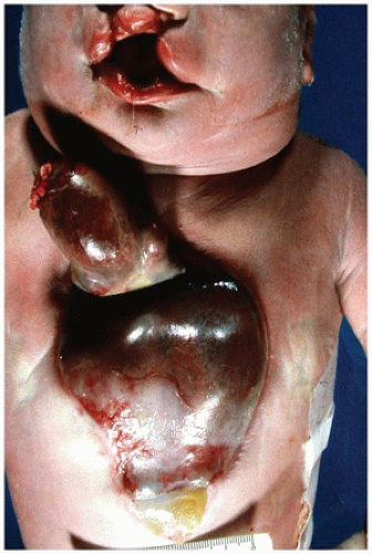
FIGURE 13-1 • Infant with multiple congenital anomalies including cleft lip seen at the top of the photograph and an anterior defect in the chest and abdomen through which the heart and liver protrude.


Ectopia Cordis

Ectopia cordis, a rare anomaly in which the heart is partially or totally outside the chest (Figure 13-1), is subclassified according to the location of the defect (14,19). Thoracic ectopia cordis, the most common type, is the result of a sternal cleft. The heart is usually located on the anterior surface of the chest without skin or a pericardial covering. Thoracoabdominal (abdominal) ectopia cordis is associated with a defect in the lower sternum, diaphragm, and abdominal wall; the heart is usually located with the abdominal viscera in a common omphalocele sac. Intracardiac defects occur frequently but are not inevitable (19).


Malformations of the Venous System


Systemic Venous Anomalies

Systemic venous blood returns to the heart via five sources: superior vena cava, coronary veins, hepatic veins, inferior vena cava, and azygos veins. With normally lateralized situs (situs solitus or situs inversus), anomalies of the systemic venous system are not uncommon but usually of little clinical significance. With situs ambiguus, complex systemic venous malformations are the rule.


Persistent Left Superior Vena Cava (LSVC)

A persistent LSVC is present in 0.3% to 0.5% of the general population and up to 10% of patients with other cardiovascular anomalies (14,20). Absence of the innominate vein serves as a clue to the presence of a persistent LSVC in approximately 40% of cases (14). The LSVC traverses the posterior surface of the left atrium to enter the coronary sinus in the AV sulcus. The wall between the coronary sinus and the left atrium becomes unroofed in approximately 8% of cases, resulting in drainage of the LSVC into the left atrium (20). This latter morphology occurs most frequently in the setting of the heterotaxy syndromes (21).


Coronary Sinus Ostium Atresia

With atresia of the coronary sinus ostium, a rare anomaly, cardiac venous drainage relies on a persistent LSVC with a patent innominate vein or other left-to-right connection. The obstructed coronary sinus ostium by itself creates few clinical problems, but ligation of the persistent LSVC should be avoided during heart surgery (21).


Interruption of the Inferior Vena Cava with Azygos Continuation

Anomalies of the inferior vena cava are much less common. Infrahepatic interruption of the inferior vena cava with azygos continuation results in an absence of the inferior vena cava between the renal and hepatic veins (14,20). The inferior vena cava below the renal veins drains via an enlarged azygos vein, which enters the thorax through the aortic hiatus and joins the superior vena cava just superior to its junction with the right atrium. This anomaly is usually associated with other cardiovascular malformations and is frequently present in the polysplenia syndrome (14,15).


Pulmonary Venous Anomalies

Pulmonary venous anomalies are listed in Table 13-3.


Partial Anomalous Pulmonary Venous Connection

Anomalous pulmonary venous connection refers to a group of conditions in which the pulmonary venous drainage is routed partially or totally to the right atrium.
In the more common anomaly, partial anomalous pulmonary venous connection, blood from one or more, but not all, of the pulmonary veins drains into a systemic vein or right atrium. This anomalous drainage is right sided in more than 80% of cases and most frequently enters the superior vena cava or the right atrium (14,22). More than 80% of cases occur in the setting of sinus venosus atrial septal defects (ASDs) as described earlier. The scimitar syndrome represents a variant of partial anomalous pulmonary venous connection characterized by anomalous pulmonary venous drainage into the inferior vena cava with a variety of associated cardiopulmonary anomalies. The most frequent associations include right lung hypoplasia, dextrocardia, systemic arterial supply to the lung, and abnormal bronchial anatomy (23).


TABLE 13-3 PULMONARY VENOUS MALFORMATIONS


Partial anomalous pulmonary venous connection


Sinus venosus ASD


Scimitar syndrome


Total anomalous pulmonary venous connection


Supradiaphragmatic


Supracardiac


Intracardiac


Infradiaphragmatic


Pulmonary vein atresia


Cor triatriatum


Total Anomalous Pulmonary Venous Connection

Total anomalous pulmonary venous connection, in which all the pulmonary veins drain to the systemic circuit, is subclassified according to the route of the abnormal venous drainage and the presence or absence of obstruction to that drainage (24,25) (Table 13-4). The most common route of drainage is through a vertical vein that arises from a confluence of the pulmonary veins posterior to the left atrium, traverses superiorly along the left side of the mediastinum, and drains into the innominate vein at its junction with the left subclavian vein (14,25). Less frequently, the common trunk drains into the superior vena cava, right atrium, coronary sinus, or subdiaphragmatic portal venous system (Figure 13-2). In up to 10% of cases, the pulmonary veins drain to multiple different sites (25,26). Drainage is obstructed in approximately 60% of cases of total anomalous pulmonary venous connection, with the cardiac sites having the lowest risk and the infracardiac the highest risk for obstruction (25,26) (Table 13-4). The venous drainage can be obstructed by intrinsically small vessels, external compression, or interposition of a capillary bed (14,25). The pulmonary venous obstruction leads to early and often severe pulmonary hypertensive changes, manifested as medial hypertrophy of the pulmonary arteries and veins combined with intimal proliferation and eventually arterialization in the pulmonary veins (25). Total anomalous pulmonary venous connection is associated with other cardiac anomalies in approximately one-third of cases, particularly with the heterotaxy syndromes (14,25). The clinical manifestations of total anomalous pulmonary venous connection vary with the degree of obstruction and the resultant PVR. With significant obstruction and high levels of resistance, cyanosis, heart failure, and death occur in the first months of life. With low resistance, infants may be asymptomatic at birth, and right-sided heart failure is the predominant manifestation (24). Surgical correction in the modern era yields more than 90% short-term survival with only rare late deaths, usually caused by pulmonary venous stenosis (24,25,26). In large series, risk factors for death include young age at surgery, cardiac or infracardiac connection sites, and preoperative pulmonary venous obstruction (26).


TABLE 13-4 TOTAL ANOMALOUS PULMONARY VENOUS CONNECTION: CLASSIFICATION


Site of Connection


% Total


% Obstructed


Supracardiac


45%


45%


Left innominate vein


25%-35%


Right SVC


10%-15%


Cardiac


25%


0%-20%


Coronary sinus


15%-20%


Right atrium


5%-15%


Infracardiac


25%


80%-90%


Portal vein


15%-25%


Mixed


5%-10%


35%-60%


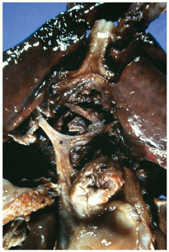
FIGURE 13-2 • Total anomalous pulmonary venous connection, infradiaphragmatic type, seen from the posterior view. A confluence of the pulmonary veins (asterisk) is isolated from the left atrium and drains into a vertical vein. This vein traverses the diaphragm to enter the portal venous system of the liver.


Pulmonary Vein Atresia/Stenosis

In pulmonary vein atresia, the entire pulmonary venous system drains into a common chamber from which there is no site for egress (14,24). In pulmonary vein stenosis, which is less severe, luminal narrowing occurs at the venoatrial junction of one or more of the pulmonary veins.


Cor Triatriatum

In cor triatriatum, the left atrium is partitioned by a fibromuscular shelf separating the pulmonary venous compartment from the atrial appendage and the mitral valve orifice compartment (14,24). The dividing membrane contains
a variably sized opening, which results in most instances in pulmonary venous obstruction. The foramen ovale may open into either compartment; when the opening is proximal to the obstruction, it can function as an escape valve for the pulmonary venous obstruction (14,27).


Septal Malformations


Malformations of the Atrial Septum

The atrial septum forms from three distinct embryonic structures: the septum primum, endocardial cushions, and septum secundum (28). In the fetus, blood flows freely between the right and the left atria via the foramen ovale, bordered by the superior right-sided septum secundum (limbus of fossa ovalis) and the inferior left-sided septum primum (valve of fossa ovalis). This opening normally fuses during the first year of life. However, in 25% to 30% of people, this fusion never occurs, leaving a “probe-patent” or “valvular-competent” foramen ovale (29).


Atrial Septal Defect

ASD can occur in one or more of four sites (Figure 13-3; Table 13-5). A secundum ASD, the most common type, manifests as multiple perforations of, a deficiency in, or absence of the fossa ovalis flap valve (14,30). In patients with a probepatent fossa ovalis, a secondary functional secundum ASD may appear following atrial dilatation. An isolated secundum ASD usually remains asymptomatic through childhood with 80% to 90% closing spontaneously, especially when of a small size (<4 mm diameter) (31). Defects greater than 8 mm, on the other hand, rarely close spontaneously, requiring surgical closure (31).

Primum defects, a form of AV septal defect, are discussed later. Sinus venosus defects result from a deficiency of the posterior superior aspect of the atrial wall that normally separates the right pulmonary veins from the superior vena cava/right atrium junction. A defect in this area therefore almost always occurs in conjunction with partial anomalous pulmonary venous return (32). Coronary sinus defects result from an unroofing or a fenestration of the coronary sinus on the posterior aspect of the left atrium and occur most often in association with a persistent left superior vena cava (LSVC) (21). Although most isolated ASDs occur sporadically, they can be inherited as an autosomal dominant anomaly with or without associated conduction system abnormalities (33,34).


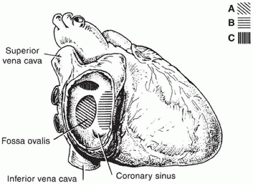
FIGURE 13-3 • The positions of various ASDs from the perspective of the right atrium, which has been opened laterally. A: Secundum defect. B: Primum defect. C: Sinus venosus defect.


TABLE 13-5 TYPES OF ATRIAL SEPTAL MALFORMATION


Septal defects


Secundum ASD


Primum ASD


Sinus venosus ASD


Coronary sinus ASD


Single atrium (cor triloculare biventriculare)


Premature closure of foramen ovale


Single Atrium

Complete absence of the atrial septum, a rare anomaly, results in a single atrial cavity, also termed common atrium or cor triloculare biventriculare. The single atrium usually accompanies other severe cardiac anomalies and is often associated with situs ambiguus (14).


Premature Closure of the Foramen Ovale

Premature closure of the foramen ovale manifests as a normally positioned but imperforate foramen ovale, an unidentifiable fossa ovalis, or an aneurysmal pouch bulging into the left atrium. The restricted mixing in utero can be complicated by hydrops fetalis and the hypoplastic left heart syndrome (14).


Malformations of the Ventricular Septum

The ventricular septum is a complex structure that can be divided into four components: inlet, trabecular, outlet, and membranous. From the right ventricular aspect, the inlet septum lies superiorly and posteriorly behind the septal tricuspid valve, the trabecular septum occupies the apex, the outlet septum sits between the crista supraventricularis and the pulmonary valve, and the membranous septum lies at the anteroseptal tricuspid commissure, where the tricuspid, mitral, and aortic valves converge (Figure 13-4). From the left ventricular aspect, the inlet septum lies posteriorly adjacent to the mitral valve, the trabecular septum occupies the apical region, the outlet septum sits beneath the right cusp of the aortic valve, and the membranous septum lies below the right and posterior aortic valve commissure (Figure 13-4).

VSDs, the most common type of congenital heart defect, can occur anywhere in the ventricular septum (35). VSDs are subclassified according to the nature of the defect rim and its anatomic position in the septum (Table 13-6).

Perimembranous (membranous, infracristal) defects account for up to 80% of VSDs (35). These defects are most easily seen from the left side, where they lie in the left
ventricular outflow tract just beneath the aortic valve (Figure 13-5). In the right ventricle, they reside beneath the crista supraventricularis and behind the papillary muscle of the conus, partially obscured by the septal leaflet of the tricuspid valve (36) (Figure 13-6).


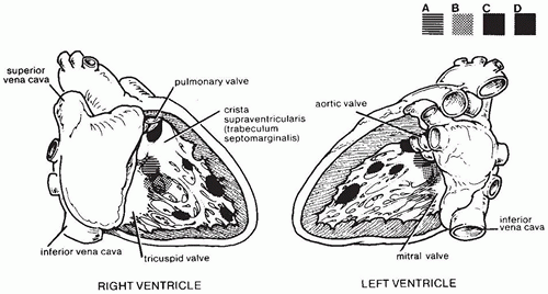
FIGURE 13-4 • The positions of the ventricular septal components and the corresponding VSTs from the lateral perspectives of the opened right and left ventricles. A: Membranous septum and perimembranous defect. B: Inlet septum and defect. C: Trabecular septum and trabecular muscular defects. D: Outlet septum and defect.

Outlet (infundibular) defects account for 5% to 7% of VSDs in the Western world but nearly 30% of VSDs in Japan and the East Asia. These defects are often roofed by pulmonary and aortic valve tissue (i.e., doubly committed subarterial) (35,36) and can be complicated by prolapse of the right coronary cusp of the aortic valve into the defect or by aortic regurgitation. Outlet and occasionally perimembranous trabecular defects may be associated with malalignment between the outlet and the trabecular portions of the ventricular septum. Anterior malalignment results in aortic override and posterior malalignment results in pulmonic override. Either can be complicated by subaortic stenosis, often with associated arch anomalies (37).


TABLE 13-6 VENTRICULAR SEPTAL DEFECTS: CLASSIFICATION


Defect Rim


Defect Location


Historical


Perimembranous


Inlet


Posterior


AV canal


Outlet


Supracristal


Infundibular


Trabecular


Membranous


Infracristal


Muscular


Inlet


AV canal


Outlet


Infundibular


Subpulmonic


Supracristal


Central trabecular


Infracristal


Remote trabecular


Muscular


Doubly committed subarterial


Outlet


Infundibular


Supracristal


AV, atrioventricular.


Inlet defects, which account for 5% to 8% of VSDs, reside beneath the septal leaflet of the tricuspid valve, but posterior and inferior to the position of perimembranous trabecular defects. Although inlet defects are similar in location to the VSD component of AV septal defects, hearts with isolated inlet defects do not show the other characteristic features of AV septal defects (35).

Muscular trabecular defects, representing 5% to 20% of VSDs, can range in size from minuscule to physiologically significant and often occur as multiples (35).

Clinical manifestations of a VSD usually first appear at the age of 2 to 6 weeks, when the normal drop in PVR results in the onset of a harsh holosystolic murmur at the left sternal border. The size of the defect and the state of the PVR rather than the anatomic location of the defect determine the nature of the symptoms (Table 13-7). In VSDs with large shunts, congestive heart failure may be resistant to therapy, and the risk for pulmonary vascular obstructive disease is substantial (38).


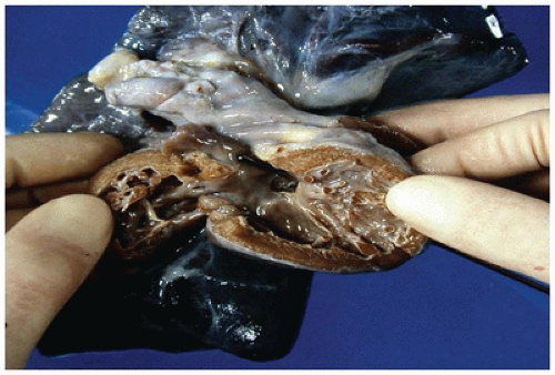
FIGURE 13-5 • Ventricular septal defect. An opened left ventricle with the free wall reflected laterally contains a perimembranous defect, visible in the outflow tract inferior to the aortic valve. The probe visible in the right ventricle in Figure 13-6 traverses the defect opening.


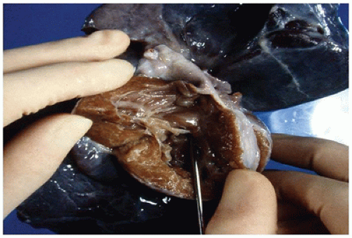
FIGURE 13-6 • Ventricular septal defect. An opened right ventricle with the free wall reflected superiorly contains a perimembranous defect hiding beneath the septal leaflet of the tricuspid valve. The probe traversing the defect is visible from the left ventricular aspect in Figure 13-5.

Spontaneous closure occurs in 25% to 40% of all VSDs and in up to 85% if the defect is small (38,39). Closure of membranous VSDs by overgrowth of fibrous connective tissue or adherence of the tricuspid valve septal leaflet can result in the formation of a ventricular septal “aneurysm.” Ventricular septal aneurysms, with or without complete defect closure, are common in patients with VSD, usually appearing after 2 years of age (40).

The penetrating and branching bundles of the conduction system traverse the membranous portion of the ventricular septum (36,41). This relationship is of particular concern during the surgical repair of perimembranous and inlet defects. Anomalies in the conduction system or an ill-placed suture can result in postoperative bundle branch block or, rarely, sudden death (36).


Malformations of the Atrioventricular Septum

The AV septum is a structure at the crux of the heart that comprises the lower atrial septum and upper ventricular septum, including the atrioventricular valves (42). In the embryo, the AV septum forms at the site of fusion of the four endocardial cushions (43). The spectrum of lesions resulting from defects of the AV septum has been traditionally called endocardial cushion defects (Figure 13-7). AV septal defect displays the following features (42):


TABLE 13-7 VENTRICULAR SEPTAL DEFECTS: CLINICAL GROUPS


Group


Defect Size


Shunt


PVR


Complications


Prognosis/Therapy


1


Small


L→R


Nl


SBE


Spontaneous closure 75%-83%


2


Moderate


L→R


Nl


SBE


Surgical closure required 15%-20%


CHF


3


Large


L→R


Nl


SBE


Surgical closure <2 years


Inc


CHF


4


Large


R→L


Inc


CHF


Inoperable


Cyanosis


PVR, pulmonary vascular resistance; L→R, left to right; R→L, right to left; Nl, normal; Inc, increased; SBE, subacute bacterial endocarditis; CHF, congestive heart failure.


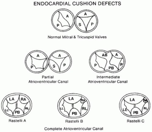
FIGURE 13-7 • Diagrammatic representation of the atrioventricular valves as viewed from the atria in a normal heart and with various atrioventricular septal defects. A, anterior leaflet; P, posterior leaflet; S, septal leaflet; AB, anterior bridging leaflet; PB, posterior bridging leaflet; LA, left anterior leaflet; RA, right anterior leaflet; RL, right lateral leaflet; LL, left lateral leaflet.


  • A decrement of tissue at the crest of the ventricular inlet, lending the inlet septum a “scooped out” appearance


  • Elongation of the left ventricular outflow tract, which creates the “gooseneck” deformity


  • Abnormal formation of the AV valves, with a characteristic “cleft” in the left-sided anterior leaflet

AV septal defects are subdivided into partial and complete forms, depending on the morphology of the AV valve leaflets.


Partial Atrioventricular Septal Defect

Partial AV septal defect is defined by the presence of two discrete AV valve annuli. Partial AV septal defects account for approximately one-third of cases (44). Most hearts in this group contain an ostium primum ASD (Figure 13-8) in conjunction with a cleft in the anterior mitral valve leaflet (42,44). The cleft mitral leaflet inserts, commissure-like, on the ventricular septum. The septal leaflet of the tricuspid valve displays variable degrees of deficiency. When both the mitral and the tricuspid valves are cleft, the
valves insert, commissure-like, onto the rim of the ventricular septum; a connecting tongue of valve tissue covers the ventricular septum and closes the ring. The size of the defect in the ventricular septum also varies in these hearts, and in some cases, the valve leaflets may be less firmly adherent to the ventricular septum so that some interventricular shunting occurs (44).


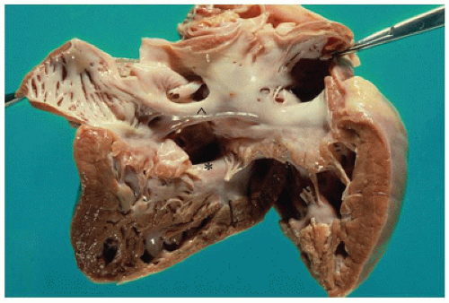
FIGURE 13-8 • Complete atrioventricular septal defect. A complete atrioventricular septal defect is readily visible centrally in this opened left atrium and ventricle. At the upper rim of the defect, a band of atrial septal tissue marked by the ˆ separates the upper secundum ASD from the lower ostium primum ASD. The lower rim of the defect marked by the * represents the upper rim of the ventricular septum. The anterior and the posterior bridging leaflets of the common AV valve extend over the defect without chordal insertion.


TABLE 13-8 ATRIOVENTRICULAR SEPTAL DEFECTS: ASSOCIATED CARDIAC ANOMALIES AND SYNDROMES


Type AVSD


Relative Incidence (% of Total)a


Associated Syndromeb


Associated CV Anomaliesc


Partial


35%


33% with trisomy 21


All AVSD:


Completed


65%


48% with trisomy 21


Subaortic stenosis


Coarctation of the aorta


Patent ductus arteriosus


Double-orifice mitral valve


Parachute mitral valve


Tetralogy of Fallot


Double outlet right ventricle


Common atrium


Rastelli A


55%-70%


Trisomy 21


Subaortic stenosis


Rastelli B


<10%


Valvular aortic stenosis


Coarctation of aorta


Hypoplastic left ventricle


Rastelli C


25%-40%


Heterotaxy


Tetralogy of Fallot


Double-outlet right ventricle


Common atrium


a See references (44,45,261,262,263,264).

b See references (44,261,263).

c See references (265,266,267,268,269).

d See references (45,265,270).


AVSD, atrioventricular septal defect; CV, cardiovascular.


Complete Atrioventricular Septal Defect

In the complete AV septal defect (complete AV canal) (Figure 13-8), the single AV orifice is guarded by five valve leaflets: posterior (inferior) bridging, right lateral, left lateral, right anterior, and left anterior (superior, bridging) (44). The complete AV septal defect is further subclassified according to the extent of septal bridging of the left anterior leaflet and the site of medial insertion:


Rastelli A: minimal bridging, attachment to right rim of septum or medial papillary muscle

Rastelli B: moderate bridging, attachment to an aberrant right apical papillary muscle

Rastelli C: marked bridging, attachment to the anterolateral papillary muscle of the right ventricle

Rastelli types A and C account for the vast majority of cases.

Additional cardiovascular anomalies occur in up to 50% of hearts with either partial or complete AV septal defects (45). The commonly associated anomalies vary in the different subtypes of AV septal defect, as outlined in Table 13-8. At least 50% of patients have trisomy 21 with a variety of other syndromes in another 25% including the heterotaxy syndromes in particular. The ventricles are “unbalanced” in approximately 10% of AV septal defects, with dominant right and dominant left ventricles occurring in nearly equal numbers (44). Clinically, the large left-to-right shunt precipitates severe congestive heart failure. Pulmonary hypertensive vascular changes can appear in the first year of life, further complicating the clinical picture (46). Early surgical repair, usually in the first year of life, is recommended.


TABLE 13-9 TYPES OF CONOTRUNCAL MALFORMATIONS


Transposition of the great vessels


Complete transposition (D-transposition)


Corrected transposition (L-transposition)


Double-outlet ventricles


Double-outlet right ventricle


Double-outlet left ventricle


Persistent truncus arteriosus


Aortopulmonary window (aortopulmonary septal defect)


D, dextro; L, levo.


Malformations of the Conus and Truncus

The conotruncal structures of the heart arise embryologically from the distal aspect of the heart tube and represent the outflow region of the developing heart. Embryologic research demonstrates the importance of cells derived from the neural crest in normal conotruncal development (47). This finding is reflected in humans by the close association between conotruncal malformations and the DiGeorge/velocardiofacial syndrome and its associated 22q11.2 chromosomal deletion (48). Table 13-9 outlines the spectrum of malformations that occur in the conotruncal region.


Transposition of the Great Vessels

In transposition of the great vessels, the aorta arises from the right ventricle and the pulmonary artery from the left ventricle (i.e., discordant ventriculoarterial connections). The transposed aorta thus originates in an anterior position, to either the right (dextrotransposition, or D-transposition) or the left (levotransposition, or L-transposition) of the pulmonary artery. Hearts with transposition are further classified according to their AV connection, as depicted in Figure 13-9.


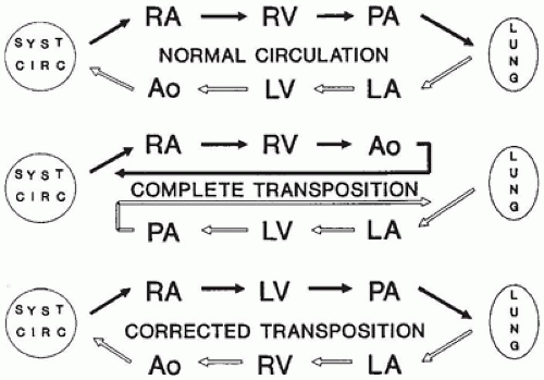
FIGURE 13-9 • Diagrammatic representation of normal blood flow (top), blood flow through complete transposition (middle), and blood flow through “corrected” transposition (bottom). Syst circ, systemic circulation; RA, right atrium; RV, right ventricle; PA, pulmonary artery; LA, left atrium; LV, left ventricle; Ao, aorta.


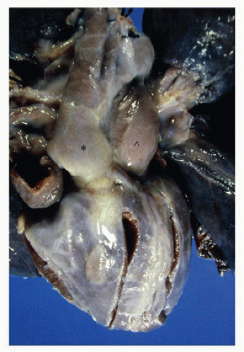
FIGURE 13-10 • Complete transposition of the great vessels from the anterior aspect of the heart. The aorta, marked with *, is situated to the right and slightly anterior to the pulmonary artery, marked with ˆ. The two vessels ascend in a parallel course.


Complete Transposition

Complete transposition, the most common form, accounts for 2.5% to 6.5% of all congenital heart malformations (see Table 13-16), with a 2:1 male predominance (49). The D-transposed aorta ascends parallel and to the right of the pulmonary artery rather than following its normal, twisted course (Figure 13-10). Internal examination reveals AV concordance with ventriculoarterial discordance (50). The aorta originates from the normally positioned morphologic right ventricle, with a muscular band separating the aortic and the tricuspid valves; the pulmonary artery arises posteriorly from the normally positioned morphologic left ventricle and is in fibrous continuity with the anterior mitral valve leaflet. The coronary arteries originate from one or both of the “facing sinuses” of the aorta (i.e., the sinuses adjacent to the pulmonary artery) (15,50). The anatomic course traversed by the coronary arteries varies considerably, a feature of significance when arterial switch surgery is planned (51,52).

A VSD accompanies the complete transposition in approximately 40% of cases, with 40% to 60% of the VSDs showing septal malalignment. Anterior (rightward) malalignment, present in 20% to 25% of cases with VSD, results in subaortic (right ventricular outflow tract) obstruction, often with associated coarctation (53). Pulmonary (left ventricular outflow tract) obstruction occurs in 25% to 30% of hearts with or without VSD, secondary to a malaligned VSD or subvalvular fibrous or fibromuscular tissue bundles (49,52,53).


Corrected Transposition

In the much less common corrected transposition, also known as L-transposition or ventricular inversion, an L-transposed aorta ascends parallel to and to the left of the pulmonary artery. Internally, both AV and ventriculoarterial discordance are present. The right atrium is in continuity with a rightsided morphologic left ventricle from which the pulmonary artery arises; the left atrium is in continuity with a left-sided morphologic right ventricle from which the aorta originates; and blood flow is thus anatomically “corrected” (54) (Figure 13-9). The defect is frequently associated with other congenital anomalies, including tricuspid valve dysplasia, pulmonary outflow tract obstruction, and VSDs (54). AV discordance can occur with other types of ventriculoarterial connections, including DORV and ventriculoarterial concordance (55).


Double-Outlet Ventricle


Double-Outlet Right Ventricle

Of the two forms of double-outlet ventricle (Table 13-9), DORV is the more common, accounting for 1% to 1.5% of congenital heart defects (2). The term DORV denotes a heart in which both great vessels originate from the right ventricle. The strictest criterion requires that both great arteries arise exclusively from the right ventricle, with no fibrous continuity between the mitral and the outflow valves (56,57). A somewhat less strict criterion requires only that both great vessels arise exclusively from the right ventricle; 50% to 60% of such hearts display at least focal fibrous continuity between the mitral and the outflow valves (58,59). A VSD almost always accompanies the DORV, serving as the only site for left ventricular outflow. The variable location of the VSD relative to the pulmonary and aortic valves serves to define pathologic subcategories (56,57,60) (Figure 13-11). The anatomic relationship between the great arteries also varies, as outlined in Table 13-10. In earlier reports, the side-by-side relationship of the great arteries was described as the most frequent one, but more recent series describe the posterior normal pattern as the most common (56,58). The wide varieties of coronary artery anomalies that are associated with side-by-side or malposed great arteries affect surgical repair options and procedures (57,61).


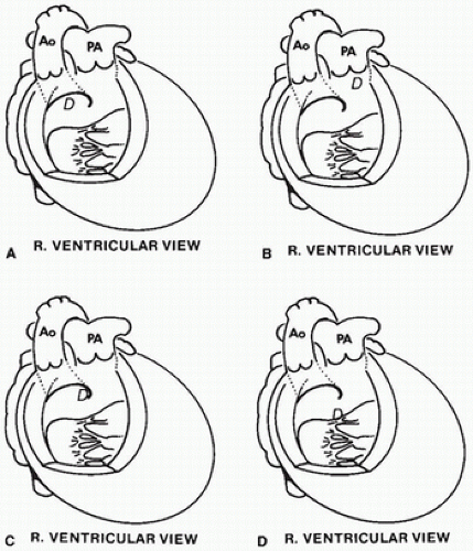
FIGURE 13-11 • The sites, D, of the VSDs in a DORV. A: Subaortic (60% to 65% of total cases). B: Subpulmonic (25% to 30%). C: Doubly committed (5% to 15%). D: Remote (10% to 15%). Ao, aorta; PA, pulmonary artery.


TABLE 13-10 DOUBLE-OUTLET RIGHT VENTRICLE: RELATIONSHIP OF GREAT ARTERIES


Root of Ao Relative to Root of PA


Descriptive Terms


Ascending Ao and PA


Posterior and right


Normal or dextroposition


Spiral


Parallel and right


Side by side


Parallel


Anterior and right


D-malposition


Parallel


Anterior and left


L-malposition


Parallel


Ao, aorta; PA, pulmonary artery; D, dextro; L, levo.


The Taussig-Bing malformation, first described in 1949 (62), is an uncommon variant of DORV in which the VSD is subpulmonic and no pulmonary stenosis is present. When strict criteria are used, fewer than 10% of DORV are of the Taussig-Bing variant (56). When broader criteria are used (“a spectrum of anomalies unified by a juxtapulmonary VSD with malalignment of the infundibular septum”), some hearts can be classified both as the Taussig-Bing variant and as transposition of the great vessels with a malaligned VSD (59,62).

Various other cardiac malformations accompany many DORVs. Pulmonary infundibular stenosis with or without valvular stenosis occurs in 40% to 70% of hearts (56,58). ASDs are not uncommon; complete AV septal defects are less common (24). Left-sided inflow and outflow obstructive lesions may be accompanied by left ventricular hypoplasia (58).

The clinical presentation of DORV depends on the location of the VSD and the presence of associated malformations, particularly pulmonary stenosis (Table 13-11). Surgical correction varies depending on the anatomic configuration of the heart (57,61) (Table 13-11).


Double-Outlet Left Ventricle

DOLV is a rare malformation in which both great vessels arise predominantly from the morphologic left ventricle; a VSD accompanies the vast majority (63,64). Similar to DORV, the DOLV is classified by the location of the VSD relative to the great vessels (63). The DOLV with subaortic VSD is frequently complicated by pulmonary outflow tract
obstruction and DOLV with subpulmonic VSD by aortic outflow tract obstruction (63,64).


TABLE 13-11 DOUBLE-OUTLET RIGHT VENTRICLE: CLINICOPATHOLOGIC CATEGORIES


Clinical Type


VSD Location


RVOTO


LVOTO


Surgical Repair


VSD


Subaortic


Absent


Absent


Intraventricular tunnel


Doubly committed


TOF


Subaortic


Present


Absent


TOF type


Doubly committed


TGV


Subpulmonic (Taussig-Bing)


Absent


Often present


Arterial switch + VSD closure


Intraventricular tunnel


Damus-Kaye-Stansel


Remote VSD


Noncommitted


Absent


Often present


Intraventricular tunnel


Single ventricle repair


VSD, ventricular septal defect; TOF, tetralogy of Fallot; TGV, transposition of the great vessels; RVOTO, right ventricular outflow tract obstruction; LVOTO, left ventricular outflow tract obstruction.


Persistent Truncus Arteriosus

Persistent truncus arteriosus is defined as a single arterial trunk that originates from a single semilunar valve and supplies the aorta, one or both pulmonary arteries, and the coronary arteries (Figure 13-12). The truncal valve is tricuspid in 50% to 70% of cases, quadricuspid in 25%, and bicuspid in most of the rest. The truncal vessel overlies and usually overrides an infundibular VSD, although occasionally, it is predominantly committed to one ventricular chamber. The truncal valve is always in fibrous continuity with the mitral valve; fibrous continuity may also be present between the truncal and tricuspid valves. The truncal valve leaflets are frequently thickened and myxomatous, with valvular insufficiency present in approximately 15% to 30%. The coronary arteries originate from the sinuses of the truncal valve in a variable pattern, with a single coronary artery present in 15% to 20% (65).


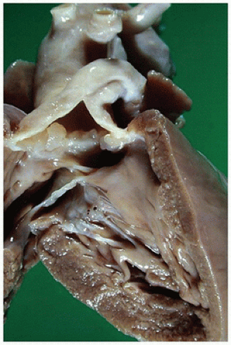
FIGURE 13-12 • Truncus arteriosus. The left ventricle free wall has been lifted to uncover the smooth-surfaced left ventricular outflow tract with a VSD opening at the top. Above the VSD lies a somewhat nodular truncus arteriosus valve. The main pulmonary artery almost immediately branches to the left from the common trunk; the aorta continues ascending posteriorly.

A classification devised by van Praagh creates a separate subtype for this latter finding and also acknowledges the rare case in which there is no VSD (66). The classification was subsequently revised by van Praagh to simplify the scheme in a surgically meaningful fashion (67) (Table 13-12).

Associated anomalies most frequently involve the aortic arch and include absent ductus arteriosus (>50%), rightsided aortic arch (20% to 35%), and interrupted aortic arch type B (10%) (65). Extracardiac anomalies, especially those related to DiGeorge syndrome, occur in 20% to 30%, and the DiGeorge syndrome-associated chromosome 22q11 deletion can be detected by fluorescence in situ hybridization (FISH) in 35% to 50% of infants with persistent truncus arteriosus (68,69).

The early clinical manifestation of congestive heart failure results from intracardiac shunting and markedly excessive pulmonary blood flow. The excessive pulmonary blood flow also produces rapidly progressive pulmonary hypertensive vascular disease; early surgical repair is therefore recommended.


Aortopulmonary Window

Aortopulmonary window, or aortopulmonary septal defect, is a rare malformation characterized by a defect in the vessel wall between the ascending aorta and the main pulmonary artery. The defect may lie proximally (just above the aortic and the pulmonary valves), distally (in the upper ascending aorta adjacent to the right pulmonary artery), or as a combined opening that involves the majority of the ascending aorta (14). Associated anomalies, present in more than 50% of cases, commonly include a VSD, interrupted aortic arch type A, and anomalous origin of one pulmonary artery from the ascending aorta (70). Although aortopulmonary window occurs in
the same general region as persistent truncus arteriosus, it is not seen in the chromosome 22q11 deletion syndromes (70).


TABLE 13-12 TRUNCUS ARTERIOSUS CLASSIFICATION


Collett and Edwards


van Praagh


Modified van Praagh


Type 1


Type 1


Large aorta type


PAs arise as a single main artery and then divide.


PAs arise as a single main artery and then divide.


TA with confluent or near confluent PAs


Type 2


Type 2


PAs arise separately but next to each other.


PAs arise separately.


Type 3


PAs arise widely separated.


Type 3


TA (large aorta type) with absence of one PA


One PA branch “absent” Arises from ductus or aorta


Type 4


Large pulmonary artery type


Aortic arch hypoplastic or interrupted


TA with IAA or severe COTA


Type A = VSD present


Type B = VSD absent


PA, pulmonary artery; VSD, ventricular septal defect; TA, truncus arteriosus; IAA, interrupted aortic arch; COTA, coarctation of the aorta.


Malformations of the Ventricular Inflow Tracts


Tricuspid Valve Malformations


Tricuspid Atresia

Tricuspid atresia, in which the only outlet to the right atrium is via a patent fossa ovalis or an ASD, accounts for 1% to 1.5% of congenital heart malformations (5). The markedly hypoplastic right ventricle, positioned along the right anterosuperior border of the heart, has no inlet segment. The markedly dilated right atrium contains no grossly identifiable valvular tissue in more than 85% of cases (18,71). A dimple in the muscular atrial floor, presumably marking the site of the missing valve, may have a fibrous attachment to the right ventricle but often is instead in continuity with the left ventricle by transillumination and pinprick studies (18). The remaining 5% to 15% of hearts display a tricuspid valve remnant in the form of an imperforate fibrous membrane. A muscular VSD, termed the outlet foramen, allows communication between the dominant left ventricular chamber and the rudimentary right ventricle; however, the VSD or the infundibular outflow tract may be restrictive (71).

Tricuspid atresia is subclassified according to the size of the VSD, concordance or discordance of the great vessels, and the presence or absence of pulmonary stenosis/atresia (Table 13-13). The clinical symptoms depend on these anatomic variables; more than 50% of cases present with cyanosis and murmur in the newborn period (72). In the vast majority of hearts, the right ventricle is too small to function adequately as a pumping chamber, and univentricular repair is required.


Ebstein Malformation

Ebstein malformation accounts for less than 1% of all cases of CHD but is the most common cause of isolated tricuspid stenosis or insufficiency (2,73). It is characterized by adherence of variable portions of the septal and posterior tricuspid valve leaflets to the right ventricular wall, with “atrialization” (i.e., downward displacement of the functional annulus) of a portion of the right ventricle (Figure 13-13) (73,74). The anterior valve annulus insertion is normally positioned, with a large, redundant, and often muscularized leaflet. The margin of the leaflet may be attached to the posteroinferior right ventricular wall and produce obstruction and in some cases complete occlusion of the AV orifice (Figure 13-14) (74). Tricuspid regurgitation occurs across the dilated AV junction (true annulus). Right-to-left shunting across a patent fossa ovalis or ASD and supraventricular arrhythmias due to accessory conduction pathways frequently complicate Ebstein malformation. A variety of
other associated cardiovascular defects, most commonly pulmonary valvular stenosis, pulmonary atresia, or a VSD, occur in 30% to 40% of cases (73,75).


TABLE 13-13 TRICUSPID ATRESIA: CLINICAL CLASSIFICATION


I.


Normally related great vessels (60%-70%)


A. Intact ventricular septum with pulmonary atresia


B. Small VSD with pulmonary stenosis


C. Large VSD without pulmonary stenosis


II.


D-transposition of the great arteries (25%-30%)


A. VSD with pulmonary atresia


B. VSD with pulmonary stenosis


C. VSD without pulmonary stenosis


II.


Malposition other than D-transposition of the great arteries (5%)


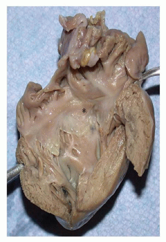
FIGURE 13-13 • Mild form of Ebstein anomaly. The opened right atrium and right ventricle display a markedly thickened ventricular wall. The septal (asterisk) and posterior leaflets of the tricuspid valve are fixed to the underlying ventricular wall.

Given the broad range of anatomic alterations encompassed by Ebstein malformation, it is not surprising to find a broad range of clinical manifestations for the disorder. Onethird to one-half of patients present in the newborn period with cyanosis and a murmur; the mortality rate among such infants is high, particularly when the malformation is associated with additional cardiac anomalies (73). In many patients, however, the diagnosis is delayed until the second decade of life or later, when arrhythmias often represent the major clinical problem (73,75).


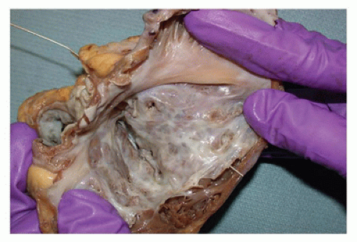
FIGURE 13-14 • Severe form of Ebstein anomaly. The opened right atrium uncovers a markedly enlarged and dysplastic anterior tricuspid leaflet attached to the apical myocardium by tiny chordae. A probe placed in the pulmonary artery traverses the remaining right ventricular cavity and appears at the base of this dysplastic valve, illustrating the severe obstruction to pulmonary inflow and outflow created by this defect.


Mitral Valve Malformations


Mitral Stenosis

The normal mitral valve apparatus is a complex structure with four primary components: annulus, anterior and posterior valve leaflets, chordae tendineae, and anterolateral and posteromedial papillary muscles. A variety of malformations affecting any or all of the valve components result in congenital mitral stenosis and insufficiency (76) (Table 13-14). A supramitral ring, a ridge of connective tissue at the atrial surface of the mitral leaflets, usually occurs with deformities of the mitral valve apparatus (77). Valve hypoplasia, in which the valve components are small but otherwise normally formed, is most commonly associated with left ventricle hypoplasia, VSDs, and coarctation of the aorta (COTA) (14,77). The “typical” mitral stenosis manifests as lesions at both the valvular and the subvalvular areas including valve dysplasia with commissure fusion, obliteration of the intrachordal spaces, and shortening of the chordae tendineae and papillary muscles. Associated malformations include tetralogy of Fallot (TOF), COTA, and subaortic stenosis with a near-normal-sized left ventricle (14,77). The double-orifice mitral valve results when excessive valve tissue bridges between the anterior and posterior valve leaflets to create two, usually unequally sized, orifices, both supported by chordal attachments that insert into often abnormally positioned papillary muscle. The double-orifice valve almost always occurs in company with other cardiac malformations, especially AV septal defects (approximately 50% of cases) or left-sided obstructive lesions (40%) (78). Two forms of cleft mitral valve without associated primum ASD or VSD have been described (78):


TABLE 13-14 MITRAL VALVE MALFORMATIONS


Supravalvular lesions


Supramitral ring


Valvular lesions


Valve hypoplasia


Valve dysplasia


Commissural fusion


Valve leaflet excess or agenesis


Double-orifice valve


Cleft mitral valve


Subvalvular lesions


Parachute deformity (single papillary muscle)


Funnel deformity (shortened fused chordae)


Arcade deformity (papillary muscle fused with valve)


Mixed


Shone syndrome


  • Associated with normally related great vessels and a shortened inlet septum (i.e., forme fruste of an AV septal defect)


  • Associated with TGA or DORV and a normal inlet septum

Parachute deformity of the mitral valve, defined as insertion of all the chordae into a single papillary muscle group, also usually occurs with other malformations of the heart, particularly VSDs and obstructive lesions of the aortic valve and arch (14,77,79). The eponym Shone syndrome denotes the association of a parachute mitral valve with a supramitral ring, subaortic stenosis, and COTA (79,80).


Mitral Atresia

Mitral atresia, defined as the absence of a left AV connection, is marked on the left atrial aspect by muscular atrial floor with or without a visible dimple or, less commonly, by an imperforate membrane (14,81,82). The microscopic examination of hearts with no grossly obvious membrane between the left atrium and the left ventricle uniformly reveals a fibrous connection at the presumed site of the absent valve (81). The outlet for pulmonary venous return is by way of a patent fossa ovalis or less commonly an ASD (14). Rarely, the fossa ovalis is prematurely closed, and pulmonary venous return is shunted to the right side of the heart by anomalous venous connections (14,83). When the great vessels are normally related, mitral atresia is most commonly associated with aortic atresia and, as such, is included in the hypoplastic left heart syndrome. The left ventricle exists as a diminutive chamber lined by translucent endocardium, which in some cases is evident only on microscopic examination of the posterosuperior aspect of the hypertrophic right ventricle (14,82). Less often, a VSD is present and a patent aortic valve arises from either the right (DORV) or the left ventricular chamber (14,82).


Floppy Mitral Valve

Floppy mitral valve represents the central defect in the floppy mitral valve (FMV)/mitral valve prolapse (MVP)/mitral valve regurgitation (MVR) triad. The primary defect in the “floppy” valve is deposition of acid mucopolysaccharides and dissolution of the collagen in the pars spongiosa and fibrosa of the valve (84). The accumulation of myxoid material leads to thickened and enlarged valve leaflets often with increased chordal insertions on the ventricular surface, elongation of the chordae tendineae, and dilatation of the valve annulus (84). With prolapse, the valve becomes “hooded,” defined as the presence of ballooning to a height of at least 4 mm and involving at least one-half of the anterior or two-thirds of the posterior mitral leaflets (85). The myxomatous degeneration in the valve leaflets is without inflammation and does not lead to fusion of the valve commissures, distinguishing these valvular changes from those of rheumatic fever (RF). Similar myxomatous changes occur elsewhere in the heart, including the conduction system, a feature that likely explains the associated arrhythmias and the conduction defects.

The reported incidence of FMV/MVP/MVR varies considerably, with less than 1% to 5% of children exhibiting clinical or echocardiographic features of MVP (86). In the pediatric population, the incidence increases with age; MVP is extremely rare before 2 years of age. Most children are asymptomatic, presenting with the characteristic late systolic “click” on auscultation; an occasional child presents with chest pain of unclear etiology (86). Skeletal anomalies, especially pectus excavatum, are common (87). The 2:1 female predominance described in adults is also observed in some but not all groups of children studied (86). Progressive MVR, a major problem in adults with FMV, occurs rarely during childhood. Other complications, including infectious endocarditis, thromboembolism, arrhythmias, and even sudden death, do occur occasionally in the pediatric population (86).


Univentricular Atrioventricular Connection

The term univentricular AV connection denotes the connection of the AV valves to a single ventricular cavity (82). Table 13-15 lists at least some of the terms previously used for this condition and outlines the range of anomalies encompassed. The anatomy of such hearts can be highly complex and variable, so that sequential segmental analysis is an essential tool for accurate classification (82).


Double-Inlet Ventricle

In double-inlet left ventricle, the most common of the doubleinlet malformations, both the left and right AV valves open into a dominant left ventricular cavity (88). The rudimentary right ventricle occupies the right anterosuperior border of a normally related, D-looped left ventricle or the left anterosuperior border of an inverted, L-looped left ventricle. The rudimentary right ventricle communicates with the dominant left ventricle via a variably sized VSD. The great vessels are
transposed in the vast majority of cases but may be normally related, atretic, or in a double-outlet configuration.


TABLE 13-15 UNIVENTRICULAR ATRIOVENTRICULAR CONNECTION


Common synonyms


Single ventricle


Common ventricle


Holmes heart


Univentricular heart


Cor triloculare biatriatum


Primitive ventricle


Anatomic subtypes


Double-inlet ventricle


Double-inlet left ventricle


Double-inlet right ventricle


Double-inlet ventricle of mixed morphology (absent ventricular septum)


Double-inlet ventricle of indeterminate morphology


Single-inlet ventricle


Mitral atresia


Tricuspid atresia


Common-inlet ventricle


Overriding atrioventricular valves


Common-Inlet Ventricle

In common-inlet ventricle, both atria communicate with a single ventricle via a common AV valve (88). Many of these hearts represent the extreme form of unbalanced AV septal defect and thus include a dominant right or left ventricular cavity and a rudimentary second ventricle. Less often, the common AV valve communicates with a single ventricular chamber of indeterminate type without an identifiable rudimentary ventricle. These hearts frequently have abnormal ventriculoarterial connections as well.


Straddling and Overriding Atrioventricular Valves

An AV valve annulus may override, or its chordal insertion may straddle, the ventricular septum (88,89). In hearts with valve annulus override, the AV connection is assigned to the ventricle to which more than 50% of the valve annulus is attached. Straddling of the chordae without valve annulus override does not change the AV connection designation.


Malformations of the Ventricular Outflow Tracts


Pulmonary Outflow Tract and Valve Malformations


Tetralogy of Fallot

TOF accounts for 3.5% to 10.5% of all CHD and represents the most common cyanotic CHD. In approximately 33% of cases, four components comprise the TOF: infundibular pulmonic stenosis, VSD, aortic valve dextroposition, and right ventricular hypertrophy (Figure 13-15). All of these anatomic components are a consequence of a single embryologic abnormality, anterosuperior malalignment of the outlet septum (Figures 13-15 and 13-16). The morphologic detail surrounding these four components can vary considerably (14,90,91). Infundibular pulmonic stenosis leads to decreased pulmonary blood flow with an associated small pulmonary artery (Figure 13-16). Over time, the stenosis becomes exacerbated by hypertrophy of the infundibular septum or cristal structures (91). The invariably large and nonrestrictive VSD is perimembranous in 75% of cases, located in the muscular outlet in 20%, and subarterial only rarely (90,91). The degree of aortic override varies from 15% to 95%. In the extreme situation, the differentiation of TOF from DORV depends on the presence of the characteristic infundibular stenosis and fibrous continuity between the aortic and mitral valves; some investigators classify hearts with more than 50% aortic override as TOF with DORV (59).

The pulmonary valve is abnormal in 66% to 75% of cases. It is most often bicuspid but may be unicuspid or stenotic by virtue of thickened dysplastic valve leaflets (14,90,91). The 20% to 25% of cases with an imperforate pulmonary valve orifice are classified as pulmonary atresia with VSD, discussed in more detail later. The pulmonary arteries show a range of accompanying abnormalities that include localized stenosis at the origin of the pulmonary artery branches, central pulmonary artery discontinuity, absent left pulmonary artery branch, and pulmonary hilar artery hypoplasia (91,92). When the pulmonary artery stenosis is severe, pulmonary artery hypertension may develop after surgical repair of the TOF (93). In 3% to 6% of cases, the pulmonary valve is absent and the pulmonary arteries are dilated; this dilatation may be significant.


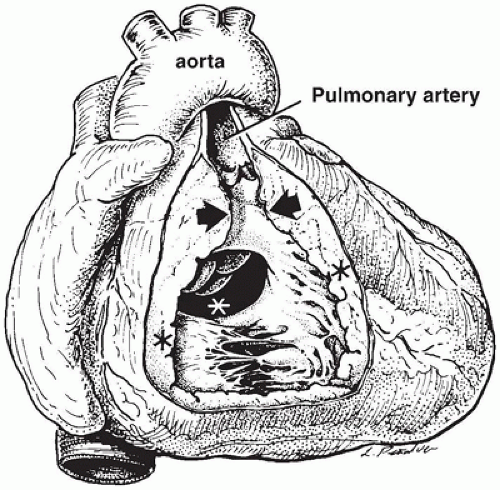
FIGURE 13-15 • An opened anterior right ventricle illustrates the four primary features of TOF: marked narrowing of the pulmonary infundibulum (between arrows); a large perimembranous VSD (white asterisk); dextroposed overriding aorta, visible through the VSD; and hypertrophy of the right ventricular myocardium (black asterisk).

A variety of other cardiovascular defects occur with TOF. TOF occurs as part of a recognizable syndrome, most commonly DiGeorge syndrome or trisomy 21 (92). Commonly associated anomalies include right-sided aortic arch (20% to 30%) and absent ductus arteriosus (20% to 25%) (92). Although a patent fossa ovalis occurs commonly in infants with TOF, a true ASD is present in only 20% to 25%.
A complete AV septal defect accompanies TOF in 1% to 2% of cases, most often in children with trisomy 21.


FIGURE 13-16 • Heart and lungs removed at autopsy with an unrepaired TOF. An incision through the anterior right ventricle ends at the base of a small pulmonary artery. The markedly enlarged aorta arises behind and to the right of the pulmonary artery.

Hypoxia and cyanosis are the principal symptoms of TOF, their severity varying with the degree of pulmonary obstruction (92). In the presence of marked stenosis or atresia, cyanosis is evident in the neonatal period. More commonly, cyanosis appears in the first 6 months of life, associated with increasing infundibular stenosis.


Pulmonary Atresia with Ventricular Septal Defect

Although the designation pulmonary atresia (PA) with VSD (PA/VSD) is frequently used as a synonym for TOF with PA, not all hearts with PA/VSD display the hypoplastic right ventricular infundibulum characteristic of TOF (94). Most commonly, a dimple (more rarely recognizable fused bicuspid valve cusps) marks the site of the pulmonary valve. Abnormalities of the pulmonary arteries include a connection of confluent, bilateral pulmonary arteries to the right ventricle by an atretic cord, absence of the left pulmonary artery, and absence of all intrapericardial pulmonary arteries (95). This latter group was classified as truncus arteriosus type IV in the past. The ductus arteriosus supplies blood to one or both of the pulmonary arteries in 40% to 65% of cases. The lungs also receive blood via collateral arteries that originate from the descending aorta and supply the pulmonary arteries via intrapulmonary, hilar, or extrapulmonary anastomoses (95). Chromosome 22q11 deletion increases the likelihood of an absent ductus arteriosus and a major aortopulmonary collateral artery supply. With a major aortopulmonary collateral artery supply, hypoplasia and arborization of the pulmonary arteries make surgical management problematic (96).


Absent Pulmonary Valve

Absent pulmonary valve is a rare anomaly usually associated with TOF (97). At the site of the expected valve, a narrow valve annulus is rimmed by rudimentary, nodular, gelatinous tissue with massive poststenotic dilatation (97). More complex pulmonary artery anomalies, including discontinuity of the main pulmonary arteries, anomalous origin of the pulmonary arteries, and absence of the left pulmonary artery, occur less often (97,98). The ductus arteriosus is frequently absent (50). Dilated pulmonary arteries can compress the adjacent bronchi and cause respiratory compromise; abnormalities of the intrapulmonary arteries and bronchi may exacerbate these pulmonary problems (98).


Pulmonary Stenosis with Intact Ventricular Septum

Pulmonary stenosis with intact ventricular septum (IVS) accounts for 2.5% to 9.0% of all cases of CHD (Table 13-16). The obstruction is usually valvular, with secondary right ventricular hypertrophy and poststenotic dilatation of the pulmonary trunk. The valve is most often dome-shaped with fused cusps and a single central orifice, but it may be unicuspid, bicuspid, or tricuspid with partially fused commissures (99,100). Thickened dysplastic valve leaflets with nonfused cusps occur sporadically and as one form of pulmonary stenosis in Noonan syndrome. The pulmonary artery trunk usually exhibits poststenotic dilatation; pulmonary artery hypoplasia is rare even with critical stenosis. Symptoms depend on the severity of the stenosis. Critical stenosis, presenting as cyanosis in infancy, requires early intervention (100). More commonly, infants are asymptomatic; stenosis develops and worsens in early childhood in approximately 15%, and many remain asymptomatic into adulthood. Secondary infundibular stenosis due to right ventricular hypertrophy can further complicate the course over time (101).


TABLE 13-16 PREVALENCE OF CONGENITAL HEART DEFECTS


Type


Range (%)a


M:F


VSD


30-52


1:1


PDA


2.5-8.5


1:2


TOF


3.5-10.5


1:1


COTA


4.5-6.5


3:2


TGA


2.5-6.5


2:1


ASD


6.0-8.0


1:2


PS-IVS


2.5-9.0


1:1


AVSD


1.5-9.5


2:3


HLHS


2.0-5.0


3:2


AS


3.0-6.0


2:1


a Percentage of total for ten most common defects; see references (39,271,272,273,274,275,276,277,278).


VSD, ventricular septal defect; PDA, patent ductus arteriosus; TOF, tetralogy of Fallot; COTA, coarctation of the aorta; TGA, transposition of the great arteries; ASD, atrial septal defect; PS-IVS, pulmonary stenosis with intact ventricular septum; AVSD, atrioventricular septal defect; HLHS, hypoplastic left heart syndrome; AS, aortic stenosis.


Subvalvular Stenosis

Pulmonary subvalvular or infundibular stenosis, which accounts for fewer than 10% of cases of pulmonary stenosis with IVS, occurs when fibrous thickening at the junction of the trabecular and outlet segments divides the right ventricle into two chambers; it may also be caused by tubular hypoplasia of the infundibulum (14). Double-chamber right ventricle is a closely related anomaly in which hypertrophied muscle bands cross the right ventricular cavity just proximal to the infundibulum and divide a high-pressure proximal chamber from a low-pressure infundibular chamber. The majority of hearts with double-chamber right ventricle exhibit other anomalies, most often (65% to 75%) a VSD (102).


Supravalvular and Peripheral Pulmonary Artery Stenosis

Supravalvular or pulmonary artery stenosis occurs as a localized area of narrowing in the pulmonary trunk or branch or as multiple areas of narrowing throughout the pulmonary artery tree (103,104). The stenosis is subclassified into four types, based on the site of the obstruction, with type III accounting
for approximately one-third of cases (104) (Table 13-17). Associated cardiac malformations, present in approximately 66% of cases, include VSD, ASD, valvular pulmonary stenosis, and TOF (104,105). A variety of malformation syndromes include pulmonary artery stenosis (Table 13-18).


TABLE 13-17 SUPRAVALVULAR AND PERIPHERAL PULMONARY ARTERY STENOSIS


Type I Single central stenosis of:


A Main pulmonary artery


B Right main pulmonary artery


C Left main pulmonary artery


Type II Bifurcation stenosis


A Short, localized stenosis


B Long, narrow segments of stenosis


Type III Multiple stenoses of peripheral segmental arteries


Type IV Multiple stenoses of peripheral and central arteries


Pulmonary Atresia with Intact Ventricular Septum

PA with IVS accounts for 1% to 3% of all cases of CHD. The atresia is usually valvular, with a fibrous membrane containing commissural lines present at the expected site of the valve (14,106). The pulmonary artery is funnel-shaped and usually only mildly to moderately hypoplastic (14,106). The right ventricular myocardium is hypertrophied and the cavity of variable size, with the size of the tricuspid valve directly related to the size of the right ventricle. The right ventricle and tricuspid valve are usually small, often with associated pulmonary infundibular stenosis or atresia (106,107). Right ventricle-coronary artery fistulas develop in more than 50% of hearts with small ventricular cavities; in a subgroup of these hearts, the volume of flow through the fistula results in right ventricle-dependent coronary blood flow. Accompanying coronary artery luminal stenosis and vessel atrophy proximal to the fistula site can result in myocardial ischemia and sudden death (108). At the opposite extreme, the right ventricular cavity may be dilated, with the tricuspid valve showing dysplasia and often Ebsteinization (106,107).


TABLE 13-18 PULMONARY ARTERY STENOSIS-ASSOCIATED MALFORMATION SYNDROMES


Syndrome


Location of Stenosis


References


Rubella


Peripheral pulmonary arteries


(279)


Williams


Peripheral pulmonary arteries


(267,280)


Alagille


Peripheral pulmonary arteries


(281)


Noonan


Main pulmonary artery


(282)


PA with IVS is a severe form of CHD in which pulmonary blood flow depends on a patent ductus arteriosus. The definitive surgical management varies according to the degree of right ventricular hypoplasia, the presence and severity of coronary artery fistula, and the status of the tricuspid valve with options including a variety of outflow tract (“biventricular”) repairs, univentricular repair, or transplantation (106,109).


Aortic Outflow Tract and Valve Malformations


Aortic Valvular Stenosis

The left ventricular outflow tract may be obstructed at any level, but the most common form of obstruction is aortic valvular stenosis. The spectrum of abnormal valvular morphology is similar to that in pulmonary valvular stenosis, but a bicuspid valve is the most common form (14,110). Bicuspid aortic valves are not congenitally stenotic and many remain asymptomatic throughout childhood, with a significant subset progressing quickly to significant stenosis requiring intervention. Unicommissural and the less common tricuspid dysplastic and dome-shaped valves are stenotic from birth and therefore more likely to be symptomatic in early childhood. With congenitally stenotic valves, the left ventricle may be dilated, normal in size, or hypoplastic. In infants with severe stenosis, endocardial fibroelastosis (EFE) and subendocardial ischemic damage often further complicate the picture (110). COTA commonly accompanies a congenitally malformed aortic valve in two clinical settings: critical aortic stenosis (110) and bicuspid valves related to Turner syndrome.


Subvalvular Aortic Stenosis

Discrete subvalvular aortic stenosis most frequently takes the form of a fibroelastic diaphragm just beneath the base of the aortic valve; less common forms include a thickened diaphragm with a muscular base or a tunnellike narrowing of the outflow tract (14,111). Commonly associated heart defects, present in 50% to 75% of cases, include a malaligned VSD, aortic valvular stenosis or regurgitation, aortic coarctation, and AV septal defects (110,111). These subaortic lesions rarely present in infancy, and current theories consider them to be acquired progressively, perhaps on the framework of an underlying subtle deformation of the outflow tract (111,112).

Only gold members can continue reading. Log In or Register to continue

Stay updated, free articles. Join our Telegram channel

Sep 23, 2016 | Posted by in PATHOLOGY & LABORATORY MEDICINE | Comments Off on The Cardiovascular System

Full access? Get Clinical Tree

Get Clinical Tree app for offline access