Examination of the Abdomen
General observation: note
- is the patient in pain?
- evidence of weight loss
Inspect
- tongue, mouth, teeth and throat
- limbs for evidence of IV drug use
Examine the hands for
- clubbing
- leukonychia, koilonychia
- palmar erythema
- spider naevi
- Dupuytren’s contracture
Inspect the eyes and conjunctivae for
- anaemia
- jaundice
- xanthelasmata
Palpate for lymphadenopathy
- neck
- supraclavicular fossae
- axillae
- groins
A scheme for examination of the abdomen is shown in Fig. 5.1. Lie the patient flat (one pillow) with arms by the sides. Look before palpation, have warm hands and palpate gently so as to gain the patient’s confidence and to avoid hurting them. Ask the patient to let you know if you are hurting them. Check this by looking at the patient’s face periodically during palpation, especially if you elicit guarding or rebound tenderness.
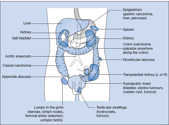
Figure 5.1 Examination of the abdomen: (1) gently palpate all quadrants for tenderness and masses; (2) feel for enlarged liver, spleen or kidneys, and for masses arising from the bowel (in the left and right iliac fossae), the epigastrium (stomach and pancreas), the aorta (aneurysm), or the pelvis (uterus, bladder and ovaries); (3) percuss and auscultate over enlarged organs or masses; (4) examine hernial orifices and external genitalia in men; and (5) perform a rectal examination.

Observe the abdomen for
- general swelling with eversion of the umbilicus in ascites
- visible enlargement of internal organs: liver, spleen, kidneys, gall bladder, stomach, urinary bladder and pelvic organs
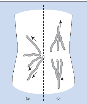
- abnormally distended veins: usually in cirrhosis with the direction of flow away from the umbilicus (portal hypertension). The flow is upwards from the groin in inferior vena cava obstruction (Fig. 5.2)
- scars of previous operations, striae, skin rashes and purpura
- pigmentation
- visible peristalsis
Figure 5.2 Blood flow through dilated anterior abdominal wall veins. Portal vein and inferior vena caval occlusion can be distinguished by the different directions of flow below the umbilicus. (a) Direction of flow in portal vein occlusion. (b) Direction of flow in inferior vena caval occlusion.

Palpate and percuss
- For internal organs and masses: start palpation in the right iliac fossa and work upwards towards the hepatic and splenic areas, first superficially and then deeper.
- Percuss the liver and spleen areas to avoid missing the lower border of a very large liver or spleen.
Liver
- The upper border is in the fourth to fifth intercostal space on percussion.
- The liver moves down on inspiration.
- Percussion over the liver is dull.
- If enlarged, the liver edge may be tender, regular or irregular, hard, firm or soft.
- Pulsatility suggests tricuspid incompetence.
- The liver may be of normal size but low because of hyperinflated lungs in chronic obstructive airway disease.
Spleen
- Smooth rounded swelling in left subcostal region, usually with a distinct lower edge.
- The spleen enlarges diagonally downward and across the abdomen in line with the ninth rib.
- The examining hand cannot get above the swelling.
- Percussion over the spleen is dull.
- There is a notch on the lower border of the spleen
- The spleen may be more easily palpated with the patient lying on the right side with the left leg flexed and abducted.
Kidneys
- Palpated in the loins bimanually, i.e. most easily felt by pushing the kidney forwards from behind on to the anterior palpating hand.
- They move slightly downwards on inspiration.
- The examining hand can easily get between the swelling and the costal margin.
- Percussion is resonant over the kidneys.
- The lower pole of the right kidney can often be felt in thin normal persons.
Abnormal Masses
- Palpate for abnormal masses particularly in the epigastrium (gastric carcinoma) and suprapubic region (bladder distension, ovarian and uterine masses). Describe in terms of their size and margins.
- Note colonic masses. The descending colon is commonly palpable in the left iliac fossa.
- The abdominal aorta is pulsatile, bifurcates at the level of the umbilicus and is easily palpable in thin and lordotic patients. Abdominal aortic aneurysms are expansile.
- Check for ascites: examine for shifting dullness by noting a change in percussion note with the patient supine and lying on their left side. A fluid thrill may be demonstrable in large, tense effusions.
Complete the examination
- femoral pulses
- hernial orifices
- leg oedema
- external genitalia
- digital rectal examination
Auscultate for
- bowel sounds
- renal and femoral bruits
Notes
Splenomegaly
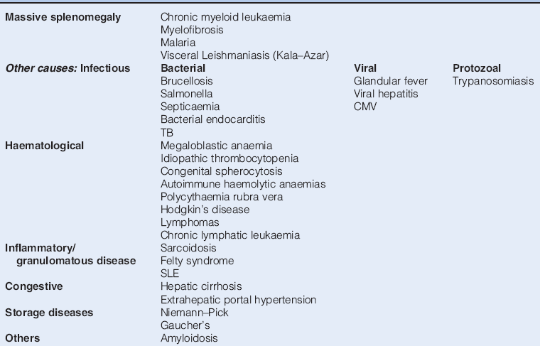
The major causes of splenomegaly are haematological and infectious (Table 5.2.)
Table 5.2 Causes of Splenomegaly

Hepatomegaly
The causes of hepatomegaly are shown in Table 5.3.
Table 5.3 Causes of Hepatomegaly
| Common | Congestive cardiac failure |
| Secondary carcinomatous deposits | |
| Cirrhosis (usually alcoholic) | |
| Other causes:Infectious | Glandular fever |
| Viral hepatitis | |
| HIV/AIDS | |
| Amoebic cysts | |
| Hydatid cysts | |
| Haematological | Leukaemia |
| Reticuloendothelial disorders | |
| Malignant | Hepatoma |
| Granulomatous disease | Sarcoidosis |
| Storage diseases | Gaucher’s |
| Others | Primary biliary cirrhosis |
| Haemochromatosis | |
| Amyloidosis |
Hepatosplenomegaly
The causes are similar to those for splenomegaly alone:
- chronic leukaemias
- cirrhosis with portal hypertension
- lymphoproliferative disorders
- myelofibrosis
Palpable Kidneys
The left kidney is nearly always impalpable, but the lower pole of a normal right kidney may be felt in thin people.
Unilateral enlargement
- renal cell carcinoma
- hydronephrosis
- cysts
- hypertrophy of a single functioning kidney
Bilateral enlargement
- polycystic kidney disease
- bilateral hydronephrosis
- amyloidosis
Following renal transplantation a kidney may be palpable in one iliac fossa and the patient may appear cushingoid as a result of steroid therapy. Look for scars of previous surgery and forearm arteriovenous shunts.
Mass in Right Subcostal Region
Consider
- liver (including Riedel’s lobe)
- colon
- kidney
- gall bladder (rarely)
Investigations
- ultrasound: identifies solid or cystic masses
- abdominal CT scan
- liver function tests
- urine for haematuria and proteinuria
- stool for occult bleeding
As appropriate
- sigmoidoscopy, barium enema and colonoscopy
- MRI scan
Suprapubic Mass
Consider
- bladder distension
- pregnancy
- uterine masses (usually benign fibroid tumours, rarely malignancy)
- ovarian tumours
Ascites
Clinical features
- abdominal distension
- dullness to percussion in the flanks
- shifting dullness
- fluid thrill
Causes
- intra-abdominal neoplasia including gynaecological pathology
- hepatic cirrhosis with portal hypertension
- congestive cardiac failure
- constrictive pericarditis
- nephrotic syndrome
- low albumin states
- tuberculous peritonitis
Diagnostic paracentesis should be performed and examined for:
- protein content (>30 g/l suggests an exudate, <30 g/l suggests a transudate)
- microscopy and bacterial culture (including acid-fast bacilli)
- cytology.
Paracentesis may occasionally be required for the relief of severe symptoms; repeated paracentesis leads to excessive protein loss and should be avoided if possible.
Dysphagia
Dysphagia includes both difficulty with swallowing and pain on swallowing. The former symptom is more prominent in obstruction and the latter with inflammatory lesions.
Causes
Common
- carcinoma of the oesophagus
- carcinoma of the gastric fundus
- peptic oesophagitis (with pain) proceeds to stricture with difficulty in swallowing
Rare
- achalasia of the cardia
- external pressure: carcinoma of the bronchus, retrosternal goitre
- dysmotility syndrome
- neurological disease: myasthenia gravis, bulbar palsies
- Plummer–Vinson syndrome – iron deficiency anaemia, koilonychia and glossitis with a post-cricoid oesophageal web
Investigations
- haemoglobin, serum iron and ferritin
- barium swallow (Fig. 5.4)
- upper GI endoscopy with biopsy to differentiate between benign peptic stricture and carcinoma
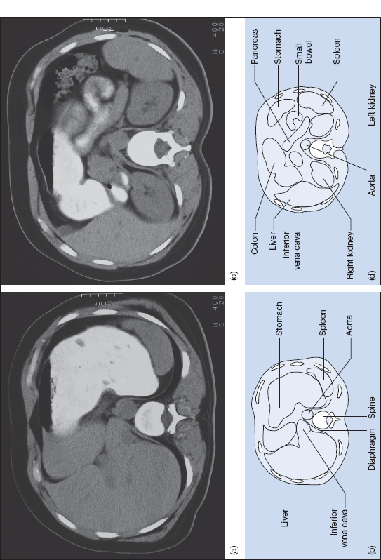
Figure 5.3 (a) CT and (b) diagrammatic representation at the level of T11 vertebra. (c) CT and (d) diagrammatic representation at the level of L1 vertebra.

Stay updated, free articles. Join our Telegram channel

Full access? Get Clinical Tree


