Gastric and Duodenal Ulceration
Aetiology
Infection with Helicobacter pylori and the use of anti-inflammatory drugs, both steroidal and non-steroidal (including aspirin), are the most common precipitating factors. Smoking increases the rate of ulcer recurrence and slows ulcer healing. Very rarely, ulceration is associated with Zollinger–Ellison syndrome (p. 134), multiple endocrine neoplasia (MEN) Type 1 syndrome (p. 226), hyperparathyroidism (p. 264) and stress (e.g. extensive burns – Curling’s ulcer).
Helicobacter pylori colonises the mucus layer overlying the gastric epithelium. Infection is often asymptomatic, although a chronic superficial gastritis invariably affects the underlying mucosa. H. pylori infection is associated with peptic ulceration and an increased incidence of gastric cancer. Production of urease and cytotoxins and disruption of the gastric mucosal barrier are thought to contribute to disease production.
There is an association between H. pylori infection and the development of B-cell gastric lymphomas of mucosa-associated lymphoid tissue (MALT). Early stage MALT lymphomas may respond to eradication of H. pylori.
Clinical Presentation
It is usually impossible, on the basis of history and examination alone, to differentiate between non-ulcer dyspepsia, duodenal ulceration, benign ulceration of the stomach and carcinoma of the stomach, but carcinoma is much less common.
Pain may be retrosternal or epigastric or occur anywhere in the anterior upper abdomen. Anorexia, vomiting and weight loss are more frequent and severe in carcinomatous ulcers of the stomach than in benign peptic ulceration.
Examination
The patient characteristically puts the hand over the upper abdomen when asked where the pain is, and there may be epigastric tenderness. The presence of an epigastric mass suggests a carcinoma. A gastric splash (or succussion) indicates the rare pyloric obstruction caused by benign duodenal stricture or due to carcinoma of the pyloric antrum.
Complications
- bleeding (p. 135)
- perforation (usually duodenal ulceration)
- pyloric stenosis
Investigation
Endoscopy with gastric biopsy is important in establishing the diagnosis and allows identification of H. pylori infection (see Box 12.1).
 Box 12.1 Tests for H. pylori
Duodenal ulcers are virtually always benign. Gastric carcinomas are more common on the greater curve and in the antrum, but lesser curve ulcers may, nevertheless, be malignant. The size of the ulcer is no guide to whether a carcinoma is present. Carcinomas may have a rolled edge. Biopsy can give histological confirmation. Repeat endoscopy after 4 weeks of treatment should show healing of a gastric ulcer. If this has not occurred the presence of a carcinoma becomes more likely.
Barium meal should be performed if dysphagia is present.
Management
Antacids and diet often ameliorate symptoms but do not hasten healing. The patient should stop smoking.
Patients with proven H. pylori infection are given eradication therapy, usually with a 7-day course of triple therapy comprising two antibiotics (chosen from clarithromycin, metronidazole and amoxicillin) in combination with a proton-pump inhibitor such as omeprazole. Approximately 10% of patients fail treatment due to either poor compliance or antibiotic resistance to metronidazole or to a lesser extent clarithromycin.
H2-receptor antagonists reduce gastric acid output. Maintenance treatment prevents ulcer relapse. In patients with a history of bleeding duodenal ulcer, long-term treatment with H2-antagonists appears safe and effective in preventing recurrent haemorrhage.
H+/K+-ATPase (proton-pump) inhibitors cause a profound reduction in gastric acidity.
Misoprostol, a synthetic prostaglandin analogue, is effective in reducing gastrointestinal damage induced by non-steroidal anti-inflammatory drugs (NSAIDs).
Indications for Surgery
Duodenal ulcer
Acute indications include:
- perforation
- pyloric obstruction
- persistent haemorrhage.
Failed medical management
- a common indication in the past, this is now rare.
Gastric ulcer
- acute indication:
 persistent haemorrhage
- non-acute indications:
 carcinoma
 failed medical treatment, either if there is a possibility of carcinoma or for persistent symptoms.
Gastric Carcinoma
Gastric carcinoma is a leading cause of cancer mortality worldwide. It is associated with H. pylori infection. It affects mainly the pylorus and antrum. Symptoms are those of a gastric ulcer in the early stages, but dysphagia may occur. Occasionally the patient complains of no more than weight loss.
The prognosis is poor. Resection with removal of the primary tumour and regional lymph nodes is the most effective treatment.
Hiatus Hernia and Gastro-Oesophageal Reflux
Aetiology
Weakness of the diaphragmatic sphincter allows the lower oesophagus and cardia of the stomach to rise into the thorax. Gastro-oesophageal reflux may occur in the presence or absence of a hiatus hernia and is aggravated by smoking and alcohol.
Symptoms
Retrosternal burning pain, usually episodic, with acid regurgitation into the throat and flatulence that may give relief; worse on lying flat or bending. It is relieved by milk and antacids. Bleeding may give positive occult blood tests and anaemia. Oesophagitis may lead to ulceration and/or stricture.
Investigations
If persistent and symptoms are severe or if associated with dysphagia (to exclude benign or malignant stricture) or weight loss (to exclude oesophageal or gastric carcinoma), barium swallow or endoscopy will reveal the hernia and the presence of gastric acid reflux.
Management
- Weight reduction and stop smoking. Avoid clothes that constrict and increase intra-abdominal pressure, and avoid foods that induce symptoms if recognised. Sleep propped up (raise head of the bed).
- Antacids for symptoms. Metoclopramide increases oesophageal sphincter contraction and increases gastric emptying. It is a dopamine antagonist and may induce acute dystonic reactions which respond to procyclidine. A course of an H2– receptor antagonist or a proton-pump inhibitor usually relieves symptoms if severe.
- Surgery for hiatus hernia is very rarely indicated in the absence of stricture formation as it is a major procedure and the results are uncertain. In Barrett’s oesophagus, reflux is associated with columnar metaplasia of the normal stratified squamous epithelium of the lower oesophagus. It can progress to low-grade dysplasia, high-grade dysplasia and carcinoma. Surveillance allows earlier treatment by endoscopic resection or ablation, or oesophagectomy.
Inflammatory Bowel Disease (Ulcerative Colitis and Crohn’s Disease)
Aetiology
In ulcerative colitis and Crohn’s disease environmental factors are thought to trigger inflammation of the bowel in genetically prone individuals.
Ulcerative Colitis
Ulcerative colitis is a distal non-transmural inflammatory disease of the rectum (proctitis) with a variable extension proximally up the large bowel. Although it is restricted to the large bowel, ileal inflammation (backwash ileitis) can occur. Genome-wide association studies have suggested links with multiple loci, including variants in the immunosuppressive cytokine IL-10.
Clinical Features (Table 12.1)
Ulcerative colitis may occur at any age, but usually presents in the 20- to 40-year age group with bloody diarrhoea, passage of mucus or pus and abdominal pain. In severe colitis, fever, tachycardia, marked abdominal tenderness, anaemia and weight loss are usually present, and marked dilatation of the colon (toxic megacolon) may lead to perforation. The disease follows a chronic relapsing–remitting course, with variation in the activity and extent of the disease changing in individual patients over the years. At any one time about 50% of patients are in remission.
Table 12.1 Clinical Features of Ulcerative Colitis and Crohn’s Disease
| Clinical feature | Ulcerative colitis | Crohn’s disease |
| Rectal bleeding | + | +/− |
| Passage of mucus or pus | + | +/− |
| Disease confined to large bowel | + | − |
| Bowel obstruction | +/− | + |
| Fistula formation | − | + |
| Extra-intestinal manifestations | + | + |
| Transmural inflammation | − | + |
| Granulomas | − | + |
| Antineutrophil cytoplasm antibodies | + | + |
| Anti-Saccharomyces cerevisiae antibodies | +/− | +/− |
| + = yes; +/- = uncommon; – = no. | ||
Diagnosis
This is suggested by the clinical picture and may be confirmed, except in the very ill, by sigmoidoscopy with biopsy. The extent of disease is confirmed by colonoscopy or imaging studies. Infectious causes should be excluded.
Sigmoidoscopy
The rectal mucosa is always abnormal in ulcerative colitis. Abnormal appearances, in order of severity, are:
- granular mucosa with loss of normal vascular pattern
- presence of pus and blood
- visible ulceration with contact bleeding at the rim of the sigmoidoscope.
Histology
Histology shows superficial inflammation with chronic inflammatory cells infiltrating the lamina propria with crypt abscesses, with little involvement of the muscularis mucosa and with reduction of goblet cells.
Imaging
Imaging by barium enema, CT or MR shows loss of normal haustral pattern with shortening of the large intestine (Fig. 12.1). The bowel takes on the appearance of a smooth tube (hosepipe appearance). Undermined ulcers and pseudopolypi may be seen. Stricture formation or carcinoma produces fixed areas of narrowing.
Figure 12.1 Long-standing ulcerative colitis. Widespread shallow ulceration leads to shortening and narrowing of the colon.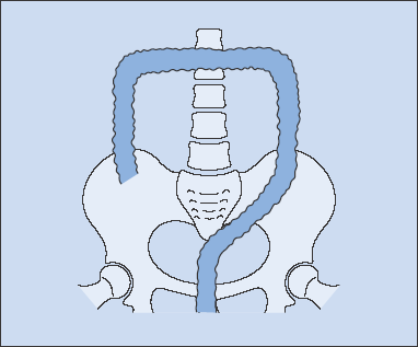
Plain abdominal film will show acute dilatation when present, and bowel gas may outline mucosal ulceration. Barium enema examination in such circumstances may produce perforation.
Differential Diagnosis
- Carcinoma of the colon, which may present with bloody diarrhoea.
- Infective enteritis. The acute case may resemble Campylobacter enteritis or bacillary dysentery, and the chronic case amoebic colitis (these should be excluded by stool examination).
- Antibiotic-associated pseudomembranous colitis (PMC) follows within 3 weeks of taking antibiotics. It is caused by toxins of Clostridium difficile when it colonises the colon following antibiotic-induced suppression of the normal bacterial flora of the gut. On sigmoidoscopy, characteristically there are patchy yellowish areas of necrotic mucosa. Histology shows mucosal destruction with characteristic exudation of fibrin and inflammatory cells in the cross-sectional shape of a mushroom. C. difficile and its toxin may be found in the stool. The condition responds to oral metronidazole or vancomycin.
- Very rarely, acute ischaemic colitis may occur and affect the rectosigmoid junction.
- Irritable bowel syndrome.
Treatment
Oral 5-aminosalicylic acid compounds are used to induce remission in mild to moderate colitis. Sulphasalazine is a combination of 5-aminosalicylic acid (5-ASA) and sulphapyridine, which acts as a carrier to deliver 5-ASA to its site of action in the colon. Mesalazine is 5-ASA by itself, and olsalazine is two linked molecules of 5-ASA that separate in the lower bowel. These newer aminosalicylates lack sulphonamide-related side effects, although their benefit over sulphasalazine in ulcerative colitis is unclear. 5-ASA compounds can be delivered by suppositor in proctitis. In moderate colitis that does not respond to 5-ASA or severe colitis, oral steroids should be started, and azathioprine can be added for its steroid-sparing effect. Rectal steroids can be used in proctitis. In patients refractory to 5-ASA and steroids, the anti-TNF (tumour necrosis factor) agent infliximab can induce remission and reduce the need for colectomy in the short term.
Treatment can usually be tapered once remission is achieved, but all of the above agents have been used to maintain remission.
Patients with severe colitis should be admitted to hospital for intravenous steroids and fluids and managed jointly by the gastroenterologist and surgeon. Patients who do not respond to intravenous steroids may respond to ciclosporin, tacrolimus or infliximab, but the need for colectomy should be continuously reviewed.
Surgery
Surgery (proctocolectomy, or total colectomy with ileal J pouch–anal anastomosis) is indicated if there is:
- severe haemorrhage
- perforation
- acute toxaemia with dilatation of the colon which fails to respond within 24–48 h to high-dose steroids.
Elective surgery is indicated if regular colonoscopy shows high-grade dysplasia or cancer, or in patients who are intolerant of or refractory to long-term medical treatments.
Crohn’s Disease
Crohn’s disease is a relapsing inflammatory disease that can affect any site in the alimentary tract from mouth to anus.
Aetiology
Current evidence suggests that genetic and environmental factors contribute to an abnormal mucosal immune response that is facilitated by the gut microflora and epithelial cell abnormalities. Nucleotide-binding oligomerisation domain 2/caspase recruitment domain-containing protein (NOD2/CARD15) was identified as the first susceptibility gene in Crohn’s disease in 2001. NOD2 contains an intracellular receptor for components of microbial pathogens, and influences inflammatory responses by regulating activation of the transcription factor NFκB. Genome-wide association studies have since implicated many other genes including the interleukin-23 receptor (IL23R) and autophagy-related 16-like 1 (ATG16L1) genes.
Clinical Features (Table 12.1)
Peak incidence is between ages 10 and 40. The terminal ileum is most frequently diseased, followed by the colon and less commonly the upper gastrointestinal tract. It usually presents as intermittent abdominal pain with diarrhoea, sometimes with passage of blood or mucus. Less commonly it presents as an ‘acute abdomen’ with signs of acute appendicitis with or without a palpable mass or obstruction. A mass in the right iliac fossa from terminal ileitis must be differentiated from a caecal carcinoma and an appendix abscess. Amoebic abscess and ileocaecal tuberculosis are less common causes.
The granulomatous inflammatory process affects short lengths of the intestine, leaving normal bowel between skip lesions. The wall is thickened and the lumen narrowed. Mucosal ulceration and regional lymphadenopathy are present. The characteristic microscopic features are of submucosal inflammation, less marked than in ulcerative colitis. There are numerous fissures down to the submucosa with or without chronic granulation tissue, consisting of non-caseating granulomas not unlike those found in sarcoid.
Imaging (Fig. 12.2)
Barium Enema
The terminal ileum is most commonly involved and may produce incompetence of the ileocaecal valve. Mucosal ulceration may be deep and ‘spikes’ of barium may enter deep into the bowel wall (rose thorn). Lesions may be multiple with normal bowel between (skip lesions). Coarse cobblestone appearance of the mucosa appears early. Later in the disease, fibrosis produces narrowing of the intestine (string sign) with some proximal dilatation.
Figure 12.2 Crohn’s disease. Patchy involvement of the bowel with: (1) deep ulcers which may lead to abscess or fistula formation; (2) strictures; (3) normal intervening bowel. Areas of disease with normal intervening bowel are known as skip lesions.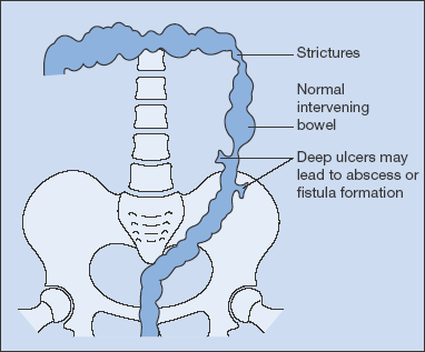
Small-Bowel enema
There may be mucosal ulceration, luminal narrowing or pooling of barium in irregular clumps at the site of an inflammatory mass.
Cross-sectional imaging with multi-slice CT, MR with oral contrast and MR enteroclysis, in which contrast is inserted through a naso-duodenal tube, provide alternative approaches.
Indium-labelled white cell scanning is helpful in localising active inflammatory bowel disease.
Histology
In Crohn’s disease the characteristic microscopic features are of submucosal inflammation, less marked than in ulcerative colitis. There are numerous fissures down to the submucosa with or without chronic granulation tissue consisting of non-caseating granulomas not unlike those found in sarcoid.
Complications
Fever, anaemia and weight loss. Hypoalbuminaemia results from loss of protein and in small-bowel disease malabsorption. Fistulae, perianal fissures and sepsis and intestinal sepsis may all complicate Crohn’s disease.
Treatment
Aminosalicylates and corticosteroids have been used to induce remission. The newer aminosalicylates may be of more benefit in treating Crohn’s disease. Mesalazine suppositories can be useful for localised rectal disease. Budesonide that is formulated to be released in the terminal ileum and colon can be effective with fewer side effects than conventional steroids. It is a steroid that is rapidly metabolised in the liver after absorption. Enteral nutrition has been used to induce remission but is less effective that steroids. Anti-TNF treatment with infliximab, adalimumab or certolizumab pegol are usually reserved for patients who do not enter remission with mesalazine or steroids. Methotrexate or ciclosporin may be of value in patients refractory to these treatments. Attention to nutritional deficiencies (p. 253) and electrolyte imbalance is essential.
Azathioprine, anti-TNF therapies and enteral nutrition have been shown to be effective in maintaining remission (see Trials Box 12.1. Smoking cessation is important in maintainence of remission.
Antibiotics (ciprofloxacin and metronidazole) are widely used for the treatment of fistulas in Crohn’s disease. Azathioprine may be effective, but anti-TNF treatments with infliximab and adalimumab are increasingly used to heal fistulas.
 Trials Box 12.1 Maintenance of Remission in Crohn’s Disease
Stay updated, free articles. Join our Telegram channel

Full access? Get Clinical Tree


