QUESTION 47.2
A. Choriocarcinoma
B. Classic seminoma
C. Embryonal carcinoma
D. Spermatocytic seminoma
E. Teratoma
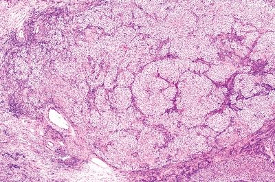
3. A testicular biopsy in a 40-year-old patient shows a neoplasm consisting of cells with conspicuous nucleoli and clear cytoplasm arranged in a nesting pattern, as shown in this picture. Fibrous septae between neoplastic nests contain numerous lymphocytes. Mitotic figures are rare. Tumor cells are immunoreactive for placental alkaline phosphatase (PLAP). Which of the following is the most likely diagnosis?
QUESTION 47.3
A. Anaplastic seminoma
B. Embryonal cell carcinoma
C. Lymphoma
D. Seminoma
E. Spermatocytic seminoma
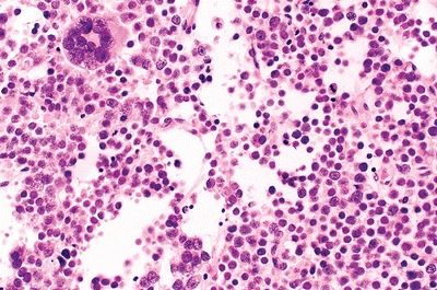
4. A 70-year-old man undergoes orchiectomy for a testicular tumor. Histologically, the tumor consists of a polymorphic population of small, intermediate, and giant cells, as seen in this picture. Mitotic activity is present. Which of the following features is typical of this tumor?
QUESTION 47.4
A. Associated with cryptorchidism
B. PLAP immunoreactivity is usually detected
C. May occur in extratesticular sites
D. Metastatic spread is rare
E. Orchiectomy and radiation are the recommended treatments
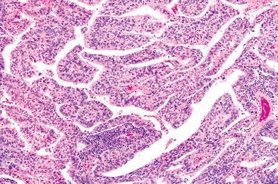
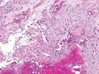
5. A testicular tumor from a 30-year-old man consists of highly anaplastic epithelial cells with poorly defined borders arranged in papillary structures, as seen in this picture. Multifocal coagulative necrosis is noted. Neoplastic cells express CK8, CK18, and CD30. Which of the following is true about this tumor?
QUESTION 47.5
A. In its pure form accounts for 30% of testicular neoplasms
B. Mitotic activity is low or absent
C. Most commonly associated with yolk sac or trophoblastic components
D. Most frequent in children under 3 years
E. Uniformly positive for placental alkaline phosphatase (PLAP)
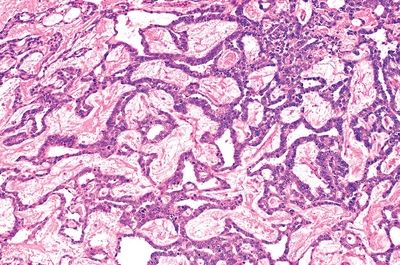
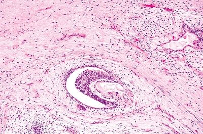
6. The most common testicular neoplasm in children less than 3 years of age is characterized by a spongy texture on the surface. Microscopically, its most typical pattern is characterized by fibrovascular cores “festooned” by tumor cells, as shown in this picture. Which of the following is it?
QUESTION 47.6
A. Choriocarcinoma
B. Embryonal carcinoma
C. Seminoma
D. Teratoma
E. Yolk sac tumor
7. A testicular teratoma:
A. Behaves as a malignant tumor in children and as a benign tumor in adults
B. Consists of a cyst filled with keratin and hair
C. Is usually associated with other germ cell tumors in adults
D. Is usually immature in prepubertal age
E. Occurs more frequently in pure form in adult patients
8. Which of the following testicular tumors is a specialized form of teratoma?
A. Carcinoid tumor
B. Dermoid cyst
C. Epidermoid cyst
D. PNET
E. All of the above
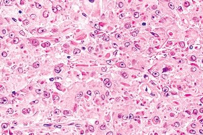
9. An 18-year-old male is evaluated for the acute onset of right hemiplegia. A CT scan shows a well-demarcated hemorrhagic mass in the left frontal lobe. The mass is removed and reveals the neoplasm shown in this photomicrograph. Further investigations lead to the discovery of a testicular tumor. Which of the following is the most likely diagnosis?
QUESTION 47.9
A. Choriocarcinoma
B. Embryonal carcinoma
C. Seminoma
D. Teratoma
E. Yolk sac tumor
10. Histologic analysis of an orchiectomy specimen reveals a neoplasm whose microscopic appearance is shown in this picture. Which of the following is the diagnosis?
QUESTION 47.10
A. Burn-out germ cell tumor
B. Diffuse embryoma
C. Polyembryoma
D. Placental site trophoblastic tumor
E. Yolk sac tumor
11. Histologic examination of a 2-cm testicular tumor from a 40-year-old man with gynecomastia shows sheets of polygonal cells with eosinophilic cytoplasm. Many rod-shaped intracytoplasmic crystals are identified, as shown in this picture. Electron microscopy reveals abundant smooth endoplasmic reticulum, mitochondria with tubular cristae, lipid droplets, and cytoplasmic elongated crystalloids. Which of the following is the most likely diagnosis?
QUESTION 47.11
A. Adrenogenital syndrome
B. Choriocarcinoma
C. Leydig cell hyperplasia
D. Leydig cell tumor
E. Sertoli cell tumor
12. Which of the following histologic patterns is most characteristic of Sertoli cell tumors?
A. Glandular arrangement
B. Mixture of cytotrophoblast and syncytiotrophoblast
C. Round nests between lymphocyte-rich septa
D. Stromal fibrovascular core surrounded by double-cell layer
E. Structures reminiscent of immature seminiferous tubules
13. Which of the following is a mixed germ cell–sex cord–stromal tumor that develops in patients with gonadal dysgenesis?
A. Diffuse embryoma
Stay updated, free articles. Join our Telegram channel

Full access? Get Clinical Tree


