T-Cell Neoplasms


T-Cell Neoplasms


Diane C. Farhi


The T-cell neoplasms are relatively rare compared to B-cell neoplasms, but they nevertheless consist of many different entities. In this chapter, we have included the WHO 2008 classification criteria; however, it should be noted that other names for some of these entities persist. The diseases described here are presented in older terminology (Tables 26.1 and 26.2). The reader is encouraged to study the WHO 2008 publication to match the two terminologies (see Appendix A).

Those neoplasms which have been reported in the bone marrow are discussed below. NK-cell neoplasms are discussed separately in Chapter 27.


PRECURSOR T-CELL NEOPLASM


T-Cell Lymphoblastic Lymphoma

T-cell lymphoblastic lymphoma (T-LBL) arises de novo and as a therapy-related malignancy, a blast crisis of chronic myeloid leukemia, and a component of 8p11 myeloproliferative disorder (Fig. 26.1) (1, 2, 3, 4, 5, 6, 7, 8, 9, 10, 11, 12, 13, 14, 15, 16, 17, 18, 19, 20, 21, 22, 23, 24, 25, 26, 27). It accounts for 85% to 90% of all cases of lymphoblastic lymphoma. The disease has a slightly more mature immunophenotype and a different gene expression profile than percursor T-cell acute lymphoblastic leukemia. It is believed to arise from a thymic precursor T-cell as opposed to a bone marrow precursor T-cell.

Patients are often young and males are affected more than females. Patients may present with involvement of the mediastinum, pleural and pericardial cavities, cervical lymph nodes, breast, central nervous system, and other sites. The disease typically pursues an aggressive course, with eventual development of disseminated disease and a leukemic phase. One case has been reported with an unusually indolent course.

Peripheral blood and bone marrow smears may show blasts up to 19% of nucleated cells. Myeloid hyperplasia may also be found. Cases with 20% or more blasts in the peripheral blood and’or bone marrow at the time of diagnosis are classified as precursor T-cell ALL (see also Chapter 18).

Histologic sections of the bone marrow may show focal or patchy disease, as opposed to the uniform involvement usually seen in acute lymphoblastic leukemia. The blasts show round to ovoid, sometimes cleaved nuclei and scant agranular cytoplasm.

Immunophenotyping usually shows expression of CD2, cytoplasmic CD3, CD5, CD7, and terminal deoxynucleotidyl transferase (TdT). CD10 is expressed in about 50% of cases, and surface CD3 in about 40% of cases. Either CD4 or CD8 may be expressed; dual expression of CD4 and CD8 is common. CD13, CD33, and CD79a are often expressed. CD117 is infrequently expressed. Other markers which may be expressed include CD19, CD20, CD34, CD38, CD43, CD45, CD56, CD57, CD99, and TCR α-β. Compared with normal T cells, expression of pan-T-cell markers (CD2, CD3, CD5, CD7) may be decreased. Some cases express TCR γ-δ instead of TCR α-β. In some cases, coexpression of myeloid or B-cell antigens may warrant interpretation as biphenotypic lymphoblastic lymphoma; this area is not entirely clear. In a few instances, conversion from T-cell to myeloid, histiocytic, or B-cell lineage has been documented.

Genetic studies have demonstrated various clonal karyotypic anomalies in more than 70% of cases. These often involve the TCR γ-chain gene located on chromosome 7p15, the TCR β-chain gene located on chromosome 7q35, and the TCR α- and δ-chain genes located on chromosome14q11. TCR gene rearrangements are found in approximately 75% to 80% of cases, involving (in decreasing order) the TCR δ-, γ-, and β-chain genes. Concomitant rearrangement of the immunoglobulin heavy chain gene is found in approximately 10% of cases. BCR’ABL1 rearrangement may be found.

T-LBL has been reported with Langerhans cell histiocytosis.

The differential diagnosis includes other clonal T-cell disease, especially T-cell ALL; B-cell and natural killer cell neoplasms; and nonhematolymphoid small round cell tumors.


MATURE T-CELL NEOPLASMS


Adult T-Cell Leukemia’Lymphoma

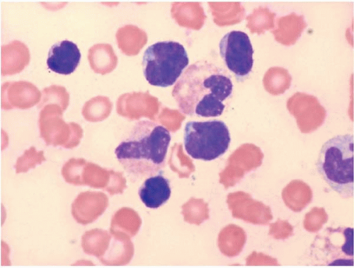
Adult T-cell leukemia’lymphoma (ATLL) arises as a consequence of human lymphotropic virus I (HTLV-I) infection (Figs. 26.2 and 26.3) (28, 29, 30, 31, 32, 33, 34, 35, 36, 37, 38, 39, 40, 41, 42, 43, 44, 45, 46, 47, 48, 49, 50, 51, 52, 53, 54, 55, 56, 57, 58, 59, 60, 61, 62, 63, 64, 65, 66, 67, 68, 69, 70, 71, 72, 73, 74, 75). The clinical course of the HTLV-I infection ranges from smoldering disease to chronic disease, lymphoma, and leukemia.


TABLE 26.1 WHO 2008 Classification of T-Cell Neoplasms


Precursor T-Cell Neoplasm


T-cell lymphoblastic leukemia’lymphoma


Mature T-Cell Neoplasms


T-cell prolymphocytic leukemia


Systemic EBV-positive T-cell lymphoproliferative disease of childhood


Hydroa vaccine form-like lymphoma


Adult T-cell leukemia’lymphoma


Enteropathy-associated T-cell lymphoma


Hepatosplenic T-cell lymphoma


Subcutaneous panniculitis-like T-cell lymphoma


Mycosis fungoides


Sézary syndrome


Primary cutaneous CD30+ T-cell lymphoproliferative disorders


Peripheral T-cell lymphoma, NOS


Angioimmunoblastic T-cell lymphoma


Anaplastic large cell lymphoma ALK positive


Aggressive NK cell leukemia


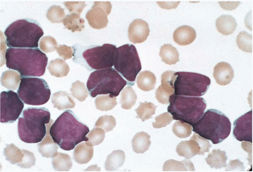
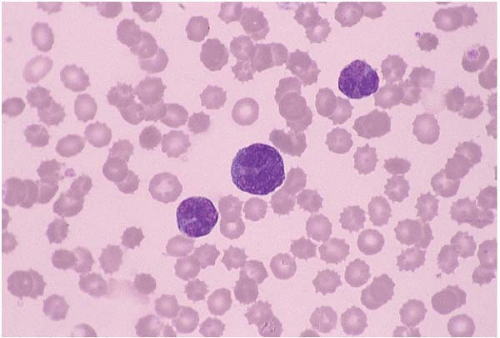
Patients with ATLL may present with disease involving lymph nodes, liver, spleen, peripheral blood, and bone marrow. Skin lesions, hypercalcemia, and opportunistic infections may be present. ATLL occurs both in children and adults. The clinical course is usually aggressive, and the prognosis is poor. Spontaneous remission has been reported but is usually transient.

Laboratory studies may show anemia, neutrophilia, thrombocytopenia, monocytosis, eosinophilia, and’or basophilia. The absolute lymphocyte count is variable.

Peripheral blood and bone marrow smears show abnormal lymphocytes. These are typically a heterogeneous population of cells, consisting of a range of small and large cells. Nuclei range from small and irregular to large and prominently lobated. Nuclear hypersegmentation has prompted the term “flower cells.” Nucleoli may be present. The cytoplasm is scant to moderately abundant and basophilic and may contain inclusions, vacuoles, and’or granules. Tartrate-resistant acid phosphatase activity may be present.


TABLE 26.2 Differential Features of Mature T-Cell Neoplasms


TPL


T-LGL


PLL


ATLL


HSTL


MF’SS


PTCL


Leukemia


+


+


+


+


+’−


+’−


+’−


Cyto gran


Rare


+


+’−


Rare


Rare


CD4


+


Rare


+’−


+’−


+


+’−


CD8


Rare


+


−’+


−’+


−’+


−’+


TCR α’β


+


+’−


+’−


+


Rare


+


+’−


TCR γ’δ


−’+


−’+


+


−’+


ATLL, adult T-cell leukemia’lymphoma; T-LGLL, T-cell large granular lymphocytic leukemia; cyto gran, cytoplasmic granules; HSTL, hepatosplenic T-cell lymphoma; MF, mycosis fungoides’Sézary syndrome; PLL, T-cell prolymphocytic leukemia; PTCL, peripheral T-cell lymphoma; TCR α’β, T-cell receptor α’β; TCR γ’δ, T-cell receptor γ’δ; +, positive; −, negative; +’−, usually positive, sometimes negative; −’+, usually negative, sometimes positive.


Figure 26.1 T-cell lymphoblastic lymphoma, peripheral blood. Numerous blasts, many with deep nuclear cleaves, are present in this specimen from a patient with leukemic spread of a mediastinal tumor. The cells are indistinguishable from precursor T-cell acute lymphoblastic leukemia.

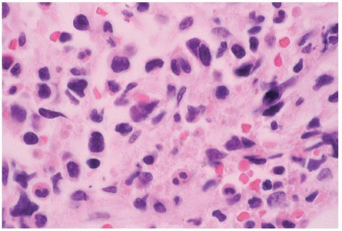
Histologic sections of the bone marrow show abnormal lymphocytes distributed as a patchy or diffuse interstitial infiltrate.

Many morphologic variants are recognized. ATLL may resemble Burkitt lymphoma’leukemia, lymphoblastic lymphoma, T-cell chronic lymphocytic leukemia, anaplastic large cell lymphoma, mycosis fungoides’Sézary syndrome, and angioimmunoblastic T-cell lymphoma.

Other findings include hemophagocytic syndrome, which may obscure the underlying disease, and osteitis fibrosa cystica, owing to tumor cell secretion of parathormone-related protein.


Figure 26.2 Adult T-cell leukemia’lymphoma, peripheral blood. The neoplastic cells vary in size and show pleomorphic nuclei in this specimen from a patient with positive serology for human T-cell lymphotropic virus type II.

Immunophenotyping usually shows expression of CD2, CD3, CD4, CD7, CD25, and TCR α-β. CD4 and CD25 are consistently expressed. Compared to normal T-cells, expression of pan-T-cell antigens (CD2, CD3, CD5, CD7) may be reduced or absent. CD3, in particular, shows low expression. Some cases express both CD4 and CD8, CD8 only, or neither CD4 nor CD8. In CD8+ cases, granzyme B and TIA-1 (but not perforin) may be expressed. CD20, CD30, CD56, epithelial membrane antigen, and S100 protein expression have been reported.

Genetic studies have shown various clonal karyotypic anomalies, especially del(6q), and rearrangements and deletions involving the TRC genes. Clonal integration of human T-cell lymphotropic virus type 1 (HTLV-1) proviral DNA into tumor cells is consistently found.


Figure 26.3 Adult T-cell leukemia’lymphoma, bone marrow biopsy. The hematopoietic tissue is replaced by neoplastic cells of variable size and shape embedded in fibrous tissue in this specimen from a patient with positive serology for human T-cell lymphotropic virus type II.


Figure 26.4 Anaplastic T-cell lymphoma, bone marrow biopsy. The hematopoietic tissue is replaced by a population of large, abnormal lymphocytes with clear cytoplasm and prominent nucleoli.

ATLL has been reported with disseminated xanthogranulomas, myelodysplastic syndrome, acute myeloid leukemia, and various B-cell neoplasms.

The differential diagnosis includes other T-cell neoplasms, clonal B-cell and NK-cell disorders, and acute leukemia.


Anaplastic Large T-Cell Lymphoma

Anaplastic large cell lymphoma (ALCL) arises de novo; as a complication of immunosuppressed states, including human immunodeficiency virus infection and solid organ transplantation; and as a transformation of mycosis fungoides’Sézary syndrome and T-cell prolymphocytic leukemia (Figs. 26.4 and 26.5) (76, 77, 78, 79, 80, 81, 82, 83, 84, 85, 86, 87, 88, 89, 90, 91, 92, 93, 94, 95, 96, 97, 98, 99, 100, 101, 102, 103, 104, 105, 106, 107, 108, 109, 110, 111, 112, 113, 114, 115, 116, 117, 118, 119, 120, 121, 122, 123, 124, 125, 126, 127, 128, 129, 130, 131). ALCL may show B-cell, T-cell, or null-cell phenotype. T-cell and null-cell ALCL are discussed here.


Figure 26.5 Anaplastic T-cell lymphoma, small cell anaplastic variant, peripheral blood. The patient is a 2-year-old boy.


Patients may present at any age, from infancy to adulthood. Either lymph node or skin involvement predominates. The bone marrow is affected in approximately 90% of cases at diagnosis. Rarely, the bone marrow is the only site of disease. Cases characterized by nodal involvement tend to have an aggressive course, with peripheral blood and bone marrow disease. Leukemia is especially common in the small cell variant of T-cell ALCL.

Laboratory studies may show anemia, neutropenia or neutrophilia, and absolute lymphocytosis. Neutrophilic leukemoid reaction has been reported.

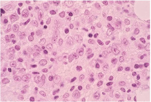
Peripheral blood and bone marrow smears show abnormal lymphocytes which range from small, relatively nondescript tumor cells to large anaplastic cells with irregular to convoluted nuclei and prominent nucleoli. Either small cells or large cells may predominate. Smaller cells may show small, relatively bland nuclei and scanty cytoplasm. Larger cells show anaplastic features such as large, folded, lobated, bean-shaped, and embryo-shaped nuclei; wreath-shaped arrangements of multiple nuclei; and abundant vacuolated cytoplasm. A leukemic phase is particularly prominent in the small cell variant of ALCL. Abnormal lymphocytes may be difficult to identify among hematopoietic cells.

Histologic sections of the bone marrow show intrasinusoidal, interstitial, and’or diffuse lymphocytic infiltration. The abnormal lymphocytes are often admixed with inflammatory cells and fibrous tissue and may be difficult to identify among hematopoietic cells. Morphologic variants have been described, consisting predominantly of small cells, large nonanaplastic cells, and neoplastic cells admixed with numerous neutrophils, eosinophils, or histiocytes.

Other findings include myelodysplasia, apparently the result of abnormal cytokine secretion; and hemophagocytic syndrome, which may obscure the underlying malignancy.

Immunophenotyping usually shows a cytotoxic T-cell phenotype, with expression of CD2, CD3, CD5, CD7, CD30, CD45, S100 protein, ALK-1 (p80, NPM’ALK fusion protein), epithelial membrane antigen, granzyme B, perforin, and TIA-1. Compared to normal T-cells, expression of pan-T-cell antigens (CD2, CD3, CD5, CD7) may be reduced or absent. Either CD4 or CD8 may be expressed. CD11c, CD13, CD14, CD15, CD25, CD33, CD43, CD56, CD68, CD99, and epithelial membrane antigen may be expressed. ALK-1 may not be expressed. Antigen expression may vary between the peripheral blood and bone marrow. Cases expressing no recognizable T-cell or B-cell antigens are immunophenotypically null and diagnosed as anaplastic null-cell large cell lymphoma.

Genetic studies show, in most but not all cases, t(2;5)(p23;q35) involving ALK (anaplastic lymphoma kinase gene) at chromosome 2p23 and NPM at chromosome 5q35. Genetic variants involving ALK have been described. TCR and IgH gene rearrangements may also be present. Other genetic abnormalities have been reported.

The differential diagnosis includes drug reaction, anaplastic large B-cell lymphoma, Hodgkin lymphoma, acute leukemia, histiocytic neoplasms, and nonhematopoietic malignancies.


ANGIOIMMUNOBLASTIC T-CELL LYMPHOMA

Angioimmunoblastic T-cell lymphoma (AITL) occurs as a de novo disease and as a complication of Epstein-Barr virus (EBV) infection, human herpesvirus 6 infection, and systemic sclerosis (132, 133, 134, 135, 136, 137, 138, 139, 140, 141, 142, 143, 144, 145, 146, 147, 148, 149, 150, 151, 152, 153).

Only gold members can continue reading. Log In or Register to continue

Stay updated, free articles. Join our Telegram channel

Jun 19, 2016 | Posted by in PATHOLOGY & LABORATORY MEDICINE | Comments Off on T-Cell Neoplasms

Full access? Get Clinical Tree

Get Clinical Tree app for offline access