Myelodysplastic Syndromes


Myelodysplastic Syndromes


Diane C. Farhi


GENERAL FEATURES


Definition and Classification

Myelodysplastic syndromes (MDS) are a group of clonal disorders of hematopoietic stem cells, characterized by dysplastic morphology, cytogenetic aberrations, and—in most cases—cytopenia (1, 2, 3, 4). MDS typically involves the main hematopoietic lineages: granulocytes, monocytes, erythroid cells, and megakaryocytes. Less often, the morphologic and genetic abnormalities affect other related lineages: mast cells, dendritic cells, and lymphocytes.

Hematopoietic stem cells show increased proliferation while retaining cellular differentiation. One or more cell lines show dysplasia, increased apoptosis, and ineffective hematopoiesis, leading to the coexistence of peripheral cytopenia and bone marrow hypercellularity. The World Health Organization (WHO) 2001 classification and criteria for MDS are given in Table 20.1, and the WHO 2008 classification without criteria in Table 20.2 (see Appendix A for WHO 2008 classification).

In some cases, proliferation outstrips apoptosis, and increased marrow cellularity coexists with increased peripheral cell counts. Such cases are termed myelodysplastic’myeloproliferative neoplasms (MDS’MPN). The WHO 2001 classification and criteria for MDS’MPN are given in Table 20.3, and the WHO 2008 classification without criteria in Table 20.4.

The WHO 2008 criteria for MDS and MDS’MPN are not available at the time of writing. Readers are encouraged to consult the WHO 2008 publication for current criteria.


Clinical Course

Myelodysplastic syndrome (MDS) occurs predominantly in older adults (median age 70 years), but also in children (5-23). MDS occurs de novo and as a transformation of aplastic anemia, pure red cell aplasia, paroxysmal nocturnal hemoglobinuria, and myeloproliferative neoplasms. MDS occurring as a complication of therapy for another neoplastic disease is termed therapy-related or secondary MDS, and is dicussed with acute leukemia in Chapter 18.

MDS progresses in about 30% of patients to acute leukemia, usually acute myeloid leukemia (AML) but sometimes acute lymphoblastic leukemia. The risk of leukemic transformation depends on age, the number and severity of cytopenias, the extent and degree of dysplasia, the percentage of blasts, cytotoxic drug resistance, number and type of genetic abnormalities, and other factors. Like AML, MDS may undergo spontaneous remission and remission following colony-stimulating factor and immunosuppressive therapy.

Autoimmune disorders occur in more than 10% of patients, owing, at least in part, to clonal lymphoid differentiation and the production of self-reactive CD5’19-positive B cells. These disorders include immune-mediated arthritis and vasculitis, autoimmune cytopenias, Behçet disease, relapsing polychondritis, inflammatory bowel disease, and others. Tuberculosis may be more common in MDS patients.

Acquired hemoglobinopathies and other hematopoietic disorders have been reported in MDS, such as thalassemia, increased hemoglobin F, aplastic anemia, Behçet disease, paroxysmal nocturnal hemoglobinuria, and erythropoietic protoporphyria.

Clonal B-cell, T-cell, and plasma cell disorders reported in MDS include chronic lymphocytic leukemia, Hodgkin lymphoma, other malignant lymphomas, clonal gammopathies, multiple myeloma, systemic amyloidosis, T-cell lymphoblastic lymphoma, and other T-cell lymphomas. These neoplasms may be either clonally related or unrelated.

Histiocytic proliferations and disorders reported with chronic myelomonocytic leukemia (CMML) include xanthomas, Langerhans histiocytosis, and nodal histiocytic proliferation. Clonal identity of CMML and histiocytic lesions is possible but unproven.


Peripheral Blood and Bone Marrow Findings

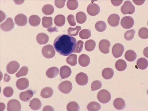
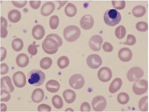
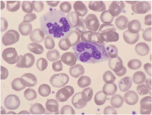
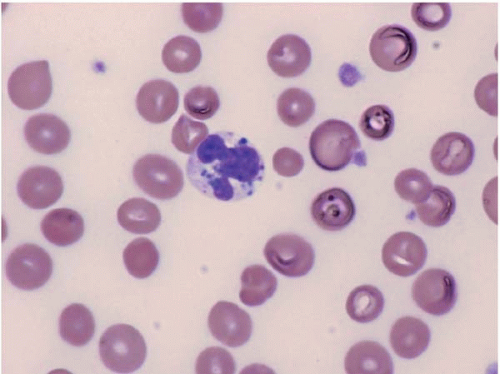
The morphologic hallmark of MDS is myelodysplastic change in hematopoietic cells, affecting one or more cell lines (Figs. 20.1, 20.2, 20.3, 20.4, 20.5, 20.6, 20.7, 20.8, 20.9, 20.10and 20.11) (24, 25, 26, 27, 28, 29, 30, 31). However, dysplastic features may be absent or rare in some cases, especially in MDS with an isolated cytopenia.

Laboratory studies often show macrocytic or normocytic anemia, neutropenia, and thrombocytopenia. Cytopenias may be cyclic. The mean corpuscular hemoglobin contration
is usually normal. The mean corpuscular volume may be macrocytic or normal. The red cell distribution width is often increased. Numerous acquired abnormalities may be found, resembling their constitutional counterparts, such as thalassemia.


TABLE 20.1 WHO 2001 Classification and Criteria for Myelodysplastic Syndromes


Disease


Blood Findings


Bone Marrow Findings


Refractory anemia


Anemia, rare blasts


Erythroid dysplasia only, <5% blasts, <15% ringed sideroblasts


Refractory anemia with ringed sideroblasts


Anemia, no blasts


Erythroid dysplasia only, <5% blasts, ≥15% ringed sideroblasts


Refractory cytopenia with multilineage dysplasia


Cytopenias (bi-‘pancytopenia), rare blasts, no Auer rods, <1 × 10⁹ monocytes


Dysplasiaa in two or more cell lines, <5% blasts, <15% ringed sideroblasts, no Auer rods


Refractory cytopenia with multilineage dysplasia and ringed sideroblasts


Cytopenias (bi-‘pancytopenia), rare blasts, no Auer rods, <1 × 10⁹ monocytes


Dysplasiaa in two or more cell lines, <5% blasts, ≥15% ringed sideroblasts, no Auer rods


Refractory anemia with excess blasts-I


Cytopenias, <5% blasts, no Auer rods, <1 × 10⁹ monocytes


Uni-‘multilineage dysplasia, 5%-9% blasts, no Auer rods


Refractory anemia with excess blasts-II


Cytopenias, 5%-19% blasts,b ±Auer rods,b <1 × 10⁹ monocytes


Uni-‘multilineage dysplasia, 10%-19% blasts,b ±Auer rodsb


Myelodysplastic syndrome, unclassified


Cytopenias, rare blasts, no Auer rods


Unilineage dysplasia, <5% blasts, no Auer rods


Myelodysplastic syndromes associated with isolated del(5q)


Anemia, <5% blasts, platelets normal or increased


< 5% blasts, isolated del(5q) cytogenetic abnormality, no Auer rods, normal to increased megakaryocytes with hypolobated nuclei


a Dysplasia is defined as the presence of dysplastic features in 10% or more of the cells in a given lineage.

b Patients with 5% to 19% blasts in the blood or with Auer rods are classified as refractory anemia with excess blasts-II even if bone marrow blast counts are less than 10%.


From Jaffe ES, Harris NL, Stein H, et al, eds. World Health Organization classification of tumors: pathology and genetics of tumours of haematopoietic and lymphoid tissues. Lyon: IARC Press, 2001


Peripheral blood smears usually show abnormal red blood cells (RBC), including subtle variations in size and form. Target cells, teardrop cells, and RBC fragments may be present. Delayed maturation of erythrocytes may cause pseudoreticulocytosis. Other RBC abnormalities include coarse basophilic stippling and Howell-Jolly bodies. Nucleated RBCs may show megaloblastic change and abnormal nuclear contours, consisting of altered shape and lobation.


TABLE 20.2 WHO 2008 Classification of Myelodysplastic Syndromes


Refractory cytopenia with unilineage dysplasia


Refractory anemia


Refractory neutropenia


Refractory thrombocytopenia


Refractory anemia with ring sideroblasts


Refractory cytopenia with multilineage dysplasia


Refractory anemia with excess blasts


Myelodysplastic syndromes associated with isolated del(5q)


Myelodysplastic syndromes, unclassifiable


Refractory cytopenia of childhood


From Swerdlow SH, Campo E, Harris NL, et al, eds. WHO Classification of Tumours of Haematopoietic and Lymphoid Tissues. Lyon: IARC Press, 2008.


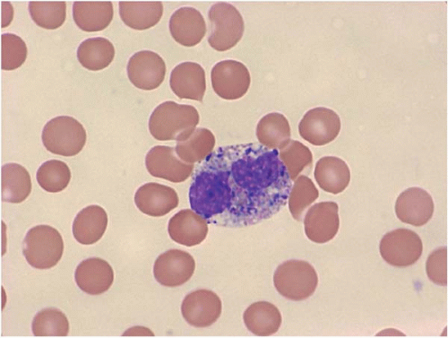
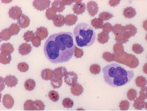
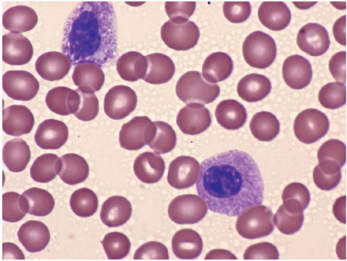
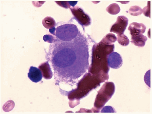
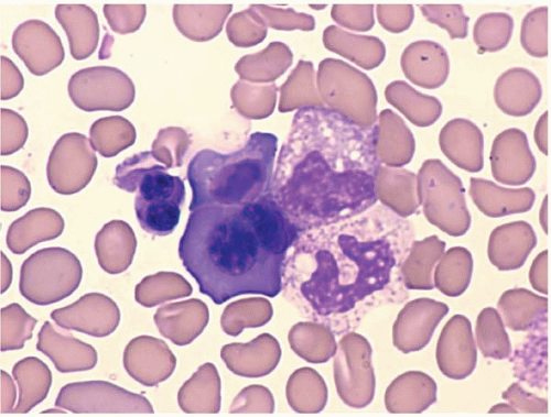
Dysplastic neutrophils are usually found. These cells may show increased size, abnormal nuclear lobation, and abnormal granularity, usually hypogranularity but occasionally abnormally large granules. Monocytes are less often recognizably dysplastic. They may show abnormal nuclear morphology and immature characteristics.


TABLE 20.3 WHO 2001 Classification and Criteria for Myelodysplastic’Myeloproliferative Diseases


Disease


Diagnostic Criteria


Chronic myelomonocytic leukemia


  1. Persistent monocytosis >1 × 10⁹‘L


  2. No Philadelphia chromosome or BCR’ABL1 fusion gene


  3. <20% blasts (including promonocytes) in blood and bone marrow


  4. Dysplasia in one or more lineages or clonal cytogenetic abnormality or monocytosis for ≥3 mo with other causes excluded


Atypical chronic myeloid leukemia


  1. Leukocytosis owing to increased neutrophils


  2. Prominent dysgranulopoiesis


  3. No Philadelphia chromosome or BCR’ABL1 fusion gene


  4. Neutrophilic precursors ≥10% of WBCs


  5. Minimal basophilia (<2% of WBCs)


  6. Minimal monocytosis (<10% of WBCs)


  7. Hypercellular marrow with granulocytic proliferation and dysplasia


  8. <20% blasts in blood and bone marrow


Juvenile myelomonocytic leukemia


  1. Persistent monocytosis >1 × 10⁹‘L


  2. No Philadelphia chromosome or BCR’ABL1 fusion gene


  3. <20% blasts (including promonocytes) in blood and bone marrow


  4. Two or more of the following:


    Hemoglobin F increased for age


    Immature granulocytes in the peripheral blood


    WBCs >10 × 109‘L


    Clonal cytogenetic abnormality (including monosomy 7)


    Granulocyte-monocyte colony-stimulating factor hypersensitivity of myeloid progenitors in vitro


Myelodysplastic’myeloproliferative disease, unclassifiable


  1. Features of MDS with <20% blasts and features of MPD (i.e., increased cell counts), and no history of MDS; MPD; cytotoxic treatment; Philadelphia chromosome or BCR’ABL1 fusion gene; del(5q), t(3;3)(q21;q26) or inv(3)(q21q26) or


  2. Mixed MDS’MPD features and cannot be assigned to other category of MDS, MPD or MDS’MPD


MDS, myelodysplastic syndrome; MPD, myeloproliferative disease; WBCs, white blood cells.


From Jaffe ES, Harris NL, Stein H, et al, eds. World Health Organization classification of tumors: pathology and genetics of tumours of haematopoietic and lymphoid tissues. Lyon: IARC Press, 2001:45-59.


TABLE 20.4 WHO 2008 Classification of Myelodysplastic’Myeloproliferative Neoplasms


Chronic myelomonocytic leukemia


Atypical chronic myeloid leukemia, BCR-ABL1 negative


Juvenile myelomonocytic leukemia


Myelodysplastic’myeloproliferative neoplasm, unclassifiable


Refractory anemia with ring sideroblasts associated with marked thrombocytosis


From Swerdlow SH, Campo E, Harris N, et al, eds. WHO Classification of Tumours of Haematopoietic and Lymphoid Tissues. Lyon: IARC Press, 2008.


FIG. 20.1 Dysplastic nucleated red blood cell, peripheral blood. The nucleus shows an abnormal shape and size.


FIG. 20.2 Dysplastic nucleated red blood cells, peripheral blood. Abnormal nuclear shape and basophilic stippling are present.


FIG. 20.3 Dysplastic neutrophils, peripheral blood. The cells are nearly agranular.


FIG. 20.4 Dysplastic neutrophil, peripheral blood. The granules are reduced and those remaining are unusually large and poorly dispersed. The nuclear segmentation is also abnormal.


FIG. 20.5 Dysplastic granulocyte with both eosinophilic and basophilic granules.


FIG. 20.6 Dysplastic neutrophils, one with an abnormally large nucleus.


FIG. 20.7 Dysplastic neutrophils, peripheral blood. Acquired Pelger-Heut anomaly is present.


FIG. 20.8 Dysplastic megakaryocytes, bone marrow aspirate. The megakaryocytes and small and hypolobated.


FIG. 20.9 Dysplastic megakaryocyte, bone marrow aspirate. The cytoplasm is mature, but the cell is abnormally small and shows hypolobation and separation of the nuclei. Myelodysplastic syndrome, bone marrow aspirate. Erythroid hyperplasia with dysplastic maturation is shown.


FIG. 20.10 Dysplastic red blood cells and neutrophils, peripheral blood. Dysplasia may be present in one to three lineages.


FIG. 20.11 Myelodysplastic syndrome, bone marrow biopsy. The bone marrow is hypercellular and shows small, dysplastic megakaryocytes.


FIG. 20.12 Myelodysplastic syndrome, bone marrow aspirate. The cells are constrained by reticulin fibers, which gives the impression of cellular streaming.


Platelets usually show no definite morphologic changes. Uncommonly, they may show a smaller size or larger size than normal, or hypogranularity. The large, irregular platelets and megakaryocytic fragments seen in myeloproliferative neoplasms are not usually found.

Bone marrow aspirate smears show variable cellularity. The myeloid:erythroid ratio is also variable, and often decreased. A shift to more immature forms may also be found. Blasts may be increased, but by definition constitute less than 20% of nucleated cells.

Erythroid precursors may show large size, nuclear multilobation, nuclear budding, and other abnormal forms. The cytoplasm may show basophilia, vacuolization, perinuclear rings of dark mitochondria (identified as ringed sideroblasts with iron staining), and coarse or fine periodic acid-Schiffpositive granules.

Granulocytic precursors show dysplastic features, visible as abnormally large size, abnormal nuclear shape and cytoplasmic granularity.

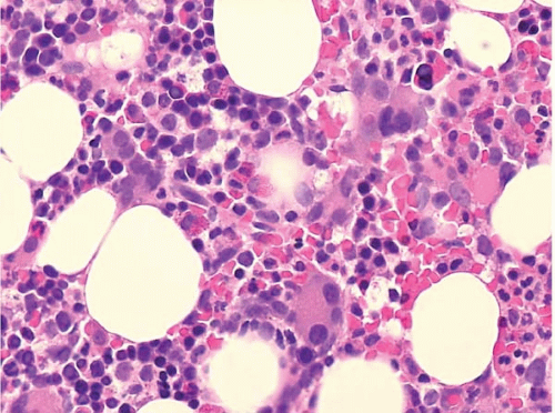
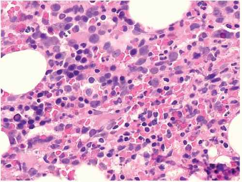
The bone marrow biopsy may show decreased or increased cellularity, depending on the net effect of hematopoietic proliferation, maturation, and intramedullary destruction through apoptosis. Cellularity should be judged by the expected cellularity in healthy subjects of the same age. The normal pattern of erythroid colonies and neutrophil proliferation and maturation is often changed to a disorganized mix of hematopoietic precursors, and blasts, termed ‘atypical localization of immature precursors’. Megakaryocytes may be increased or decreased, and may show smaller size, smaller nuclei with fewer lobes, and separated nuclear lobes.

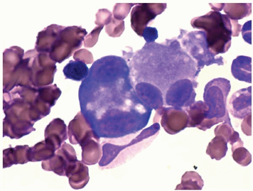
Other bone marrow findings include reactive lymphocytosis and mastocytosis, lymphoid aggregates, fibrosis, increased histiocytes, pseudo-Gaucher histiocytes (Fig. 20.12), and hemophagocytic syndrome.

The differential diagnosis includes nonclonal myeloplastic syndromes (see Chapter 16), myeloproliferative neoplasms, and acute myeloid leukemia.


Cytochemistry

MDS cells with myeloid and monocytic differentiation show expression of myeloid markers and monocytic enzymes, respectively. These include α1-antitrypsin, α1-antichymotrypsin, lysozyme, myeloperoxidase, chloroacetate esterase, and nonspecific esterase. As in acute erythroid leukemia, some cases show aberrant periodic acid-Schiff staining of erythroid precursors.


Immunophenotyping

Immunophenotyping may be done be immunostaining and by flow cytometry. Compared to normal bone marrow samples, MDS cases usually show larger populations of immature myeloid cells (32, 33, 34, 35, 36, 37, 38, 39, 40, 41, 42). In most cases, flow cytometry shows myeloid cells have at least two antigens with abnormal features. Among CD34-positive myeloid precursors, expression of CD7, CD13, CD33, CD34, CD117, HLA-DR, and p53 is increased. Expression of CD10 (in mature granulocytes), CD11b, CD64, and myeloperoxidase is decreased. B cell precursors are decreased.

Only gold members can continue reading. Log In or Register to continue

Stay updated, free articles. Join our Telegram channel

Jun 19, 2016 | Posted by in PATHOLOGY & LABORATORY MEDICINE | Comments Off on Myelodysplastic Syndromes

Full access? Get Clinical Tree

Get Clinical Tree app for offline access