Metaplasia → dysplasia → carcinoma sequence


Top Differential Diagnoses
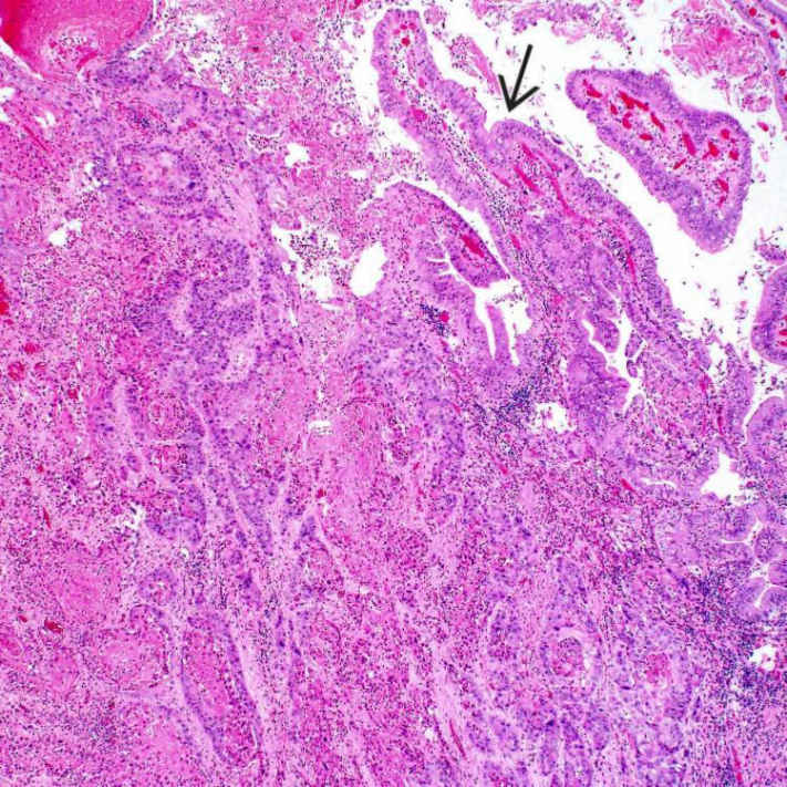
This primary squamous cell carcinoma was resected from a 84-year-old man who presented with acute cholecystitis. A 4.6-cm intraluminal mass was found in the fundus in cholecystectomy specimen. Extensive sampling did not reveal glandular component. Note the presence of partially preserved gallbladder mucosa
 over tumor.
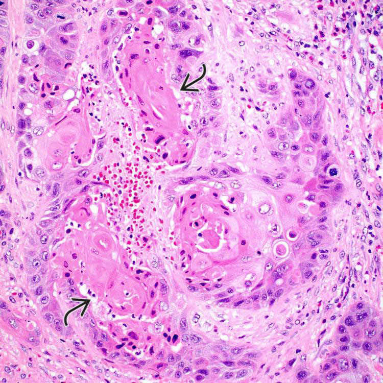
This case of primary squamous cell carcinoma of the gallbladder shows islands of malignant cells with prominent keratinization
 .
.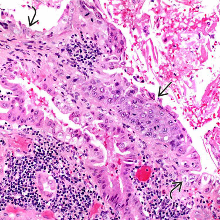
The gallbladder mucosa adjacent to invasive squamous cell carcinoma shows a focus of squamous metaplasia with dysplastic changes
 . Note that the focus is flanked by preserved gallbladder mucosa showing reactive changes
  .
.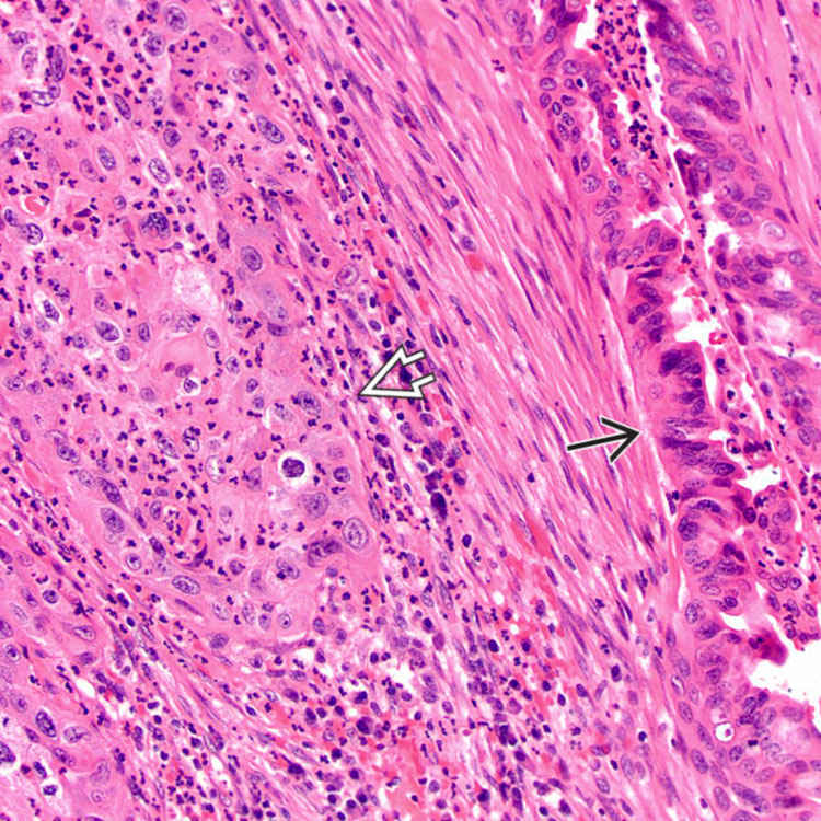
 adjacent to a malignant gland
  .
.ETIOLOGY/PATHOGENESIS
Histogenesis
• Neoplastic transformation from squamous metaplasia of gallbladder mucosa


Stay updated, free articles. Join our Telegram channel
Full access? Get Clinical Tree
