Predominantly composed of atypical squamous cells, with scattered melanocytes


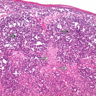
Squamomelanocytic tumor is a dermal-based tumor composed of 2 populations of cells: Atypical epithelioid melanocytic cells
 and larger squamoid cells
  . (Courtesy P. Amerio, MD, PhD.)
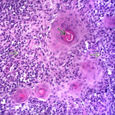
Squamomelanocytic tumor shows squamoid cells with pink cytoplasm in islands, forming keratin pearls
 , surrounded by smaller melanocytic cells
  with less distinctive cytoplasm. (Courtesy P. Amerio, MD, PhD.)
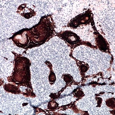
Cytokeratin AE1/AE3 strongly highlights the islands of squamoid cells forming keratin. (Courtesy P. Amerio, MD, PhD.)
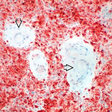
Melan-A staining is positive in the melanocytic cells surrounding the negative islands of squamoid cells
 . (Courtesy P. Amerio, MD, PhD.)
ETIOLOGY/PATHOGENESIS
Field Cancerization Theory
Tumor Collision Theory
• Arguing against this theory is presence of identical molecular alterations and immunohistochemical staining patterns in both squamous and melanocytic components of some squamomelanocytic tumors
Stay updated, free articles. Join our Telegram channel

Full access? Get Clinical Tree


