QUESTION 18.1
A. Arteriolar thrombi
B. Ceroid-laden histiocytes in the red pulp
C. Congested red pulp cords
D. Hemophagocytosis
E. Immunoblastic proliferation
2. According to the most recent studies, which of the following is the most common primary lymphoproliferative disorder of the spleen?
A. Hairy cell leukemia
B. Large B-cell lymphoma
C. Mantle cell lymphoma
D. Marginal zone lymphoma
E. Small lymphocytic lymphoma
3. This photomicrograph shows a lymphomatous proliferation in the spleen. Neoplastic lymphocytes express CD5 and CD23, but not cyclin D1 or tartrate. Which of the following is the most likely diagnosis?
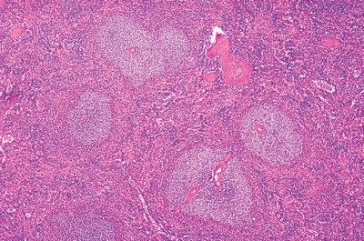
QUESTION 18.3
A. Hairy cell leukemia
B. Lymphoplasmacytic lymphoma
C. Mantle cell lymphoma
D. Marginal zone lymphoma
E. Small lymphocytic lymphoma
4. These low- and high-power photomicrographs (A and B) show the characteristic infiltration pattern and cellular morphology of a splenic lymphoma bearing a t(11;14) chromosomal translocation. Morphologically, this lymphoma is similar to small lymphocytic lymphoma (SLL). Which of the following markers allows differentiation from SLL?

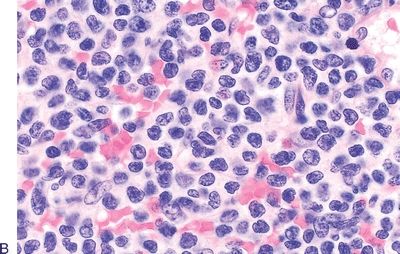
QUESTION 18.4
A. CD5
B. CD10
C. CD23
D. CD43
E. Cyclin D1
5. A 70-year-old man presents with abdominal pain and fullness in the left upper quadrant. On physical examination, severe splenomegaly is found. Laboratory studies shows pancytopenia and paraproteinemia. He undergoes splenectomy, which reveals white pulp expansion as shown in this photomicrograph. Infiltrating cells express CD19, CD20, and IgM. They are negative for CD5, CD10, CD23, and cyclin D1. Which of the following is the most likely diagnosis?
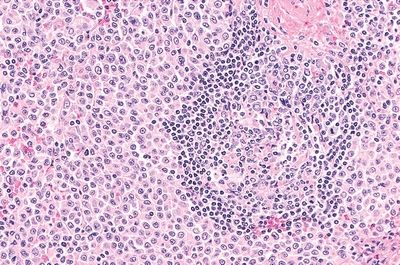
QUESTION 18.5
A. Diffuse large B-cell lymphoma
B. Follicular lymphoma
C. Mantle cell lymphoma
D. Marginal zone lymphoma
E. Small lymphocytic lymphoma
6. This photograph shows the cut surface of a very enlarged spleen. Which of the following disorders is most consistent with this gross appearance?
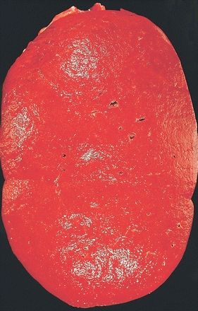
QUESTION 18.6
A. Chronic myelogenous leukemia
B. Hodgkin lymphoma
C. Large B-cell lymphoma
D. Reactive follicular hyperplasia
E. Small lymphocytic lymphoma
7. This photomicrograph shows the red pulp of a massively enlarged spleen. The cell population seen here is most compatible with which of the following disorders?
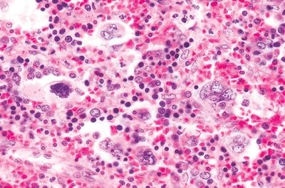
QUESTION 18.7
Stay updated, free articles. Join our Telegram channel

Full access? Get Clinical Tree


