Small cells with scant cytoplasm and small hyperchromatic nuclei; typically at periphery of tumor lobules
Larger cells with eosinophilic cytoplasm and oval, vesicular nuclei; typically in centers of tumor lobules
• Focal to diffuse duct lumen formation
• Tumor lobules associated with vascularized stroma, hemorrhage may be present
Top Differential Diagnoses
• Cylindroma
Significant overlap with spiradenoma and may have combined tumors
Cylindroma has jigsaw puzzle pattern
• Spiradenocarcinoma (malignant spiradenoma)
Associated with precursor spiradenoma
• Basal cell carcinoma
Peripheral palisading with tumor-stroma retraction
• Merkel cell carcinoma
More cytologic atypia and high mitotic rate
Positive for CK20 and neuroendocrine markers
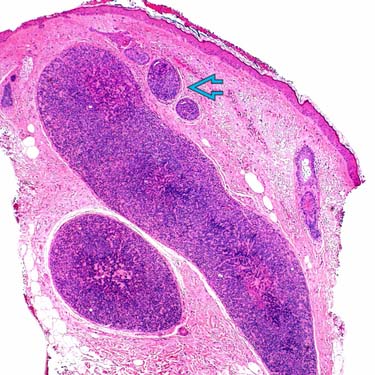
Spiradenoma at Low Magnification Low magnification of a spiradenoma shows a dermal-based tumor characterized by irregularly-shaped nodules and smaller lobules in the dermis.
Spiradenoma With Large Dermal Nodules Spiradenoma is characterized by circumscribed, cellular basophilic nodules or lobules in the dermis. The tumor lobules often have a surrounding fibrous capsule .
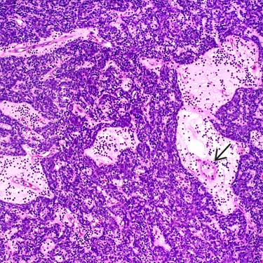
Spiradenoma With Edematous Stroma The stroma in this case is edematous and highlights the associated capillary vasculature .
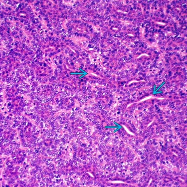
Spiradenoma at High Magnification The tumor is composed of a biphasic population of smaller basaloid cells and larger pale cells. Duct lumen formation is present and may be focal or relatively prominent, as in this case.
TERMINOLOGY
Synonyms
• Eccrine spiradenoma
Definitions
• Benign adnexal tumor composed of nodules of basaloid cells with ductal differentiation
• May have apocrine or eccrine differentiation
ETIOLOGY/PATHOGENESIS
Genetic Syndrome
• Familial cases associated with autosomal dominant Brooke-Spiegler syndrome
a.k.a. familial cylindromatosis or turban tumor syndrome
Multiple cylindromas, but can also have spiradenomas and trichoepitheliomas
CLINICAL ISSUES
Epidemiology
• Age
Most common in young adults but can present at any age
Site
• Upper 1/2 of body most commonly involved; > 75% present on ventral surface
Presentation
• Dermal mass/nodular lesion
Often tender or painful, may have bluish color
Usually solitary but may be multiple
– Multiple lesions may be part of Brooke-Spiegler syndrome
– Less frequently, may be associated with trichoblastoma and cutaneous lymphadenoma as part of morphological spectrum of Brooke-Spiegler syndrome
Treatment
• Surgical approaches
Complete surgical excision is curative
Prognosis
• Benign, but local recurrence may occur; very rare malignant transformation
MACROSCOPIC
General Features
• Dermal-based, bluish nodule
Only gold members can continue reading. Log In or Register to continue
 Larger cells with eosinophilic cytoplasm and oval, vesicular nuclei; typically in centers of tumor lobules


 in the dermis.
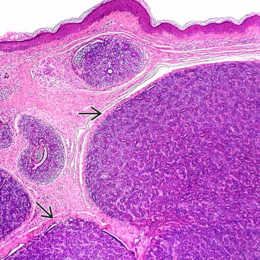
 .
.
 .
.
 is present and may be focal or relatively prominent, as in this case.


