Spindle Cell Hemangioma
Steven D. Billings, MD
Key Facts
Terminology
Formerly called spindle cell hemangioendothelioma
Clinical Issues
Subcutaneous mass
Usually acral location
Sometimes multifocal
Associated with Mafucci syndrome in some cases
Local recurrence in 50-60%
No metastatic potential
Microscopic Pathology
Dilated vessels often with focal thrombi
Solid spindled cell areas
Vacuolated endothelial cells, so-called “blister cells”
Negative for HHV8 latent nuclear antigen
Top Differential Diagnoses
Kaposi sarcoma
Kaposiform hemangioendothelioma
Epithelioid hemangioendothelioma
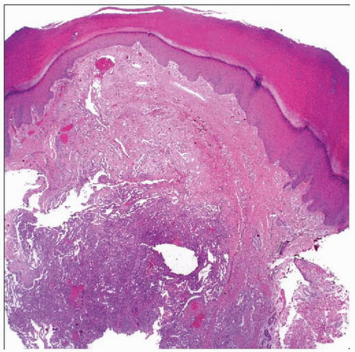 Hematoxylin & eosin shows a noncircumscribed lesion in the dermis and subcutis composed of an admixture of thin-walled dilated blood vessels and sheets of spindle cells. |
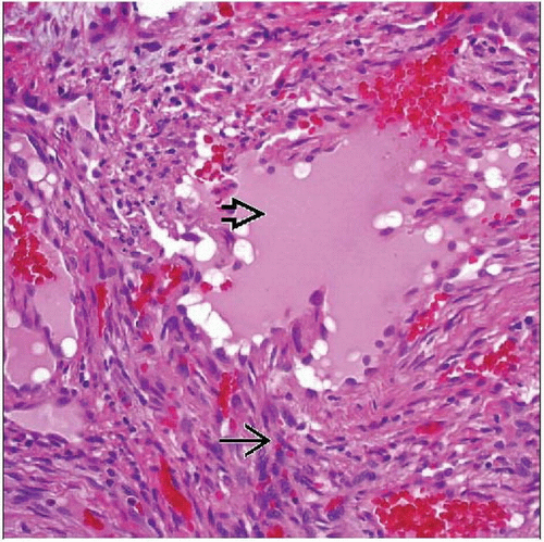 Hematoxylin & eosin shows straight/curved fascicles of uniform spindle cells  with irregularly shaped vascular spaces with irregularly shaped vascular spaces  and small areas of hemorrhage. and small areas of hemorrhage. |
TERMINOLOGY
Synonyms
Formerly called spindle cell hemangioendothelioma
Definitions
Benign vascular tumor characterized by cavernous and spindled areas
CLINICAL ISSUES
Epidemiology
Age
Young adults
Site
Distal extremities
Acral location most common


