Myxoid Liposarcoma
Thomas Mentzel, MD
Key Facts
Terminology
Malignant lipogenic neoplasm composed of primitive nonlipogenic mesenchymal cells and variable number of lipoblasts set in myxoid stroma with characteristic branching blood vessels
Clinical Issues
2nd most common type of liposarcoma
Young adults
Commonest form of liposarcoma in patients younger than 20 years
Deep soft tissue of extremities
May present initially with synchronous or metachronous multifocal neoplasms
Increased rate of local recurrences
Metastases develop in ˜ 30-40% of cases
Presence of round cell areas is of prognostic importance
> 5% round cell differentiation is associated with unfavorable outcome
Microscopic Pathology
Uniform, round to oval-shaped, primitive mesenchymal cells
Small, univacuolated, signet ring lipoblasts
Prominent myxoid stroma
Delicate, arborizing, “chicken wire” capillary vasculature
Ancillary Tests
Most frequently t(12;16)(q13;p11)
S100 protein can be positive
MDM2(-) and CDK4(-)
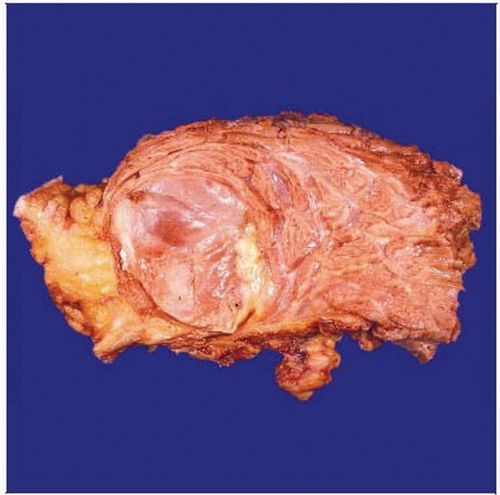 Gross photograph shows a myxoid/round cell liposarcoma with myxoid cut surfaces and small, indurated, gray-white areas corresponding to tumor areas with increased cellularity and round cell morphology. |
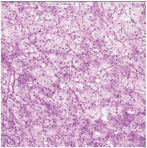 Low-grade myxoid liposarcoma is composed of small, undifferentiated, mesenchymal tumor cells associated with lipoblasts set in a prominent myxoid stroma with thinwalled, branching capillaries. |
TERMINOLOGY
Abbreviations
Myxoid liposarcoma (MLS)
Synonyms
Myxoid/round cell liposarcoma
Round cell liposarcoma
Definitions
Malignant lipogenic neoplasm composed of primitive nonlipogenic mesenchymal cells and a variable number of lipoblasts set in myxoid stroma with characteristic branching blood vessels
CLINICAL ISSUES
Epidemiology
Incidence
2nd most common type of liposarcoma
Accounts for more than 1/3 of all liposarcomas
Accounts for ˜ 10% of all sarcomas arising in adults
Age
Young adults
Peak incidence in 4th and 5th decade
Rare in children
Commonest form of liposarcoma in patients younger than 20 years
Gender
No gender predilection
Site
Deep soft tissue of extremities
2/3 of cases arise within musculature of thigh
Rare in subcutaneous location
Extremely rare in dermal location
Presentation
Painless mass
May present initially with synchronous or metachronous multifocal neoplasms
Deep-seated soft tissue neoplasms
Natural History
Locally aggressive growth
Increased rate of local recurrences
Metastases develop in ˜ 30-40% of cases
Tends to metastasize to unusual soft tissue or bone locations
Treatment
Surgical approaches
Complete excision with wide tumor-free margins
Prognosis
Presence of round cell areas is of prognostic importance
> 5% round cell differentiation is associated with unfavorable outcome
Large tumor size (> 10 cm) is associated with unfavorable outcome
Tumor necrosis associated with unfavorable outcome
p53 overexpression and p53 mutations associated with unfavorable outcome
Loss of p27 associated with unfavorable outcome
Prognosis of multifocal neoplasms is poor independent of morphology
Molecular variability has no prognostic influence
MACROSCOPIC FEATURES
General Features
Well-circumscribed, multinodular neoplasms
Gelatinous cut surfaces in low-grade neoplasms
Round cell areas correspond to indurated, gray-white tumor areas
MICROSCOPIC PATHOLOGY
Histologic Features
Nodular growth pattern
May show enhanced cellularity at periphery of tumor lobules
Uniform, round to oval-shaped, primitive mesenchymal cells
Small, univacuolated, signet ring lipoblasts
May show “maturation” with lipoma or atypical lipomatous tumor-like areas
Prominent myxoid stroma
Mucin pooling
Delicate, arborizing, “chicken wire” capillary vasculature
Interstitial hemorrhages
May contain hibernoma-like cells
No nuclear pleomorphism
No significant mitotic activity
Progression to hypercellular, round cell areas
Increased cellularity
Nests or solid sheets of back-to-back located round cells
Round cells have high nuclear:cytoplasmic ratio
Enlarged nuclei that may show overlapping
Rare hypercellular, spindle cell areas
Rare heterologous differentiation (cartilaginous, rhabdomyoblastic, leiomyomatous, osseous)
Reported dedifferentiation may represent rather mixed-type liposarcomas
Predominant Pattern/Injury Type
Circumscribed
Predominant Cell/Compartment Type
Adipose
Lipoblast
Mesenchymal, adipose cell
ANCILLARY TESTS
Cytogenetics
Most frequently t(12;16)(q13;p11)


