Deep Leiomyoma
Cyril Fisher, MD, DSc, FRCPath
Key Facts
Clinical Issues
Rare: 4% of benign soft tissue tumors
Slow-growing mass
Usually painless
Deep subcutis or subfascial in limbs in either sex
Retroperitoneum, especially in females
Range of appearances like uterine leiomyoma
Macroscopic Features
Well circumscribed
Retroperitoneal tumors can reach very large size
No necrosis
No hemorrhage
Focal myxoid change and cyst formation
Foci of calcification
Microscopic Pathology
Short fascicles of benign smooth muscle cells
Focal epithelioid cell morphology
No nuclear pleomorphism
No necrosis
No mitoses except in female retroperitoneum
Ancillary Tests
Positive for actin-sm, desmin, HCAD
ER, PR diffusely in female retroperitoneum
Top Differential Diagnoses
Leiomyosarcoma
Any necrosis or pleomorphism
Any mitoses in deep somatic soft tissue leiomyomas
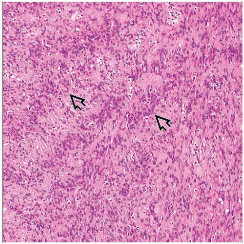
 Hematoxylin & eosin shows short fascicles of uniform spindle cells with blunt- or round-ended nuclei and eosinophilic cytoplasm. There is a fibrous stroma with scattered mast cells  . . |
 Hematoxylin & eosin shows nuclear palisading  . This is more common in retroperitoneal leiomyomas in females, which resemble those of the uterus. This should not be misinterpreted as a schwannoma. . This is more common in retroperitoneal leiomyomas in females, which resemble those of the uterus. This should not be misinterpreted as a schwannoma. |
TERMINOLOGY
Definitions
Benign smooth muscle tumor involving deep soft tissue of limbs or retroperitoneum
CLINICAL ISSUES
Epidemiology
Incidence
Rare: 4% of benign soft tissue tumors
Age
4th-6th decade of life
Gender
M<F
Site
Deep subcutis or subfascial in limbs
Retroperitoneum, especially in females
Resemble uterine leiomyomas
Presentation
Painless mass
Slow growing
Treatment
Simple excision
Prognosis
Benign tumor
Correctly diagnosed cases do not recur or metastasize
MACROSCOPIC FEATURES
General Features
Well circumscribed
Gray-white cut surface
Myxoid areas
Foci of calcification
Rare cystic change
Necrosis absent
Size
5-20 cm
Retroperitoneal tumors can reach very large size
MICROSCOPIC PATHOLOGY
Predominant Pattern/Injury Type
Sheets
Predominant Cell/Compartment Type
Smooth muscle
Key Microscopic Features
Short fascicles or nests of uniform spindle-shaped smooth muscle cells
Nontapered nuclei
Abundant eosinophilic cytoplasm
Focal epithelioid cell morphology
No nuclear pleomorphism
Occasional degenerative atypia seen; no nucleoli
No necrosis
No mitoses except in retroperitoneal tumors in females
Not exceeding 5 per 50 high-power fields (HPF)
Stay updated, free articles. Join our Telegram channel

Full access? Get Clinical Tree


