QUESTION 55.1
A. Adult-type granulosa cell tumor
B. Embryonal carcinoma
C. Juvenile-type granulosa cell tumor
D. Sertoli–Leydig cell tumor
E. Thecoma
2. Which of the following immunostains is positive on most cases of sex cord–stromal tumors?
A. Calretinin
B. Inhibin
C. Melan-A
D. SF-1
E. All of the above
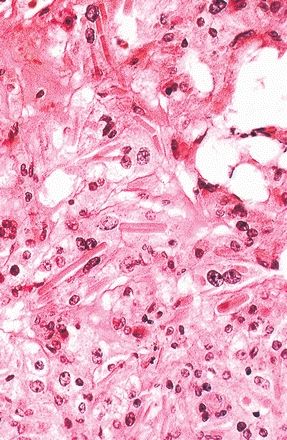
3. An 8-year-old girl presents with precocious puberty, and a pelvic ultrasound reveals a right ovarian mass. The histologic appearance of the tumor is shown in this picture. What is the diagnosis?
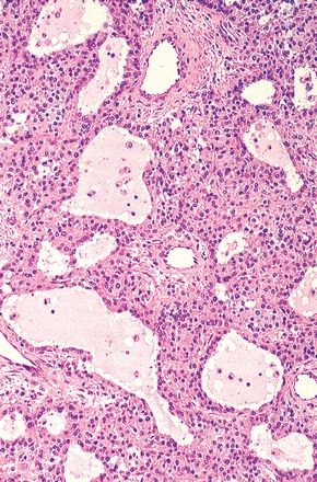
QUESTION 55.3
A. Adult-type granulosa cell tumor
B. Clear cell carcinoma
C. Juvenile-type granulosa cell tumor
D. Sertoli–Leydig cell tumor
E. Thecoma
4. A syndrome characterized by an ovarian tumor, ascites, and right pleural effusion is typically associated with:
A. Fibroma
B. Granulosa cell tumor
C. Leydig cell tumor
D. Sclerosing stromal tumor
E. Thecoma
5. This photomicrograph shows an ovarian neoplasm from a postmenopausal woman presenting with abnormal uterine bleeding. Which of the following immunohistochemical or special stains is most useful in differentiating this neoplasm from adult granulosa cell tumor?
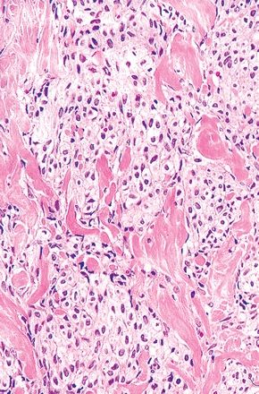
QUESTION 55.5
A. FOXL2 immunostain
B. Inhibin immunostain
C. Masson trichrome stain
D. Reticulin stain
E. SF-1 immunostain
6. A 25-year-old woman with menstrual irregularities undergoes resection of an ovarian tumor whose histologic appearance is shown in this picture. These features are most consistent with:
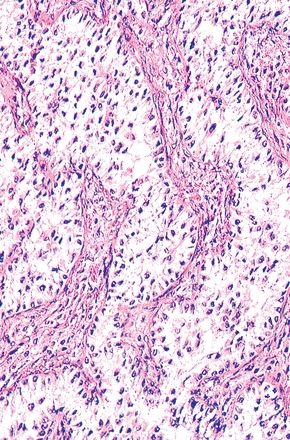
QUESTION 55.6
A. Leydig cell tumor
B. Sclerosing stromal tumor
C. Sertoli cell tumor
D. Sertoli–Leydig cell tumor
E. Sex cord tumor with annular tubules
7. The ovarian tumor shown in this picture is most frequently associated with production of:
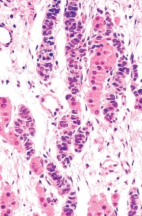
QUESTION 55.7
A. AFP
B. Androgens
C. HCG
D. Estrogens
E. PLAP
8. This photomicrograph demonstrates the most significant histopathologic findings in a 4-cm tumor located in the ovarian hilus and associated with clinical signs of virilization. Which of the following is the most likely diagnosis?
QUESTION 55.8
A. Leydig cell tumor
B. Luteinized thecoma
C. Sertoli cell tumor
D. Steroid cell tumor, NOS
E. Stromal luteoma
9. An ovarian dysgerminoma contains scattered isolated foci of glandlike structures immunoreactive for alpha-fetoprotein (AFP) and keratin. This immunohistochemical pattern is evidence of differentiation toward:
Stay updated, free articles. Join our Telegram channel

Full access? Get Clinical Tree


