Sebaceous Hyperplasia
Christine J. Ko, MD
Key Facts
Clinical Issues
Commonly on face
Yellow to flesh-colored to slightly pink papule
Often there is central dell
Often biopsied to rule out basal cell carcinoma
Microscopic Pathology
Lobules of sebocytes arranged around infundibulum of central hair follicle
1 layer of basaloid cells compressed at periphery of sebocytes
No cytologic atypia
Top Differential Diagnoses
Sebaceous adenoma
Ectopic sebaceous glands in other sites (e.g., nipple)
Phymatous rosacea
Sebaceous trichofolliculoma
Folliculosebaceous (cystic) hamartoma
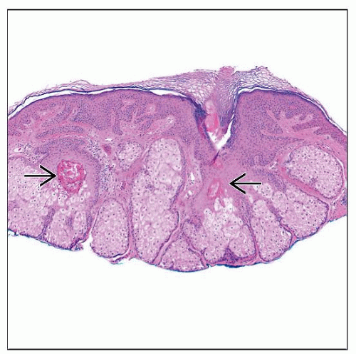 H&E shows low-magnification view of sebaceous hyperplasia. There are normal-appearing lobules of sebocytes surrounding invaginations of epidermis that resemble the infundibulum of hair follicles  . . |
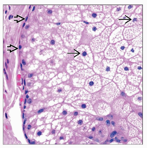 High-magnification view of the edge of a lobule of sebocytes shows that central sebocytes  with round to scalloped nuclei and bubbly cytoplasm are rimmed by a compressed layer of small, basaloid cells with round to scalloped nuclei and bubbly cytoplasm are rimmed by a compressed layer of small, basaloid cells  . . |
TERMINOLOGY
Definitions
Benign
Hyperplasia (overgrowth) of sebaceous glands
Plump lobules of sebaceous glands arranged around central follicles
CLINICAL ISSUES
Site
Commonly on face
Rarely on the trunk or other sites
Presentation
Yellow to flesh-colored to slightly pink papule
Often there is central dell
Telangiectasias may be present
Often biopsied to rule out basal cell carcinoma
Laboratory Tests
Generally not performed
Stay updated, free articles. Join our Telegram channel

Full access? Get Clinical Tree


