• 3 types described: Solid (most common), venous, and cavernous
• Well-differentiated smooth muscle component
• Prominent component of medium- to large-sized blood vessels with dilated or compressed lumina
Smooth muscle cells can show concentric, perivascular accentuation in some cases
• No significant nuclear atypia or mitotic activity
• Mature adipose tissue, stromal hyalinization, myxoid changes, or calcification may be present
Ancillary Tests
• Strong, diffuse SMA(+) and h-caldesmon (+)
• Desmin (+) but varies in extent
Top Differential Diagnoses
• Myopericytoma
• Hemangioma
• PEComa
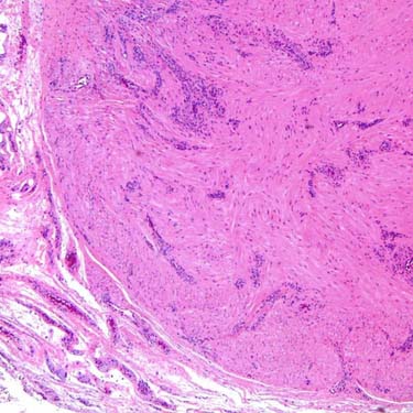
Angioleiomyoma Angioleiomyoma is a small, sharply circumscribed smooth muscle neoplasm containing a prominent component of stromal blood vessels. These vessels vary in size and may be compressed (solid variant, shown) or markedly dilated/ectatic (venous and cavernous variants).
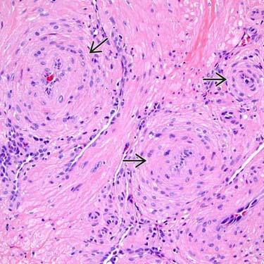
Perivascular Growth by Smooth Muscle Cells Perivascular arrangement of the lesional smooth muscle cells is a common finding in angioleiomyoma and creates morphologic overlap with myopericytoma. This finding, however, may be focal or absent in some cases.
Mature Smooth Muscle Cells The well-differentiated smooth muscle cells of angioleiomyoma show classic cytologic features including prominent eosinophilic cytoplasm and elongated, blunt, cigar-shaped nuclei. They are also typically arranged in bundles and fascicles, as seen in other smooth muscle neoplasms.
Cavernous Morphology The cavernous type of angioleiomyoma has thin-walled, dilated vascular channels arranged in small clusters within a stroma composed of smooth muscle fibers that form branching fascicles.
TERMINOLOGY
Synonyms
• Vascular leiomyoma
• Angiomyoma
Definitions
• Benign, well-circumscribed neoplasm composed of mature smooth muscle cells arranged around prominent blood vessels
Now classified as tumor of pericytic (perivascular) origin (2013 WHO classification)
Considered to exist on morphologic spectrum with myopericytoma
CLINICAL ISSUES
Epidemiology
• Age
Wide range (most common: 40-70 years)
• Sex
Male predominance in upper extremity
Only gold members can continue reading. Log In or Register to continue

 of the lesional smooth muscle cells is a common finding in angioleiomyoma and creates morphologic overlap with myopericytoma. This finding, however, may be focal or absent in some cases.
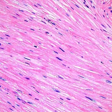
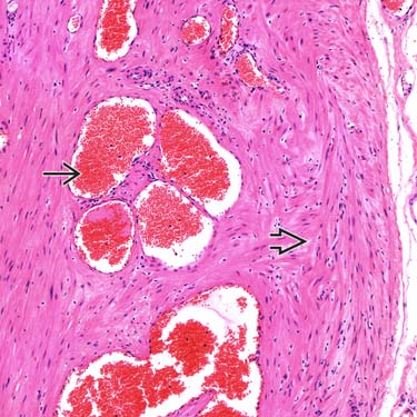
 arranged in small clusters within a stroma composed of smooth muscle fibers
  that form branching fascicles.


