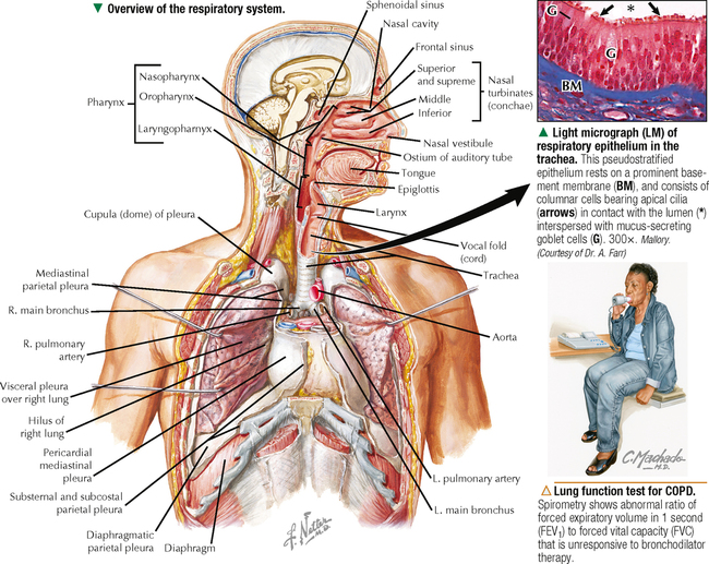
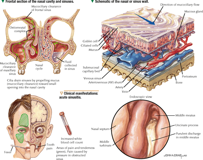
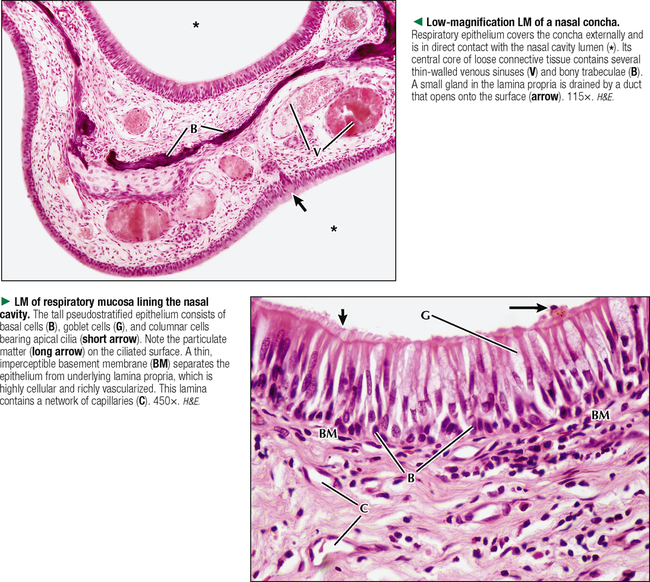
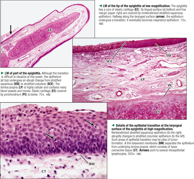
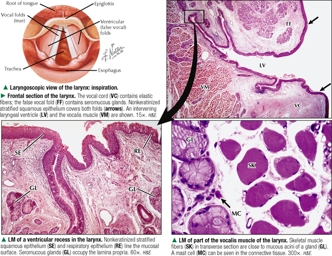
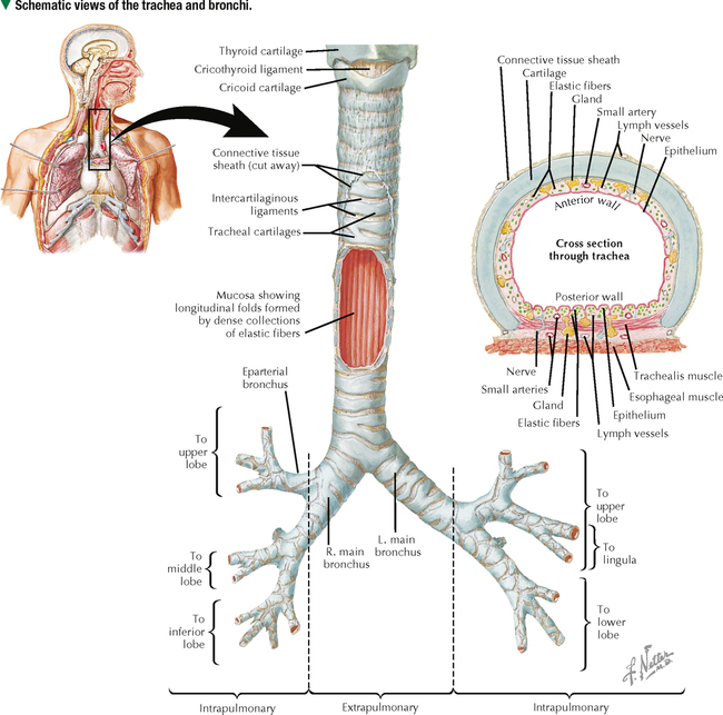
15 RESPIRATORY SYSTEM 15.1. Overview 15.2. Structure of the Nasal Cavities and Paranasal Sinuses 15.3. Histology of the Nasal Cavities and Paranasal Sinuses 15.4. Histology of the Epiglottis 15.5. Histology of the Larynx and Vocal Cords 15.6. Structure of the Trachea and Major Bronchi 15.7. Histology of the Trachea 15.8. Scanning Electron Microscopy of Tracheal and Bronchial Epithelium 15.9. Ultrastructure of Tracheal and Bronchial Epithelium 15.10. Ultrastructure and Function of Respiratory Cilia 15.11. Histology of the Bronchi 15.12. Structure of Intrapulmonary Airways 15.13. Histology of Terminal and Respiratory Bronchioles 15.14. Ultrastructure of Bronchiolar Epithelium: Clara Cells 15.15. Intrapulmonary Blood Circulation 15.16. Histology and Ultrastructure of Pulmonary Alveoli 15.17. Ultrastructure of the Blood-Air Barrier 15.18. Ultrastructure of Type II Pneumocytes 15.19. Ultrastructure of Alveolar Macrophages 15.20. Development of the Lower Respiratory System 15.1 OVERVIEW The respiratory system is divided functionally into a conducting portion that conveys air from outside the body to the lungs and a respiratory portion where exchange of gases between the air and blood occurs. The conducting airways moisten, warm, and cleanse the air, whereas the respiratory portion provides O2 obtained from the air and removes excess CO2 from the bloodstream. The conducting passageways (the cavities and tubes) consist anatomically of the nose and paranasal sinuses; pharynx, which is the passageway for both air and food; larynx, which produces the voice; trachea, which divides into bronchi and bronchioles of decreasing size; and terminal bronchioles. The respiratory portion comprises respiratory bronchioles, which branch into alveolar ducts and pulmonary alveoli, where exchange of gases with adjacent capillaries takes place. Pseudostratified ciliated columnar epithelium plus numerous mucus-secreting goblet cells line the mucous membrane of the upper airways of the conducting portion. This ciliated epithelium, commonly known as respiratory epithelium, is well suited for airway protection and cleansing and removal of particulate matter. The cilia beat in a rhythmic fashion toward the oral cavity and move debris and pathogen-laden mucus so it can be expectorated or swallowed. Subepithelial mucous and serous glands liberate their secretions onto the mucosal surface to also aid in entrapment of particulate matter, lubrication, and moistening. Accessory structures needed for proper functioning of the respiratory system include the pleurae, diaphragm, thoracic wall, and muscles that raise and lower ribs during inspiration and expiration. CLINICAL POINT Chronic obstructive pulmonary disease (COPD)—a progressive inflammatory condition of the lower airways with high morbidity and mortality worldwide—includes both emphysema and chronic bronchitis. Major symptoms are limitation of expiratory airflow, long-lasting cough, shortness of breath (dyspnea), fatigue, and copious sputum production. Mucociliary dysfunction accompanies airway inflammation (mainly neutrophil, macrophage, and CD8+ T-lymphocyte infiltration). Although most often caused by cigarette smoking, repeated childhood lung infections, genetic abnormalities (e.g., α1-antitrypsin deficiency), and long-term exposure to air pollutants and chemical irritants may also contribute to it. The diagnosis is via spirometry and other lung function tests. Depending on the disease severity, treatment includes bronchodilator medications and avoidance of respiratory irritants. 15.2 STRUCTURE OF THE NASAL CAVITIES AND PARANASAL SINUSES The nasal cavities—paired passages separated by a nasal septum—are the first structures of the conducting part of the respiratory system. Each cavity consists of an anterior vestibule and nasal cavity proper. The vestibule, which is lined by epidermis containing many sebaceous glands, sweat glands, and hair follicles, leads into the nasal cavity proper, which is lined by mucosa consisting of pseudostratified ciliated columnar epithelium interspersed with goblet cells and resting on a prominent basement membrane. The underlying lamina propria, a thick, vascular connective tissue rich in collagen and elastic fibers, attaches firmly to the periosteum and perichondrium of the bony and cartilaginous walls of the nasal cavity, which provide rigidity during inspiration. Seromucous glands are also found in the lamina propria and drain onto the epithelial surface via small ducts. Cilia on the epithelial surface beat to move surface secretions toward the nasopharynx. In the lamina propria are large venous plexuses whose major role is to warm inspired air via heat exchange. The plexuses may become engorged during an allergic reaction or nasal infection, which leads to mucous membrane swelling and restricted air passage. Paranasal sinuses—frontal, ethmoidal, sphenoidal, and maxillary—are air-filled cavities that communicate with nasal cavities. Their mucosa, consisting of respiratory epithelium with numerous goblet cells, is continuous with that of the nasal cavities, a feature that favors the spread of infection. The lamina propria is very thin and blends with the periosteum of surrounding bony tissue. A few small seromucous glands are found in the mucosa of paranasal sinuses. CLINICAL POINT Sinusitis is a common clinical condition referring to inflammation of the mucous membrane of the sinuses. Often associated with the common cold or allergies, it may be caused by bacterial, viral, or fungal infection. Acute and chronic forms affect 30–40 million people in North America annually. The mucosal lining of the nasal and paranasal sinuses produces about 750 mL of mucus daily. Inflamed sinuses become blocked with mucus and can become infected. In cases of chronic sinusitis, the drainage pathways of the sinuses are obstructed and do not function properly. The mucous glands produce thick secretions that stay in the cavities, which increases bacterial overgrowth and thickens the lining. 15.3 HISTOLOGY OF THE NASAL CAVITIES AND PARANASAL SINUSES Each nasal cavity is a narrow passage that communicates posteriorly via a small orifice, the choana, with the nasopharynx. The cavity’s surface area is dramatically increased by nasal conchae consisting of bony trabeculae covered by mucous membrane. The pseudostratified ciliated columnar epithelium of the mucous membrane has abundant, unevenly distributed mucus-secreting goblet cells. Many branched seromucous glands extend into the underlying lamina propria and are connected to the surface via small ducts. An extensive, tortuous network of venous sinuses, arteriovenous anastomoses, and capillaries characterizes the lamina propria. In certain areas of the nasal mucosa, thin-walled venous sinuses that are superficially located resemble erectile tissue and warm inspired air via heat exchange. A surface layer of mucus produced by goblet cells and seromucous glands entraps foreign particles and is constantly moved by cilia. This process, known as mucociliary clearance, sweeps particulate matter toward the nasopharynx, where it is swallowed or expectorated. The lining epithelium in the paranasal sinuses is lower than that of the nasal cavities, with fewer goblet cells than in the nasal cavities. Three types of cells characterize the respiratory epithelium: basal, ciliated, and goblet cells. Basal cells serve as reserve cells, which continuously replace other epithelial cells that are shed. They are small rounded cells in a monolayer resting on the basement membrane. Goblet cells sit on the basement membrane and extend to the surface, where they have a relatively wide apical region that appears pale or washed out because of a varying content of mucus. 15.4 HISTOLOGY OF THE EPIGLOTTIS The epiglottis is an unpaired leaf-shaped structure below the root of the tongue that covers the entrance to the larynx. It has a core of elastic cartilage, which is highly flexible and attaches to the hyoid bone. Its lingual surface is covered by a protective mucosa with a nonkeratinized stratified squamous epithelium that is directly continuous with the epithelium covering the dorsal surface of the tongue. This epithelium continues onto the laryngeal undersurface of the epiglottis. Deep along this surface, the epithelium becomes a transitional zone of stratified columnar epithelium and then pseudostratified ciliated columnar epithelium with goblet cells, commonly known as respiratory epithelium. Scattered seromucous glands are found between the plates of elastic cartilage or close to the mucosa lining the undersurface. Lamina propria of loose connective tissue underneath the epithelium contains numerous blood and lymphatic vessels, nerves, and scattered mononuclear connective tissue cells. The perichondrium surrounding the elastic cartilage attaches firmly to the lamina propria. At rest, the epiglottis is usually upright and allows air to pass into the larynx and the rest of the lower respiratory airways. During swallowing, it folds back like a flap to cover the entrance to the larynx, to prevent food and liquid from entering the trachea. A sore throat—infection or inflammation of the tonsils, pharynx, or larynx—can obstruct the trachea and make breathing more labored, which may be fatal unless promptly treated. 15.5 HISTOLOGY OF THE LARYNX AND VOCAL CORDS The larynx lies between the pharynx and trachea and is 4–5 cm long. Part of the respiratory conducting system, it plays a critical role in phonation and closes during swallowing, thereby preventing food from entering the lower airways. Its wall is made of a framework of hyaline and elastic cartilage united by connective tissue and associated with skeletal muscles. The laryngeal mucous membrane has two sets of prominent folds that project inward: false (or ventricular) folds and true vocal folds (or cords). Between the folds lies a space, the laryngeal ventricle, with narrow pouch-like invaginations known as ventricular recesses. The vocal folds contain vocal ligaments of elastic fibers to which skeletal muscle fibers of the vocalis part of the thyroarytenoid muscles attach. Contraction of the vocalis muscle relaxes elastic fibers of the vocal ligaments, thereby changing the shape of the laryngeal ventricles and allowing production of different sounds. The mucous membrane of the larynx consists mainly of respiratory epithelium, but over the vocal folds it becomes nonkeratinized stratified squamous epithelium; this change is a function of active movement of the folds and wear and tear induced by friction. The epithelium at the junction of the two types of folds is ciliated stratified columnar. Beneath the epithelium is the lamina propria of loose, highly cellular connective tissue, with lymphoid nodules near the laryngeal recesses. Mixed seromucous glands, which are invaginations of the epithelium, occur in the ventricular folds but not in the vocal cords. The larynx is unusually replete with mast cells that release histamine during allergic responses, which results in edema that may become life threatening (can occlude the airway). CLINICAL POINT Hoarseness—an abnormal, harsh, and raspy voice—is a common symptom of many disorders affecting the vocal cords. Although laryngitis caused by acute upper respiratory tract infection is the most frequent cause, persistent hoarseness may be an early sign of laryngeal cancer, which in North America accounts for nearly 15,000 new cases annually. More than 95% of laryngeal tumors are squamous cell carcinomas that arise directly from the true vocal cords. Such neoplasms are typically slow growing and are late to metastasize because of relatively sparse lymphatic drainage of the glottis. Affecting more men than women, chronic tobacco use and excessive alcohol consumption are major risk factors. Biopsy is required for diagnosis and tumor staging; treatment options include surgical resection, radiation therapy, and laryngectomy. 15.6 STRUCTURE OF THE TRACHEA AND MAJOR BRONCHI The tracheobronchial tree, a conduit for air traveling to and from alveoli in the lungs, comprises the trachea and the right and left bronchi and their subdivisions. The outer anterolateral aspect of the trachea contains 16–20 crescent-shaped rings of hyaline cartilage that provide rigidity, maintain shape, and ensure patency of the tracheal lumen. With aging, the cartilage often shows degenerative changes and may calcify. Posteriorly, the ends of the cartilage rings are spanned by a fibrous membrane containing smooth muscle fibers that constitute the trachealis muscle. Contraction of this muscle, which is mainly circular in orientation, causes the tracheal lumen to narrow. Pseudostratified ciliated columnar epithelium lines the lumen and rests on a prominent basement membrane, one of the thickest in the body. Metaplasia of the epithelium occurs in response to local friction and chronic coughing. Goblet cells interspersed in the epithelium secrete mucus, which lubricates the tracheal surface and traps foreign particulate matter such as dust and bacteria. Small seromucous glands characterize the underlying submucosa. A layer of longitudinally oriented elastic fibers Only gold members can continue reading. Log In or Register to continue Share this: Share on X (Opens in new window) X Share on Facebook (Opens in new window) Facebook Like this:Like Loading... Related Related posts: CARDIOVASCULAR SYSTEM SPECIAL SENSES THE CELL FEMALE REPRODUCTIVE SYSTEM Stay updated, free articles. Join our Telegram channel Join Tags: Netters Essential Histology Jun 18, 2016 | Posted by admin in HISTOLOGY | Comments Off on RESPIRATORY SYSTEM Full access? Get Clinical Tree
15 RESPIRATORY SYSTEM 15.1. Overview 15.2. Structure of the Nasal Cavities and Paranasal Sinuses 15.3. Histology of the Nasal Cavities and Paranasal Sinuses 15.4. Histology of the Epiglottis 15.5. Histology of the Larynx and Vocal Cords 15.6. Structure of the Trachea and Major Bronchi 15.7. Histology of the Trachea 15.8. Scanning Electron Microscopy of Tracheal and Bronchial Epithelium 15.9. Ultrastructure of Tracheal and Bronchial Epithelium 15.10. Ultrastructure and Function of Respiratory Cilia 15.11. Histology of the Bronchi 15.12. Structure of Intrapulmonary Airways 15.13. Histology of Terminal and Respiratory Bronchioles 15.14. Ultrastructure of Bronchiolar Epithelium: Clara Cells 15.15. Intrapulmonary Blood Circulation 15.16. Histology and Ultrastructure of Pulmonary Alveoli 15.17. Ultrastructure of the Blood-Air Barrier 15.18. Ultrastructure of Type II Pneumocytes 15.19. Ultrastructure of Alveolar Macrophages 15.20. Development of the Lower Respiratory System 15.1 OVERVIEW The respiratory system is divided functionally into a conducting portion that conveys air from outside the body to the lungs and a respiratory portion where exchange of gases between the air and blood occurs. The conducting airways moisten, warm, and cleanse the air, whereas the respiratory portion provides O2 obtained from the air and removes excess CO2 from the bloodstream. The conducting passageways (the cavities and tubes) consist anatomically of the nose and paranasal sinuses; pharynx, which is the passageway for both air and food; larynx, which produces the voice; trachea, which divides into bronchi and bronchioles of decreasing size; and terminal bronchioles. The respiratory portion comprises respiratory bronchioles, which branch into alveolar ducts and pulmonary alveoli, where exchange of gases with adjacent capillaries takes place. Pseudostratified ciliated columnar epithelium plus numerous mucus-secreting goblet cells line the mucous membrane of the upper airways of the conducting portion. This ciliated epithelium, commonly known as respiratory epithelium, is well suited for airway protection and cleansing and removal of particulate matter. The cilia beat in a rhythmic fashion toward the oral cavity and move debris and pathogen-laden mucus so it can be expectorated or swallowed. Subepithelial mucous and serous glands liberate their secretions onto the mucosal surface to also aid in entrapment of particulate matter, lubrication, and moistening. Accessory structures needed for proper functioning of the respiratory system include the pleurae, diaphragm, thoracic wall, and muscles that raise and lower ribs during inspiration and expiration. CLINICAL POINT Chronic obstructive pulmonary disease (COPD)—a progressive inflammatory condition of the lower airways with high morbidity and mortality worldwide—includes both emphysema and chronic bronchitis. Major symptoms are limitation of expiratory airflow, long-lasting cough, shortness of breath (dyspnea), fatigue, and copious sputum production. Mucociliary dysfunction accompanies airway inflammation (mainly neutrophil, macrophage, and CD8+ T-lymphocyte infiltration). Although most often caused by cigarette smoking, repeated childhood lung infections, genetic abnormalities (e.g., α1-antitrypsin deficiency), and long-term exposure to air pollutants and chemical irritants may also contribute to it. The diagnosis is via spirometry and other lung function tests. Depending on the disease severity, treatment includes bronchodilator medications and avoidance of respiratory irritants. 15.2 STRUCTURE OF THE NASAL CAVITIES AND PARANASAL SINUSES The nasal cavities—paired passages separated by a nasal septum—are the first structures of the conducting part of the respiratory system. Each cavity consists of an anterior vestibule and nasal cavity proper. The vestibule, which is lined by epidermis containing many sebaceous glands, sweat glands, and hair follicles, leads into the nasal cavity proper, which is lined by mucosa consisting of pseudostratified ciliated columnar epithelium interspersed with goblet cells and resting on a prominent basement membrane. The underlying lamina propria, a thick, vascular connective tissue rich in collagen and elastic fibers, attaches firmly to the periosteum and perichondrium of the bony and cartilaginous walls of the nasal cavity, which provide rigidity during inspiration. Seromucous glands are also found in the lamina propria and drain onto the epithelial surface via small ducts. Cilia on the epithelial surface beat to move surface secretions toward the nasopharynx. In the lamina propria are large venous plexuses whose major role is to warm inspired air via heat exchange. The plexuses may become engorged during an allergic reaction or nasal infection, which leads to mucous membrane swelling and restricted air passage. Paranasal sinuses—frontal, ethmoidal, sphenoidal, and maxillary—are air-filled cavities that communicate with nasal cavities. Their mucosa, consisting of respiratory epithelium with numerous goblet cells, is continuous with that of the nasal cavities, a feature that favors the spread of infection. The lamina propria is very thin and blends with the periosteum of surrounding bony tissue. A few small seromucous glands are found in the mucosa of paranasal sinuses. CLINICAL POINT Sinusitis is a common clinical condition referring to inflammation of the mucous membrane of the sinuses. Often associated with the common cold or allergies, it may be caused by bacterial, viral, or fungal infection. Acute and chronic forms affect 30–40 million people in North America annually. The mucosal lining of the nasal and paranasal sinuses produces about 750 mL of mucus daily. Inflamed sinuses become blocked with mucus and can become infected. In cases of chronic sinusitis, the drainage pathways of the sinuses are obstructed and do not function properly. The mucous glands produce thick secretions that stay in the cavities, which increases bacterial overgrowth and thickens the lining. 15.3 HISTOLOGY OF THE NASAL CAVITIES AND PARANASAL SINUSES Each nasal cavity is a narrow passage that communicates posteriorly via a small orifice, the choana, with the nasopharynx. The cavity’s surface area is dramatically increased by nasal conchae consisting of bony trabeculae covered by mucous membrane. The pseudostratified ciliated columnar epithelium of the mucous membrane has abundant, unevenly distributed mucus-secreting goblet cells. Many branched seromucous glands extend into the underlying lamina propria and are connected to the surface via small ducts. An extensive, tortuous network of venous sinuses, arteriovenous anastomoses, and capillaries characterizes the lamina propria. In certain areas of the nasal mucosa, thin-walled venous sinuses that are superficially located resemble erectile tissue and warm inspired air via heat exchange. A surface layer of mucus produced by goblet cells and seromucous glands entraps foreign particles and is constantly moved by cilia. This process, known as mucociliary clearance, sweeps particulate matter toward the nasopharynx, where it is swallowed or expectorated. The lining epithelium in the paranasal sinuses is lower than that of the nasal cavities, with fewer goblet cells than in the nasal cavities. Three types of cells characterize the respiratory epithelium: basal, ciliated, and goblet cells. Basal cells serve as reserve cells, which continuously replace other epithelial cells that are shed. They are small rounded cells in a monolayer resting on the basement membrane. Goblet cells sit on the basement membrane and extend to the surface, where they have a relatively wide apical region that appears pale or washed out because of a varying content of mucus. 15.4 HISTOLOGY OF THE EPIGLOTTIS The epiglottis is an unpaired leaf-shaped structure below the root of the tongue that covers the entrance to the larynx. It has a core of elastic cartilage, which is highly flexible and attaches to the hyoid bone. Its lingual surface is covered by a protective mucosa with a nonkeratinized stratified squamous epithelium that is directly continuous with the epithelium covering the dorsal surface of the tongue. This epithelium continues onto the laryngeal undersurface of the epiglottis. Deep along this surface, the epithelium becomes a transitional zone of stratified columnar epithelium and then pseudostratified ciliated columnar epithelium with goblet cells, commonly known as respiratory epithelium. Scattered seromucous glands are found between the plates of elastic cartilage or close to the mucosa lining the undersurface. Lamina propria of loose connective tissue underneath the epithelium contains numerous blood and lymphatic vessels, nerves, and scattered mononuclear connective tissue cells. The perichondrium surrounding the elastic cartilage attaches firmly to the lamina propria. At rest, the epiglottis is usually upright and allows air to pass into the larynx and the rest of the lower respiratory airways. During swallowing, it folds back like a flap to cover the entrance to the larynx, to prevent food and liquid from entering the trachea. A sore throat—infection or inflammation of the tonsils, pharynx, or larynx—can obstruct the trachea and make breathing more labored, which may be fatal unless promptly treated. 15.5 HISTOLOGY OF THE LARYNX AND VOCAL CORDS The larynx lies between the pharynx and trachea and is 4–5 cm long. Part of the respiratory conducting system, it plays a critical role in phonation and closes during swallowing, thereby preventing food from entering the lower airways. Its wall is made of a framework of hyaline and elastic cartilage united by connective tissue and associated with skeletal muscles. The laryngeal mucous membrane has two sets of prominent folds that project inward: false (or ventricular) folds and true vocal folds (or cords). Between the folds lies a space, the laryngeal ventricle, with narrow pouch-like invaginations known as ventricular recesses. The vocal folds contain vocal ligaments of elastic fibers to which skeletal muscle fibers of the vocalis part of the thyroarytenoid muscles attach. Contraction of the vocalis muscle relaxes elastic fibers of the vocal ligaments, thereby changing the shape of the laryngeal ventricles and allowing production of different sounds. The mucous membrane of the larynx consists mainly of respiratory epithelium, but over the vocal folds it becomes nonkeratinized stratified squamous epithelium; this change is a function of active movement of the folds and wear and tear induced by friction. The epithelium at the junction of the two types of folds is ciliated stratified columnar. Beneath the epithelium is the lamina propria of loose, highly cellular connective tissue, with lymphoid nodules near the laryngeal recesses. Mixed seromucous glands, which are invaginations of the epithelium, occur in the ventricular folds but not in the vocal cords. The larynx is unusually replete with mast cells that release histamine during allergic responses, which results in edema that may become life threatening (can occlude the airway). CLINICAL POINT Hoarseness—an abnormal, harsh, and raspy voice—is a common symptom of many disorders affecting the vocal cords. Although laryngitis caused by acute upper respiratory tract infection is the most frequent cause, persistent hoarseness may be an early sign of laryngeal cancer, which in North America accounts for nearly 15,000 new cases annually. More than 95% of laryngeal tumors are squamous cell carcinomas that arise directly from the true vocal cords. Such neoplasms are typically slow growing and are late to metastasize because of relatively sparse lymphatic drainage of the glottis. Affecting more men than women, chronic tobacco use and excessive alcohol consumption are major risk factors. Biopsy is required for diagnosis and tumor staging; treatment options include surgical resection, radiation therapy, and laryngectomy. 15.6 STRUCTURE OF THE TRACHEA AND MAJOR BRONCHI The tracheobronchial tree, a conduit for air traveling to and from alveoli in the lungs, comprises the trachea and the right and left bronchi and their subdivisions. The outer anterolateral aspect of the trachea contains 16–20 crescent-shaped rings of hyaline cartilage that provide rigidity, maintain shape, and ensure patency of the tracheal lumen. With aging, the cartilage often shows degenerative changes and may calcify. Posteriorly, the ends of the cartilage rings are spanned by a fibrous membrane containing smooth muscle fibers that constitute the trachealis muscle. Contraction of this muscle, which is mainly circular in orientation, causes the tracheal lumen to narrow. Pseudostratified ciliated columnar epithelium lines the lumen and rests on a prominent basement membrane, one of the thickest in the body. Metaplasia of the epithelium occurs in response to local friction and chronic coughing. Goblet cells interspersed in the epithelium secrete mucus, which lubricates the tracheal surface and traps foreign particulate matter such as dust and bacteria. Small seromucous glands characterize the underlying submucosa. A layer of longitudinally oriented elastic fibers Only gold members can continue reading. Log In or Register to continue Share this: Share on X (Opens in new window) X Share on Facebook (Opens in new window) Facebook Like this:Like Loading... Related Related posts: CARDIOVASCULAR SYSTEM SPECIAL SENSES THE CELL FEMALE REPRODUCTIVE SYSTEM Stay updated, free articles. Join our Telegram channel Join Tags: Netters Essential Histology Jun 18, 2016 | Posted by admin in HISTOLOGY | Comments Off on RESPIRATORY SYSTEM Full access? Get Clinical Tree