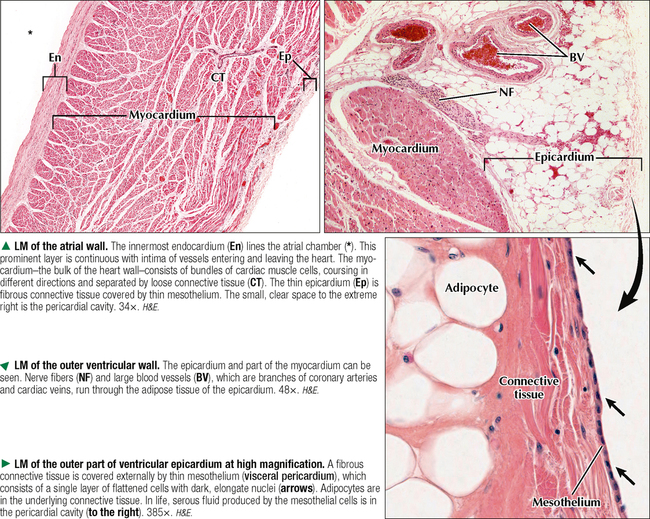
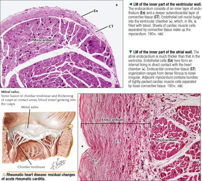
CARDIOVASCULAR SYSTEM

8


CARDIOVASCULAR SYSTEM


image


8.1 OVERVIEW


The cardiovascular system consists of the heart—a muscular pump—and closed vessels through which blood circulates in the body. Arteries leave the heart, branch repeatedly, and have smaller diameters as they course toward the periphery. They deliver blood to capillaries, which are the thinnest vessels and are closest to body cells. Blood in capillaries is returned to the heart via veins. The blood circulatory system consists of two functional parts: pulmonary (which conducts blood to and from lungs for gas exchange) and systemic (which delivers blood to and from other parts of the body). Closely associated with this circulatory system is a large network of lymphatic vessels that collects excess fluid from body tissues and returns it as lymph to the blood circulation. The cardiovascular system consists of tubular structures, the heart itself being a cone-shaped tube with dilated segments reflected on itself. Continuous, simple squamous epithelium known as cardiac and vascular endothelium lines the whole system internally. Capillaries are made almost entirely of a single layer of endothelial cells and associated cells called pericytes. All other vessels have added tissue layers that are arranged concentrically around the endothelium. Arteries operate in a high-pressure system and veins serve a reservoir function under low pressure, so arteries usually have thicker walls than veins. Blood vessels differ in size, function, and distribution, but they share a histologic plan, with structural differences reflecting functions in various parts of the system. Walls of blood vessels above the capillary level have three layers, or tunics: inner tunica intima (closest to the lumen), middle tunica media, and outer tunica adventitia.


image


8.2 HISTOLOGY AND FUNCTION OF THE HEART WALL AND PERICARDIUM


The heart develops embryonically from a simple blood vessel and thus retains the three concentric tunics of vessel walls. In the heart wall, the organization and tissue composition of these layers—endocardium, myocardium, and epicardium—are modified to reflect the heart’s main function as a four-chambered muscular pump. The inner endocardium, homologous to the tunica intima, is in contact with blood, which fills the heart chambers. This layer consists of an endothelium and underlying connective tissue. The myocardium substitutes for the tunica media of vessels. Forming the bulk of the heart wall, it consists mostly of cardiac muscle. The outer layer, analogous to the tunica adventitia, is the epicardium. Unlike the adventitia, the epicardium has two layers: Deeper loose, fatty connective tissue is covered externally by mesothelium. One layer of squamous to cuboidal mesothelial cells—mainly secretory cells resembling mesothelial cells lining pleural and peritoneal cavities—rests on a basal lamina and makes up the mesothelium, which also forms the visceral layer of the pericardium. The pericardium, the fibroelastic, fluid-filled sac that holds the heart, consists of an outer parietal layer that reflects onto the heart surface as a visceral layer (epicardium). Mesothelial cells lining these two parts of the pericardium secrete a thin film of clear, serous fluid (usually less than 50 mL) into the pericardial sac. The fluid lubricates the heart’s surface during contraction to reduce friction. The epicardium contains adipose tissue to act as a shock absorber and support branches of coronary arteries; veins that drain blood from the heart wall; lymphatics; and many nerve fascicles and ganglia.


image


8.3 HISTOLOGY OF THE ENDOCARDIUM AND MYOCARDIUM


The endocardium contains several distinct layers, which may vary histologically in different parts of the heart. An innermost endothelium, derived embryonically from mesoderm, is made of one layer of endothelial cells, which are a type of simple squamous epithelium. It is continuous with endothelium of veins and arteries that enter and leave the heart. A subendothelial layer of connective tissue consists of collagen fibers, elastic fibers, and scattered smooth muscle cells. In some areas is another layer of loose fibroelastic connective tissue, the subendocardium. It may contain elements of the cardiac conduction system, such as Purkinje fibers, which are modified cardiac muscle cells (see Chapter 4). The endocardium is usually thicker in the atria than in the ventricles. The inner surface of the ventricles under the endocardium has trabeculae that project into the lumen and are composed of cardiac muscle—called papillary muscles. Although the luminal surface of the atria is relatively smooth, a small auricular appendage is trabeculated internally by muscular bands, or pectinate muscles. The much thicker ventricular myocardium compared with the atrial layer reflects differences in workload of heart chambers. The myocardium consists of interlacing bundles, or sheets, of cardiac muscle cells embedded in richly vascularized, loose connective tissue, which is the endomysium. The muscle fibers in each sheet have a complex spiral pattern that winds around the atria and ventricles. Cardiac muscle cells form a three-dimensional anastomosing network whereby intercalated discs link almost all cells and other cells insert into the cardiac skeleton of dense fibrous connective tissue.


image


8.4 HISTOLOGY OF HEART VALVES


The four heart valves are attenuated folds of endocardium that prevent backflow of blood. Two atrioventricular (AV) valves are intake valves for the right and left ventricles. The right AV valve, between right atrium and right ventricle, has three leaflets and is called the tricuspid valve. The left AV valve, between left atrium and left ventricle, has two leaflets and is the bicuspid valve, or, because it resembles a bishop’s miter, the mitral valve. The free edges of the AV valves are continuous with thin tendinous cords, the chordae tendinae, which attach to papillary muscles associated with ventricles. The two ventricles have outtake valves that guard orifices of the pulmonary artery and aorta: the pulmonary and aortic semilunar valves. The first, the valve of the right ventricle, is found where the pulmonary artery originates from the right ventricle. The outtake valve of the left ventricle, the aortic valve, lies where the aorta originates from the left ventricle. Although leaflets of the two semilunar valves are thinner than those of AV valves, all heart valves possess the same basic histologic plan. Each valve leaflet has a central core of dense fibrous connective tissue, which is covered externally on both sides by endocardium. In AV valves, the endocardium is thicker on the ventricular side than on the atrial side. The central, avascular connective tissue core of each valve is dominated by a mixture of collagen and elastic fibers but also contains fibroblasts and occasional smooth muscle cells. These cells receive nutrients and O2 from blood in the heart chambers. The heart also has a framework of dense irregular connective tissue—the cardiac skeleton—that consists of four annuli fibrosi, a septum membranaceum, and two trigona fibrosa. Annuli fibrosi support heart valves; the other two elements of the cardiac skeleton serve as attachment sites for cardiac muscle.


image


8.5 CLASSIFICATION OF ARTERIES AND VEINS


Arteries are efferent vessels that function in a high-pressure system; veins are afferent vessels that function under low pressure. Their histologic organization and tissue composition reflect physiologic conditions under which they operate. Arteries and veins are classified into types that differ mainly in size, microscopic structure, and location; the scheme is arbitrary because gradual histologic changes occur along the length of the vessels. The scheme is useful, however, as these vessels do more than merely transport blood along the circulatory route. Of three types of arteries, elastic (conducting, or conduit) arteries are closest to the heart, are the largest, and include the aorta and pulmonary, common carotid, subclavian, and common iliac arteries. With highly elastic walls, they can expand during ventricular contraction (systole) and passively recoil during ventricular relaxation (diastole) to sustain continuous blood flow despite pulsatile pumping of the heart. Muscular arteries, also called distributing arteries, regulate blood flow to organs and parts of the body by contraction and relaxation of smooth muscle in their walls. Many bear names such as femoral and brachial arteries. Arterioles, the smallest arteries at 100 μm or less in diameter, are small-resistance vessels that mainly regulate systemic blood pressure. Their walls contain one or two layers of circularly arranged smooth muscle. The three types of veins have thin walls relative to their arterial counterparts and often look collapsed in histologic sections. Large veins, such as superior and inferior venae cavae, are large-capacitance vessels that return blood under low pressure to the heart. Muscular (or medium-sized) veins commonly travel with muscular arteries. Because of low intraluminal pressure, they often have simple flap-like valves that prevent backflow of blood against gravity as it is returned to the heart. Venules, the smallest veins, accompany arterioles and have very thin walls, which are often porous to allow migration of leukocytes from the circulation, especially during an inflammatory response.


image


8.6 HISTOLOGY OF ELASTIC ARTERIES


Elastic arteries, with a large lumen relative to wall thickness, conduct blood from the heart to muscular arteries. The tunica media in the wall of elastic vessels is the most prominent of three layers. It has abundant elastic fibers organized as multiple, concentric, fenestrated laminae interspersed with scattered, circularly arranged smooth muscle cells. The number and thickness of elastic laminae vary with age: for example, newborn aortas have about 25 concentric laminae, adult aortas, 50–75. Smooth muscle cells in the media synthesize and secrete elastic fibers of the laminae as well as some collagen and other elements of extracellular matrix. Collagen confers tensile strength to arterial walls, and elastic fibers impart distensibility, which allows passive recoil under pressure. The tunica intima, at up to 20% of wall thickness, is relatively thick, with its luminal surface lined internally by an endothelium of flattened cells resting on a basal lamina. A deeper, subendothelial layer of connective tissue consists mostly of collagen and elastic fibers embedded in ground substance, plus scattered fibroblasts and occasional smooth muscle cells. Underneath the intima is a border of an internal elastic lamina, which is often difficult to discern as it merges imperceptibly with elastic laminae of the media. The tunica adventitia

Stay updated, free articles. Join our Telegram channel

Jun 18, 2016 | Posted by in HISTOLOGY | Comments Off on CARDIOVASCULAR SYSTEM

Full access? Get Clinical Tree

Get Clinical Tree app for offline access