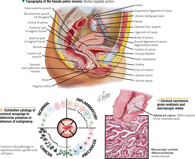
FEMALE REPRODUCTIVE SYSTEM

18


FEMALE REPRODUCTIVE SYSTEM


image


18.1 OVERVIEW


The female reproductive system consists of paired ovaries and the genital tract, including fallopian tubes (oviducts, or uterine tubes), uterus, cervix, and vagina, located in the pelvis—the internal genitalia. External genitalia consist of labia majora, labia minora, and clitoris. Mammary glands (see Chapter 2) and placenta are not classified as genital organs but are functionally associated with them. Ovaries, the center of cyclic changes in the female reproductive system, produce female germ cells (ova) and steroid hormones. Fallopian tubes are sites for fertilization of ova, and the uterus harbors fertilized ova during gestation. Like ovaries, the uterus undergoes a regular sequence of changes known as the menstrual cycle. The vagina connects the internal genitalia with the exterior. Embryonic development of the female reproductive system, as in the male, closely parallels that of the urinary system. The system derives mainly from a urogenital ridge of intermediate mesoderm in the posterior abdominal wall. At 6 weeks of gestation, primordial germ cells migrate from their origin in the yolk sac endoderm to the urogenital ridge. Gonad development proceeds with interaction of germ cells with surrounding mesenchyme and coelomic surface epithelium. Germ cells in the primitive ovary develop into oogonia; surface epithelium differentiates into follicular cells. The female genital duct system and external genitalia then develop under the influence of circulating fetal hormones. The paramesonephric (Müllerian) duct system gives rise to most of the genital duct system, and the lower part of the vagina originates from the urogenital sinus.


image


18.2 OVARIAN STRUCTURES AND DEVELOPMENT


The ovaries—solid, almond-shaped glands—are 3 cm long and 2 cm wide in adults, although their size and histologic appearance differ during menstrual cycles, pregnancy, and postmenopausal period. One side of the ovary has a mesentery—the mesovarium—which attaches the ovary at its hilum to the broad ligament. Ovaries are covered by a reflection of visceral peritoneum, originally known as germinal epithelium but better termed ovarian surface epithelium. Germinal epithelium is a misnomer, as its cells are not the source of ova but are modified mesothelial cells lining the peritoneal cavity. An ovary is divided into an outer cortex and an inner medulla, which are not clearly demarcated. Under the surface epithelium is a dense fibrous connective tissue, the tunica albuginea, which encapsulates the whole ovary. The remaining cortex is richly cellular connective tissue arranged in a whorl-like pattern and harboring oocyte-containing ovarian follicles of various sizes and at different stages of maturation and degeneration. In childhood, the cortex contains numerous primordial follicles; in sexually mature women, corpora lutea form at sites of ruptured follicles. The ill-defined medulla consists of loose connective tissue with many convoluted blood vessels, nerves, and lymphatics. Ovaries at birth hold about 400,000 primary oocytes, which developed from oogonia; by puberty, about 40,000 oocytes remain after degeneration or atresia. In women, an ovum is liberated from an ovary via ovulation about every 28 days. Like testes, ovaries have both exocrine (cytogenic) and endocrine functions: They produce the hormones estrogen and progesterone.


image


18.3 HISTOLOGY OF THE OVARIAN CORTEX


Development of ovarian follicles, which consist of an oocyte and surrounding epithelial layer of follicular cells, is complex. By birth, all oogonia have become primary oocytes, which have reached prophase of the first division of meiosis. Follicles in the cortex may be resting, or primordial; maturing (known as primary and secondary follicles); or mature (Graafian). Primordial follicles are just under the tunica albuginea and have not yet begun to develop. They contain a primary oocyte, measuring about 25 μm in diameter, that has an eccentric nucleus with a prominent nucleolus. One layer of squamous epithelial cells, the follicular cells, surrounds it. A thin basal lamina lies on the outer surface of these cells and separates them from surrounding connective tissue stroma. After puberty, about 20 primordial follicles become activated monthly during menstrual cycles. Usually, one follicle among them becomes dominant and moves to the next developmental stage by becoming a primary follicle. This follicle is slightly larger, with an oocyte, 40–45 μm in diameter, containing a large clear nucleus with distinct nucleolus. Surrounding follicular cells undergo cell division and become cuboidal. Their cytoplasm assumes a granular appearance, so the cells are now known as granulosa cells, which are surrounded by a basal lamina. Interstitial (stroma) cells adjacent to the follicle differentiate into a concentric sheath of theca interna cells.


image


18.4 HISTOLOGY OF DEVELOPING OVARIAN FOLLICLES


The follicular epithelium and surrounding stroma are involved in the maturation process of follicles and undergo both hyperplasia and hypertrophy. They form a solid multilaminar secondary follicle in which mitotically active granulosa cells become stratified and form several layers of concentrically arranged, closely packed cells. The primary oocyte diameter increases, and a homogeneous, eosinophilic extracellular layer, the zona pellucida, surrounds the cell’s plasma membrane. Both oocyte and granulosa cells synthesize the zona pellucida, which is rich in proteoglycans. As the follicle enlarges and consists of 8–12 layers of granulosa cells, small, irregular fluid-filled spaces develop among the cells, and the follicle is called a secondary (vesicular, or antral) follicle. When the growing follicle has a diameter of about 200 μm, spaces coalesce (and accumulate more fluid) to form a single cavity known as the follicular antrum. The clear, viscous fluid within the antrum—the liquor folliculi—is rich in hyaluronic acid, growth factors, and steroid hormones produced by granulosa cells. Theca interna cells become vascularized and secrete the steroid androstenedione, from which granulosa cells produce estrogens. An outer layer of theca externa cells also forms and is continuous with connective tissue cells of the stroma.


image


18.5 ULTRASTRUCTURE OF DEVELOPING OVARIAN FOLLICLES


Follicular and granulosa cells display a high mitotic activity during development of ovarian follicles. These cells are involved in synthesis and maintenance of the zona pellucida. Their cytoplasm is rich in rough endoplasmic reticulum (RER) and free ribosomes; mitochondria, lipid droplets, and lysosomes, although present, are not abundant. Junctional complexes occur between granulosa cells. Desmosomes probably reinforce the structural integrity of the follicle, zona pellucida, and corona radiata during ovulation. Gap junctions are sites of electrical and ionic communication between cells. The large round oocyte has a spherical, eccentrically placed nucleus with dispersed chromatin and an irregular nuclear envelope. The surrounding oocyte cytoplasm contains an array of organelles including closely packed cytoplasmic filaments, spherical mitochondria, free ribosomes, assorted vesicles, and profiles of endoplasmic reticulum. The zona pellucida is a thick extracellular layer between the oocyte and the granulosa cells of the follicle. Slender microvilli of the oocyte and granulosa cells extend into the zona pellucida.


image


18.6 HISTOLOGY OF MATURE GRAAFIAN FOLLICLES


After 12–14 days, the mature Graafian (tertiary) follicle is the final stage in development. With a diameter of 1.5–2.5 cm, it contains an oocyte that has reached a maximum size of about 150 μm. The primary oocyte sits in a local eccentric thickening of the granulosa cell layer, the cumulus oophorus, which projects into the antrum. One or more layers of granulosa cells are attached to the oocyte as the corona radiata and accompany it after ovulation. The antrum, the largest part of the follicle, is surrounded by multiple granulosa cell layers, which are, in turn, surrounded by thecae interna and externa. The zona pellucida is now 5–10 μm thick and anchors the oocyte to the corona radiata. The dominant follicle occupies the full breadth of the cortex and usually bulges above the ovarian surface. At their point of contact—the stigma—the tunica albuginea and the thecae become attenuated on the surface. The oocyte and corona radiata detach from the follicular wall and float freely in the fluid-filled antrum. Shortly before ovulation, the oocyte resumes meiosis to form a large secondary oocyte and a smaller polar body that disintegrates. The secondary oocyte, with a haploid number of chromosomes, is arrested in metaphase of the second meiotic division until fertilization. Increased luteinizing hormone on about day 14 of the menstrual cycle is thought to stimulate this meiotic division just before ovulation and may cause a follicle to rupture. Ovaries of young women usually have several Graafian follicles that may stay at this stage for several months. At ovulation, a follicle ruptures and releases the oocyte and corona radiata, which enter the fallopian tube infundibulum.


image


18.7 STRUCTURE AND FUNCTION OF THE CORPUS LUTEUM


After the Graafian follicle has ruptured at ovulation and the secondary oocyte is released, a temporary glandular structure—the corpus luteum (yellow body)—forms in the follicular remnant. The follicle collapses and becomes highly infolded, and its lumen fills with fibrin-containing fluid and blood. The coagulation in the antral space forms a clot that is replaced by fibrous scar tissue. The basement membrane separating granulosa cells from theca interna cells is broken down, and vascular invasion of the formerly avascular granulosa layer results. Luteinizing hormone from the anterior pituitary influences both granulosa and theca interna cells to undergo marked histologic changes and become granulosa lutein and theca lutein cells, respectively. These increase in number and size and become polyhedral. They are lightly eosinophilic; their cytoplasm accumulates numerous lipid droplets. Both cell types have features in common with steroid-secreting cells. Granulosa lutein cells synthesize and secrete the hormone progesterone, which prepares the endometrium for implantation of a fertilized ovum and stimulates growth of mammary glands. Theca lutein cells synthesize and secrete estrogen. If pregnancy occurs, the corpus luteum persists for the first 8 weeks, after which the placenta becomes the major site for steroid hormone production. If pregnancy does not occur, the corpus luteum gradually involutes, stops producing progesterone, and forms a white scar called the corpus albicans, or white body.


image


18.8 ULTRASTRUCTURE AND FUNCTION OF STEROID-SECRETING CELLS IN THE OVARY


Ovarian steroid-secreting cells include theca interna, granulosa lutein, and theca lutein cells. They share ultrastructural features with steroid-secreting cells in the male reproductive tract and in other organs producing steroid hormones. They have many unique structural features that facilitate acquisition of cholesterol and its conversion into steroid hormones. The plasma membrane on the cell surface has many microvilli and clathrin-coated pits that house receptors for low-density lipoprotein for cholesterol uptake. Underlying the microvilli is a narrow zone of cytoplasm with many tightly packed filaments extending into the microvilli. Steroidogenic organelles include smooth endoplasmic reticulum (SER) and abundant mitochondria with tubulovesicular cristae. The SER consists of highly folded, radiating cisternae

Stay updated, free articles. Join our Telegram channel

Jun 18, 2016 | Posted by in HISTOLOGY | Comments Off on FEMALE REPRODUCTIVE SYSTEM

Full access? Get Clinical Tree

Get Clinical Tree app for offline access