Chronic Obstructive Pulmonary Disease (COPD)
Definitions
The WHO-sponsored ‘Global initiative for chronic obstructive lung disease’ (http://www.goldcopd.com) defines COPD as ‘a preventable and treatable disease with some significant extrapulmonary effects that may contribute to the severity in individual patients. Its pulmonary component is characterised by airflow limitation that is not fully reversible. The airflow limitation is usually both progressive and associated with an abnormal inflammatory response of the lungs to noxious particles or gases.’
It covers many previously used labels, including:
- Emphysema – enlargement of the air spaces distal to the terminal (smallest) bronchioles with destructive changes in the alveolar wall. In centrilobular emphysema, damage is limited to the central part of the lobule around the respiratory bronchiole, whereas in panacinar emphysema, there is destruction and distension of the whole lobule. If the air spaces are > 1 cm in diameter they are called bullae.
- Chronic obstructive airways disease.
- Chronic airflow limitation.
Poorly reversible airflow limitation may also occur in bronchiectasis, cystic fibrosis, tuberculosis and some cases of chronic asthma.
- Chronic bronchitis is daily cough with sputum for at least 3 months a year for at least 2 consecutive years.
Pathogenesis
There is typically chronic inflammation throughout the airways and pulmonary vasculature. In the trachea, and bronchi and bronchioles that are >2–4 mm in internal diameter, inflammatory cells infiltrate the surface epithelium and there is hypersecretion from enlarged mucus-secreting glands and increased numbers of goblet cells. In small bronchi and bronchioles that have an internal diameter <2 mm, chronic inflammation is associated with remodelling of the airway wall, with fibrosis, narrowing the lumen and producing fixed airways obstruction.
Destruction of the lung parenchyma typically occurs as centrilobular emphysema. In mild cases lesions usually involve upper lung regions, but in advanced disease they may appear diffusely throughout the entire lung. Vascular changes occur early. Intimal thickening of the vessel wall is followed by smooth-muscle proliferation and an inflammatory cell infiltrate. These pathological changes lead to characteristic physiological changes. Mucus hypersecretion and ciliary dysfunction cause a chronic productive cough. Airflow limitation, hyperinflation and gas exchange abnormalities cause breathlessness. Pulmonary hypertension and cor pulmonale are late features.
Aetiology
- tobacco smoking
- atmospheric pollution
- α1-antitrypsin deficiency is a recessive disorder accounting for about 5% of patients with emphysema (and about 20% of neonatal cholestasis). Five percent of homozygotes tend to develop emphysema by the age of 40 years and heterozygotes are at risk. The emphysema is predominantly of the lower zones and is much worse in smokers.
Most die from one of three diseases: CHD; COPD; lung cancer (the risk of many cancers is increased by smoking).
Clinical Presentation
Often the patient has a productive morning cough and an increased frequency of lower respiratory tract infections producing purulent sputum. The organisms responsible are usually Haemophilus influenzae, Streptococcus pneumoniae and the respiratory viruses. Over years there is slowly progressive dyspnoea with wheezing, exacerbated in the acute infective episodes. There is clinical emphysema with hyperinflation of the lungs. Respiratory failure (p. 113) and chronic right heart failure (cor pulmonale) are long-term complications.
Investigation
Ventilatory Function Tests
The diagnosis of COPD is established by spirometry, which shows a post bronchodilator FEV1 : forced vital capacity (FVC) ratio < 70%. The peak expiratory flow rate (PEFR) is reduced. The airways obstruction is only partially reversible by bronchodilator (or other) therapy.
Chest X-Ray
This may be normal. Abnormalities correlate with the presence of emphysema and are caused by:
- overinflation with a low, flat, poorly moving diaphragm and a large retrosternal window on lateral X-ray
- vascular changes with loss of peripheral vascular markings but enlarged hilar vessels – the heart is narrow until cor pulmonale develops
- bullae if present.
The chest X-ray is an important investigation because it excludes other disease (carcinoma, tuberculosis, pneumonia, pneumothorax).
Arterial Blood Gas Estimations
These may be normal. In later stages the PaO2 falls and the PaCO2 rises, particularly with exacerbations.
Electrocardiogram (ECG)
This records the presence and progression of cor pulmonale (right atrial and ventricular hypertrophy).
Sputum for Bacterial Culture and Sensitivity
This is useful in acute infective episodes when infections other than Haemophilus influenzae or Streptococcus pneumoniae may be present.
Haemoglobin
This may show secondary polycythaemia.
Management (Box 11.1)
Management should include assessment and reduction of risk factors in addition to the management of stable COPD and exacerbations. There should be a stepwise increase in treatment according to disease severity.
 Box 11.1 Management of COPD
Patients benefit from rehabilitation and exercise programmes. Education can help patients to cope and achieve goals, including stopping smoking.
Bronchodilators are used to prevent or reduce symptoms: the β2-agonists, e.g. salbutamol (Ventolin), terbutaline (Bricanyl), the anticholinergic ipratropium (Atrovent) or a combination of these drugs are given by metered aerosol or nebuliser on an as-required or regular basis. Theophylline may also help.
Regular treatment with inhaled steroids can benefit patients with a documented spirometric response to steroids, or who have repeated exacerbations requiring treatment with antibiotics or oral steroids. Chronic treatment with systemic steroids should be avoided. Long-term home oxygen (> 15 h/day) increases survival in patients with chronic respiratory failure. Patients should receive annual influenza vaccination.
Exacerbations are treated with inhaled bronchodilators; theophylline and systemic steroids are effective treatments. Although a cause is often not identified, infection is a common trigger and patients with signs of infection are given antibiotics – amoxicillin or a macrolide (erythromycin or clarithromycin). Bacterial sensitivities are useful if clinical improvement has not occurred.
Non-invasive intermittent positive pressure ventilation (NIPPV) can decrease the need for intubation and mechanical ventilation.
Asthma
Asthma affects 5–7% of the population of Europe and North America. It is characterised by recurrent shortness of breath, wheeze or cough caused by reversible narrowing of the airway lumen. The principal cause of increased airways resistance is contraction of smooth-muscle cells as a result of hypersensitivity to many different stimuli such as cold air, smoke, exercise and emotion, as well as antigens. Thickening of the airways by oedema and cellular infiltrates, as well as blockage of airways by mucus and secretions, also contribute. Wheeze is not an essential feature.
Asthma is sometimes classified into extrinsic and intrinsic, although treatment is the same.
Clinical Features
Acute Attacks
These may be fairly abrupt in onset and brief in duration (hours), or longer (a week or two). Longer severe attacks are called ‘status asthmaticus’ (p. 113). In an attack the patient feels tightness in the chest and both inspiratory and expiratory effort are difficult. There may be a cough that is initially dry but later becomes productive, particularly if there is infection. The patient usually sits up with an overinflated chest, an audible expiratory wheeze, using the accessory muscles of respiration. The respiratory rate may be little altered but the pulse is invariably rapid. Acute attacks are precipitated by specific allergens (e.g. pollens or house dust mite), exertion, cold air, a respiratory infection and β-blockers.
Recurrent Asthma
Mild asthmatics (particularly with extrinsic asthma) usually have normal respiratory function between attacks, but those with long-standing severe asthma tend to develop some degree of dyspnoea and persistent airways obstruction between acute attacks.
Investigation
Investigation includes chest X-ray (for regional collapse, pneumonia, pneumothorax) and measurement of ventilatory function (FEV1 or PEFR, preferably at several times a day on several days at home) and the response to bronchodilators. Variability through the day, especially with a ‘morning dip’ in PEFR, is characteristic. Skin hypersensitivity tests performed by pricking standard allergens into the skin can help the patient recognise and avoid environmental precipitants. Bronchial reactivity may be more precise but should be tested only in carefully controlled conditions. Adrenaline (0.5 ml of 1 : 1000) must be available in case of acute anaphylactic reactions (p. 115).
Management of chronic asthma
The patient should be asked about precipitating factors, including upper respiratory tract infection, season (grass pollen and fungal spores), cold, exercise, food, house dust (contains the mite Dermatophagoides), smoke, emotion and drugs (e.g. aspirin, NSAIDs, β-blockers). Patients should be trained to use a peak flow meter reliably and to document values at home. Increasing morning dips provide an early warning of deterioration.
Most patients respond to simple therapy and may be controlled by:
- removing known allergies, e.g. feather pillows, cats
- inhaled short-acting β2-agonists, e.g. salbutamol, as required – the response is a guide to severity
- inhaled corticosteroids in a regular regimen
- inhaled sodium cromoglicate (Intal)
- inhaled antimuscarinic preparations, e.g. ipratropium (Atrovent)
- theophylline preparations, given as a slow-release preparation at night to control overnight symptoms
- leukotriene receptor antagonists, e.g. montelukast, are used as add-on therapies in the prophylaxis of moderate asthma not controlled by the above therapies
- oral steroids may be required for exacerbations
- hyposensitisation is of value in only a small number of patients who demonstrate specific allergies.
The British Thoracic Society (http://www.brit-thoracic.org.uk/) has established guidelines for a stepwise approach to the management of asthma. A cumulative drug regimen is prescribed for each step, stepping up if necessary to achieve control, and stepping down when control is good.
NB When metered aerosols are used, always check inhaler technique. A number of simple inhalers are available: spacers, discs, rotahalers, breath-actuated aerosols.
Acute Severe Asthma
Acute severe asthma is a life-threatening condition. It typically occurs in poorly controlled individuals whose condition has been deteriorating over days or weeks, but death can be sudden and sometimes unexpected, as the patient may not appear severely ill. It is this lack of recognition of severity plus inadequate early treatment that is so dangerous. Most patients have some degree of respiratory failure at presentation. The term status asthmaticus is sometimes used to describe severe asthma attacks that have not responded to conventional therapy.
Features of severe life-threatening asthma are as follows.
Clinical
- severe breathlessness (unable to complete sentence in one breath)
- exhaustion, confusion
- tachycardia, bradycardia or other arrythmia
- hypotension
- cyanosis
- respiratory rate > 25 breaths/min
- poor respiratory effort
- silent chest
Laboratory
- PaO2 < 8 kPa
- oxygen saturation < 92%
Clinical Presentation
Inability to speak or difficulty in maintaining speech is one criterion of severity. It also implies inability to drink and dehydration. Hypoxaemia is usually then present.
Very severe, life-threatening airways obstruction is indicated by absence of wheeze (because of poor ventilation), PEFR < 33% of best (PEFRs can usually not be obtained), bradycardia or hypotension, and confusion, drowsiness and exhaustion. Cyanosis is also ominous and tends to occur at a lower PaO2 than in the chronic bronchitic (because the bronchitic tends to have polycythaemia).
NB Signs of drowsiness, cyanosis, bradycardia or hypotension signify a very severe attack and vigorous treatment is essential before they are apparent.
Investigation
Arterial blood gases provide the most useful guide to the severity of the attack and to the success of treatment. The following values indicate a very severe attack: SaO2 < 92%, PaO2 < 8 kPa, PaCO2 > 5.0 kPa. A chest X-ray is useful if pneumothorax, pneumomediastinum or consolidation are suspected. It should also be performed if there is a failure to respond to treatment and if asthma is life threatening.
Management
Sedation may depress respiration further and is contraindicated.
- Continuous high-flow oxygen.
- Bronchodilators: β2-agonists (e.g. salbutamol, terbutaline) plus ipratropium by oxygen-driven nebuliser or intravenous infusion if inhaled therapy cannot be used reliably.
- Corticosteroids. Give oral prednisolone, or intravenous hydrocortisone if unable to swallow. Continue oral prednisolone 40–50 mg daily for at least five days or until recovery.
- Intravenous fluids are required to make up the initial dehydration and for as long as oral fluids are not taken. Monitor urine output.
- Bacterial infection is a rare trigger but antibiotics are given if it is present or strongly suspected. The usual organisms are Strepcococcus pneumoniae or Haemophilus influenzae.
- Mechanical ventilation may be necessary. Persistent or increasing elevation of arterial PaCO2, or worsening hypoxia especially with accompanying exhaustion, indicates the need for artificial ventilation.
Discharge from hospital after PEFR returns to > 75% of best, with day variability < 25%, with early clinic follow-up.
Respiratory Failure
Respiratory failure can be defined as a reduction in arterial PaO2 below 8 kPa with a normal arterial PaCO2 (hypoxaemic respiratory failure), or with an increase in arterial PaCO2 above 6.6 kPa (hypercapnic respiratory failure).
NB The normal range for PaO2 is about 10–13 kPa. Values of PaO2 are lower in the elderly.
The PaO2 may fall while the PaCO2 remains normal. This may occur with alveolar parenchymal lung disease: infiltrations, fibrosing alveolitis and ‘pure’ emphysema. Much more commonly, both arterial gas levels are abnormal. This occurs with ventilatory failure.
Acute
- patients with normal lungs, with upper airways obstruction (e.g. croup and acute anaphylaxis) or mechanical failure (e.g. flail chest) or central nervous system depression of respiratory drive (e.g. sleep apnoea, drug overdosage, stroke)
- patients with abnormal lungs (e.g. asthma, chronic bronchitis)
Chronic
Usually in patients with abnormal lungs, especially COPD. These patients are particularly likely to develop acute failure if infection occurs.
In restrictive disorders, lung expansion is limited by:
- lung disease, such as fibrosis collapse, oedema, consolidation
- pleural disease, such as fibrosis, effusion or mesothelioma
- chest-wall disease, such as costospinal rigidity and deformity, or abdominal splinting by obesity, ascites or pregnancy
- neuromuscular disease, such as muscular dystrophy, myasthenia or phrenic nerve paralysis.
Acute on Chronic Respiratory Failure
This usually occurs on a background of COPD.
Clinical Presentation
- peripheral vasodilatation with headache, engorged veins in the fundi, warm hands and a bounding pulse, all caused by carbon dioxide retention
- varying degrees of agitated confusion, drowsiness and coma
- increasing cyanosis
- signs of right heart failure
- flapping tremor of the outstretched hands and papilloedema are late signs
NB Unfortunately, physical signs are a poor guide to the presence and degree of respiratory failure. It is therefore necessary to measure blood gases in all patients in whom the diagnosis is suspected.
Management
This consists of the measures used for COPD as given above, with the addition of controlled oxygen therapy. The danger to life in this situation is hypoxia, but, paradoxically, relief of hypoxia may make the situation worse by causing hypercapnia. Give oxygen at 24 or 28% by Ventimask (or other controlled technique) if the arterial PaO2 or oxygen saturation is low, targeting oxygen saturation at 94–98% unless the patient is at risk of hypercapnia, when the target should be lower at 88–92%. Oxygen is given continuously until the acute situation (including infection and heart failure) has recovered. PaCO2 is monitored. For chronic respiratory failure controlled oxygen can be given continuously at home with improvement in symptoms and an increase in life expectancy (Trials Box 11.1).
NB Sedatives are absolutely contraindicated.
 Trials Box 11.1 Home Oxygen Therapy in COPD
Indications for Respiratory Support and Mechanical Ventilation
If the PaCO2 falls, conservative therapy should be continued and reassessed periodically. If the PaCO2 rises, this indicates that the patient’s ventilation is inadequate and is the prime indication for non-invasive positive pressure ventilation using a full facial or nasal mask that delivers ventilatory support from a flow generator (Trials Box 11.2), or if this fails mechanical positive-pressure ventilation. Patients should be ventilated before they become exhausted. The final decision to ventilate a patient is determined mainly on the basis of respiratory function before the acute illness: if very poor, it may not be possible to wean the patient off the ventilator.
 Trials Box 11.2 Non-invasive Positive Pressure Ventilation in Acute Respiratory Failure
Acute Anaphylaxis
This rare condition occurs following exposure to allergens such as certain foods, e.g. peanuts; drug therapy, e.g. penicillin; and insect stings. Clinical features range from mild with flushing of the face, pruritus and blotchy wheals, to severe with asthma, respiratory obstruction from oedema of the larynx (angioedema) and circulatory collapse.
Immediate treatment is with adrenaline (epinephrine) 0.5 ml of 1 : 1000 (1 mg/ml) solution, i.e. 500 μg given intramuscularly and repeated after 5 min if no improvement is observed. Oxygen, if available, is important early on. An intravenous fluid challenge is given if there is hypotension. Hydrocortisone takes several hours to act. It is given (after the first injection of adrenaline (epinephrine)) in a dose of 200 mg slowly intravenously or intramuscularly, with chlorphenamine 10 mg slowly intravenously or intramuscularly.
In patients with a history of anaphylaxis allergens should be identified and avoided. Most patients will wish to carry self-adminstration preassembled pens containing adrenaline (epinephrine) for intramuscular injection.
Hereditary angioedema is a rare condition caused by C1 esterase deficiency (autosomal dominant). It gives rise to erythema, patchy oedema and colicky abdominal pain. It responds to danazol prophylaxis and fresh frozen plasma (or if available plasma derived C1 inhibitor) to correct the deficiency during attacks.
Pneumonia
Community-acquired pneumonia affects approximately 5–10/1000 adults per year. One in 1000 requires hospitalisation, and mortality in these patients is around 10%.
Clinical Presentation
Symptoms of cough, sputum, pleurisy and dyspnoea are less common in the elderly.
Clinical findings include fever, tachypnoea, crackles and signs of consolidation. The likely causative agent cannot be predicted from clinical findings.
The most common organism in UK studies of hospitalised patients is Streptococcus pneumoniae (approximately one-third of cases in which an organism is identified), followed by Chlamydia pneumoniae and Mycoplasma pneumoniae. Haemophilus influenzae, Legionella species, Chlamydia psittaci and Staphylococcus aureus account for most of the remainder. Gram-negative bacilli, Coxiella burnetii and anaerobes are rare. Viruses (including influenza) account for 10–15% of cases.
Investigations
Investigations are performed to establish the diagnosis and assess severity.
- Chest X-ray shows infiltrates. Computed tomographic (CT) scanning is more sensitive and may be useful in detecting interstitial disease, cavitation and empyema.
- Blood gases to assess severity and guide oxygen treatment.
- Blood count – white cell count > 15 × 109/l suggests bacterial infection; white cell count > 20 × 109/l or ≤ 4 × 10⁹/l indicates poor prognosis. Haemoglobin for haemolysis.
- Creatinine and liver function tests for underlying or associated renal or hepatic disease.
- Gram staining and culture of sputum – but cough is unproductive in one-third of patients, and negative results are common, particularly if antibiotics have been given.
- Blood culture.
- Pleural fluid, if present, should be aspirated for culture.
Management
- Oxygen – to maintain PaO2 above 8 kPa and oxygen saturation > 92%.
- Antibiotics – the organism is usually unknown initially. Treatment is started immediately and should cover Streptococcus pneumoniae. In uncomplicated pneumonia, treatment is usually started with oral amoxicillin or a macrolide (erythromycin or clarithromycin). In severe pneumonia intravenous therapy is given, often using a combination of a macrolide (erythromycin) and a second- or third-generation cephalosporin (cefuroxime or cefotaxime). The choice of antibiotics should take account of local guidelines, which will take account of other factors, including the incidence of Clostridium difficile enteritis.
- Intravenous fluids may be required.
- Analgesia for pleuritic pain.
Pneumococcal pneumonia is the most common bacterial pneumonia. S. pneumoniae is a Gram-positive diplococcus. It affects all ages, but is more common in the elderly, after splenectomy, in the immunosuppressed, alcoholics, patients with chronic heart failure and those with pre-existing lung disease. It typically presents acutely with fever, pleuritic pain and rust-coloured sputum. It causes both lobar and bronchopneumonia. Treatment is with penicillin, or erythromycin in the penicillin-sensitive. A polysaccharide pneumococcal vaccine is available for those at high risk. It should be given at least 2 weeks before splenectomy and before chemotherapy.
Staphylococcal pneumonia produces widespread infection with abscess formation. It may complicate influenzal pneumonia, and this makes it relatively common during epidemics of influenza. It also occurs in patients with underlying disease, which prevents a normal response to infection, e.g. chronic leukaemia, lymphoma, cystic fibrosis. Flucloxacillin is the drug of choice, although increasingly Staphylococcus aureus is methicillin resistant (MRSA) and requires treatment wth vancomycin. Lung abscess, empyema and subsequent bronchiectasis are relatively common complications.
Legionnaire’s disease was first described in a group of American army veterans (legionnaires). The causative Gram-negative bacillus flourishes in the cooling waters of air conditioners and may colonise hot-water tanks kept at < 60°C. It begins as an influenza-like illness with fever, malaise and myalgia, and proceeds with cough (little sputum), dyspnoea and sometimes severe anorexia, marked confusion and coma. Diarrhoea and vomiting are common and renal failure may develop. Examination shows consolidation that usually affects both lung bases. X-ray changes may persist for more than 2 months after the acute illness. Erythromycin or ciprofloxacin are the antibiotics of choice, but the mortality remains high.
NB Legionnaire’s disease (and Mycoplasma pneumoniae or psittacosis) should be suspected in all patients who develop pneumonia that does not respond to standard antibiotics.
Mycoplasma pneumonia is caused by M. pneumoniae, the only mycoplasma definitely pathogenic to humans. The clinical picture resembles bacterial pneumonia, although cough and sputum are absent in one-third of cases.
Respiratory symptoms and signs and X-ray changes (patchy consolidation with small effusions) are usually preceded by several days of influenza-like symptoms. Polyarthritis occurs and may persist for months. Malaise and fatigue may persist long after the acute illness is over. The diagnosis is confirmed by a positive Mycoplasma-specific immunoglobulin M (IgM) titre. Erythromycin or tetracycline are the antibiotics of choice.
Psittacosis pneumonia is caused by Chlamydia psittaci. It is transmitted in the excrement of infected birds (the bird need not be ill). Headache, fever and dry cough may be accompanied by a rash and splenomegaly. Hepatitis, encephalitis, renal failure and haemolysis also occur. Treatment is with tetracycline or erythromycin.
Viral pneumonia in children is commonly due to the respiratory syncytial virus (RSV), so called as it is a respiratory virus which produces syncytium formation when grown in culture. Infection may be indistinguishable from acute bacterial bronchitis or bronchiolitis in children and infants. The presence of a skin rash supports the likelihood of RSV infection.
Acute viral pneumonia in adults is less common but occurs during epidemics of influenza. Fever, headache and myalgia are followed after a few days by dry cough and chest pain. Treatment is largely symptomatic (paracetamol, rest, fluids). Influenza vaccines are available for those at high risk. The most common cause of pneumonia during influenza epidemics results from secondary bacterial infection, the most serious being staphylococcal pneumonia. The viruses of measles, chickenpox and herpes zoster may directly affect the lung. The diagnosis is confirmed by a rise in specific antibody titre.
Aspiration pneumonia comes in two main varieties, differentiated from each other by the type of fluid aspirated and the circumstances in which it occurs.Aspiration of gastric contents may produce a severe chemical pneumonitis with considerable pulmonary oedema and bronchospasm (Mendelson syndrome). The acute respiratory distress and shock can be very rapidly fatal and very difficult to treat. It tends to occur in states of reduced consciousness such as general anaesthesia, drunks and when gastric lavage (for drug overdose) has been performed inexpertly.
Aspiration of bacteria from the oropharynx may follow dental anaesthesia and can occur in bulbar palsies. The bacteria, apart from Bacteroides, are nearly all penicillin-sensitive and amoxicillin (or ampicillin) with metronidazole are the antibiotics of choice until sensitivities of isolated organisms are known. Recurrent episodes occur in some oesophageal disorders, including hiatus hernia, stricture, achalasia of the cardia, and in patients with diverticula or pharyngeal pouch.
Recurrent bacterial pneumonia in the absence of chronic bronchitis arouses suspicion of:
- bronchial carcinoma preventing drainage of infected areas of lung
- bronchiectasis
- cystic fibrosis
- achalasia of the cardia, 25% of which present as chest disease, pharyngeal pouch and neuromuscular disease of the oesophagus, e.g. bulbar palsy, all producing aspiration
- hypogammaglobulinaemia and myeloma.
Opportunistic infection of the lungs occurs in patients immunosuppressed as a result of treatment, e.g. for myeloproliferative disorders or acquired immunodeficiency syndrome (AIDS, p. 343).
Lung Abscess
Aetiology
- aspiration (see above)
- bronchial obstruction, usually by carcinoma or a foreign body (especially peanuts and teeth)
- pneumonia partially resolved or treated, particularly when caused by the Staphylococcus, Klebsiella or Pneumococcus organisms
Clinical Features
There is a swinging fever and the patient is very ill. The sputum is foul and purulent and there is a high polymorph cell count. Clubbing may develop.
Investigation
Sputum is sent for Gram stain and culture, and blood for culture. Chest X-ray shows round lesions which usually have a fluid level, and serial X-rays monitor progress. It may be necessary to proceed to bronchoscopy to exclude obstruction and to obtain a biopsy and sputum trap specimen.
Treatment
Antibiotic therapy is given according to sensitivities and continued until healing is complete. Repeated postural drainage is started. In resistant cases, repeated aspiration, antibiotic instillation and even surgical excision may be required.
Bronchiectasis
Bronchiectasis means dilatation of the airways. It only becomes of clinical significance when infection and/or haemoptysis occurs within these dilatated airways. Severe forms are now rare, especially in the young.
Aetiology
- following acute childhood respiratory infection, particularly measles, whooping cough or pneumonia
- cystic fibrosis
- bronchial obstruction predisposes to bronchiectasis (e.g. peanuts)
- tuberculosis has become less common as a cause
- congenital (rare): primary ciliary dyskinesia, e.g. Kartagener syndrome (bronchiectasis, sinusitis, situs inversus)
Clinical Features
- chronic cough, often postural
- sputum, often copious, especially with acute infections
- halitosis
- febrile episodes
- haemoptysis: may be the only symptom (‘dry bronchiectasis’) and is occasionally severe
- dyspnoea, coarse basal crepitations and wheeze
- cyanosis and clubbing
- loss of weight and cor pulmonale in advanced cases
Management
Stop smoking.
The object is to get rid of chronic sepsis. Twice-daily postural drainage will help empty dilated airways and decrease the frequency of further infections. Bronchodilators will often help improve clearance of sputum. Antibiotics, as for chronic bronchitis, are given for acute infections and exacerbations. Treatment is unnecessary in the absence of symptoms.
Surgery is rarely indicated unless there is uncontrolled bleeding because the disease is seldom limited to one or two lung segments. Patients with severe disease may develop respiratory failure.
Pneumothorax
Aetiology
Spontaneous Pneumothorax
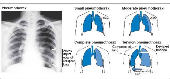
This is the most common type and usually occurs in normal, tall, thin, young, male smokers following rupture of a small subpleural bulla. There is history of sudden onset of one-sided pleuritic pain and/or dyspnoea. Dyspnoea rapidly increases in tension pneumothorax and the patient becomes cyanosed. The classical signs are diminished movement on the affected side with deviation of the trachea to the other side. There is hyperresonance to percussion and reduced pulmonary sounds (breath sounds, tactile fremitus and vocal resonance). Pneumothoraces are best diagnosed by seeing a lung edge on X-ray; it is clearest on an expiratory film (Fig. 11.1). It recurs in 25% of cases within 5 years, usually in the first year. Conditions predisposing to pneumothorax include:
- emphysematous bullae
- tuberculosis – often with a small effusion
- bronchial asthma.
Other rare causes include staphylococcal pneumonia, carcinoma, occupational lung disease and connective tissue disorders, e.g. Marfan and Ehlers–Danlos syndromes. Familial spontaneous pneumothorax is associated with mutations in the folliculin gene.
Figure 11.1 Pneumothorax. Reproduced with permission from Ward, J., Ward, J. and Leach, R. (2010) The Respiratory System at a Glance, 3rd edn. Blackwell, Oxford.
Stay updated, free articles. Join our Telegram channel

Full access? Get Clinical Tree


