Patient Story
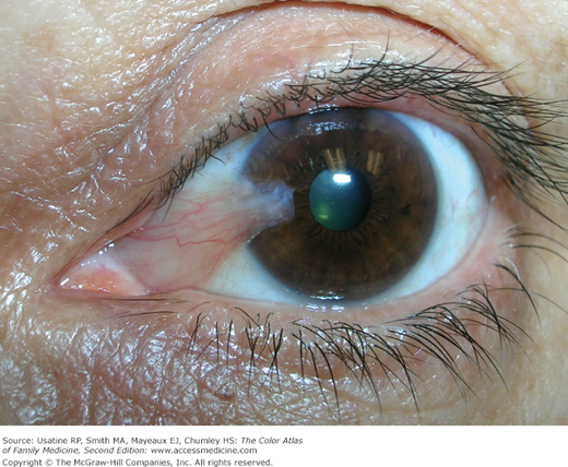
A 50-year-old man had spent most of his adult life working outdoors in southern Texas near the Mexico border. He denies any problems with his vision, but wonders what is growing on his eye and if it should be removed (Figure 12-1). His eyes are often dry and irritated. He is diagnosed with a pterygium and instructed that it does not need to be removed unless it interferes with his vision in the future. Liquid tears are suggested for his dry and irritated eyes. He is also instructed to wear wraparound sunglasses to avoid UV exposure and irritation from wind and dust.

Introduction
A pterygium is a generally benign growth of fibroblastic tissue on the eye of an adult with chronic UV exposure. Pterygia can be unilateral or bilateral, are usually located on the nasal side, and extend to the cornea. Pterygia often require no treatment, but can be removed surgically if they interfere with vision. Patients with dry eyes are prone to the development and progression of pterygia.
Epidemiology
- Pterygium most often develops between the ages 20 and 50 years.
- The frequency of pterygium increases with sun exposure and age. In a population-based study (Indonesia), it was found that the prevalence ranged from 3% (in 21- to 29-year-olds) to 18% (older than 50 years of age).1 In rural China, the prevalence was 3.76% and also increased with age.2
- In one study carried out in Australia, it was found that sun exposure is consistently the greatest risk factor, contributing 43% of the risk.3
Etiology and Pathophysiology
- A pterygium is a proliferation of fibrovascular tissue on the surface of the eye, which extends onto the cornea.
- The etiology of pterygium is incompletely understood; however, chronic UV exposure is accepted as a causative agent. Chronic inflammation and oxidative stress may also play a role in pathogenesis.
- Pterygia have features seen in malignant tissues, such as normal tissue invasion and high recurrence rate.4 Pterygia can be associated with premalignant lesions.4
Risk Factors
Diagnosis
- Redness, itching, and/or irritation of the involved eyes (some are symptom free).
- Visual blurring if the pterygium grows over the visual axis. Even if not obscuring the visual axis, the pterygium can cause poor vision by leading to irregular and high astigmatism.
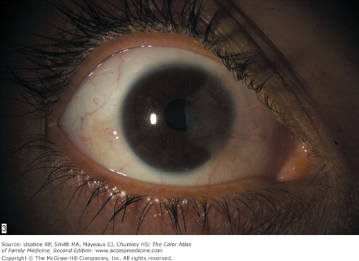
- Pterygia are diagnosed clinically by their distinctive appearance (Figures 12-1, 12-2, 12-3, and 12-4).
Stay updated, free articles. Join our Telegram channel

Full access? Get Clinical Tree


