Patient Story
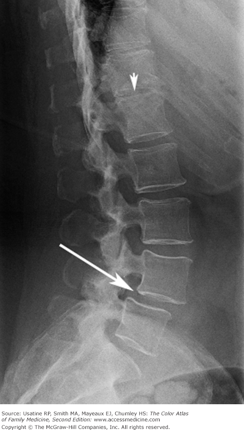
A 60-year-old woman presents with chronic low back pain that began many years ago. Her back pain waxes and wanes and she has taken acetaminophen and ibuprofen with some relief. About 3 months ago, she began to have daily pain. She recalls no trauma. Her examination is unremarkable other than some decreased flexion. Straight leg raise test is negative. As the patient is older than 55 years of age, radiographs are ordered and they demonstrate degenerative changes in her lumbar spine (Figure 99-1). She is started on scheduled acetaminophen and ibuprofen along with an exercise program.
Introduction
Back pain is one of the most common reasons that adults see their physician. Most acute back pain is a result of mechanical causes. Serious pathology can be recognized by the presence of red flags. Acute back pain is treated with reassurance, returning to activities, and acetaminophen with or without an NSAID. Psychological factors increase the risk of development of chronic pain. Chronic back pain is difficult to treat and the best outcomes are typically achieved by an interprofessional team.
Epidemiology
- Six percent of visits to primary care physicians are for back pain.1
- Low back pain 1-year incidence is 20% and 1-year prevalence is 40% in adults.2
- Thoracic back pain 1-year prevalence is 15% to 27% in adults.3
- Treatment for back and neck problems accounted for approximately $86 billion in health care expenditures in the United States in 2005.4
- Prevalence of malignancy in patients with LBP presenting to a primary care office or emergency room is 0.1% to 1.5%.5
Etiology and Pathophysiology
Risk Factors
- Older age—Prevalence of LBP increases with age into the sixth decade.2
- Low educational status.2
- Occupational factors—Manual labor, bending, twisting, and whole-body vibration.2
- Psychosocial factors increase the risk of transition from acute to chronic pain.2
- Risk factors for cancer—Previous history of cancer (positive likelihood ratio [LR+] 23.7), elevated erythrocyte sedimentation rate (ESR) (LR+ 18.0), reduced hematocrit (LR+ 18.3).5
Diagnosis
Nonspecific back pain—Pain for less than 6 weeks (acute), 6 to 12 weeks (subacute), or more than 12 weeks (chronic); negative straight-leg raise test; absence of red flags.
Radicular syndrome—LBP with radiation down leg; positive straight-leg raise test; absence of red flags.
Serious pathology—Further work-up required for presence of red flags, including age younger than 20 or older than 55 years; significant trauma; fever; unexplained weight loss; neurologic signs of cauda equina; progressive neurologic deficit.
Helpful in the presence of red flags:
- Complete blood count (CBC) to evaluate for anemia (malignancy) or leukocytosis (infection).
- Consider human leukocyte antigen (HLA)-B27 in younger patients with inflammatory symptoms.
- In acute back pain without red flags, imaging can be delayed for 6 weeks.
- Radiographs may show degenerative joint disease changes in osteoarthritis; vertebral fractures; malignancies; and findings of ankylosing spondylitis including erosions, sclerosis, syndesmophytes (see Chapter 98, Ankylosing Spondylitis).
- MRI is the best imaging test for disc herniation and imaging of the spinal cord. Emergent MRI is indicated in patients with suspected spinal cord compromise or cauda equina syndrome.
- CT myelogram is a useful alternative to evaluate disc herniation in patients who cannot undergo MRI.
Stay updated, free articles. Join our Telegram channel

Full access? Get Clinical Tree


