Presents in infants or young children as multiple small vesicular lesions
• Lymphangiomatosis
Diffuse involvement of multiple organ systems, including skin, soft tissues, bone, and internal organs
• Atypical vascular lesion (radiotherapy-related)
Presents as multiple tiny vesicles in radiation field, usually on breast
Cytologic atypia typically present
• Lymphangioma-like Kaposi sarcoma
• Angiosarcoma
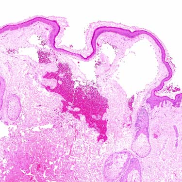
Progressive Lymphangioma Low-power examination of a progressive lymphangioma shows superficial, dermal-dilated, lymphatic spaces, some of which contain numerous red blood cells.
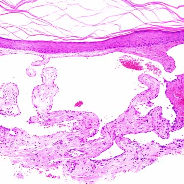
Progressive Lymphangioma at Higher Magnification Higher magnification examination of the superficial portion of a progressive lymphangioma shows widely dilated lymphatic spaces in the superficial dermis.
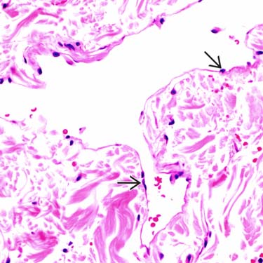
Progressive Lymphangioma: High Magnification of Endothelial Cells High-power examination shows irregular contours of lymphatic spaces and small, bland-appearing, lining endothelial cells .
Progressive Lymphangioma: High Magnification of Papillary Projection High-power examination of another section shows a large papillary projection protruding into the lymphatic space.
TERMINOLOGY
Synonyms
• Acquired progressive lymphangioma
• Benign lymphangioendothelioma
Definitions
• Benign, localized proliferation of lymphatic vessels
ETIOLOGY/PATHOGENESIS
Unknown
• May be related to trauma in some cases
CLINICAL ISSUES
Epidemiology
• Age
Middle-aged or older adults
• Sex
No predilection
Site
• Usually presents on lower extremities but may occur anywhere
Only gold members can continue reading. Log In or Register to continue


 .
.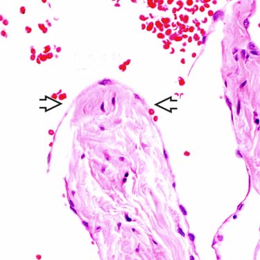
 protruding into the lymphatic space.


