Chapter Forty-Five. Postpartum haemorrhage and other third-stage problems
Introduction
Once the baby has been born, delivery of the placenta and membranes may seem an anticlimax. However, the third (3rd) stage of labour is hazardous for the mother because of the risk of haemorrhage and other complications. The management of the 3rd stage should be aimed at minimising these possible serious complications but interfering as little as possible with the physiological process (discussed in Chapter 40) and the mother’s enjoyment of her baby (Enkin et al 2000). A major role of the midwife is to explain the need for active interventions, such as the giving of an oxytocic drug or commencing an intravenous infusion, to the mothers, prior to labour so that women are enabled to make informed choices should the need suddenly arise.
Postpartum haemorrhage
Definition
Postpartum haemorrhage (PPH) is defined as excessive bleeding from the genital tract following the birth of the child. PPH occurs in the period extending from the time of birth to the end of the puerperium. If bleeding occurs in the first 24 h, it is called primary PPH (Mousa & Alfirevic 2007) and complicates about 6% of labours. If the bleeding occurs after the first 24 h and before the end of the 6th week, it is called secondary PPH (Alexander et al 2008), a much less common occurrence that complicates less than 1% of deliveries.
Postpartum haemorrhage is also classified according to the site of bleeding. Most commonly, the bleeding is from the placental site and there is poor tone of the uterine muscle. This is atonic haemorrhage. Bleeding may also be traumatic due to a laceration of the genital tract. In primary PPH, bleeding is said to be excessive if the amount exceeds 500 ml or is sufficient to cause deterioration in the woman’s condition. Because of the diuresis and haemoconcentration that follow delivery, smaller amounts of blood loss are detrimental in secondary PPH.
Primary PPH is one of the most serious complications of labour that a midwife has to deal with until medical aid arrives. At term, maternal circulating blood flow to the uterus is 450–700 ml/min, where 80% is perfusion for the placenta and 20% is perfusion for the myometrium (Murray 2003). The blood loss may be rapid and devastating if the bleeding is not controlled. PPH is still a significant cause of maternal mortality, especially following a caesarean section (CS) (Lewis 2007). The most recent CEMACH Report stipulates that there were 14 maternal deaths related to obstetric haemorrhage and 9 of these were due to PPH (Lewis 2007). Measuring blood loss at delivery can be difficult. It is important to remember that blood soaks into sheets and towels and that it separates into clot and serum. Any clot placed in a jug and measured will only be 40% of the total loss so that it is easy to underestimate the total loss by up to 50%.
Primary postpartum haemorrhage from the placental site
Causes
Uterine atony, which is failure of the uterine muscle fibres to contract and retract to compress the blood vessels is the most common cause (Mousa & Alfirevic 2007). Risk factors are:
• A history of previous postpartum haemorrhage.
• High parity: para 3 or more.
• Overdistension of the uterus in multiple pregnancy, polyhydramnios and a large fetus.
• Fibroids may interfere with efficient contraction and retraction.
• Antepartum haemorrhage: the bleeding that occurs into the muscle during placental abruption will reduce the fibres’ ability to contract and retract and in placenta praevia there is little contractile ability in the lower uterine segment.
• Prolonged labour with weak or uncoordinated contractions.
• Atony caused by drugs such as antihypertensives, general anaesthesia and tocolytics.
• Retained placenta.
• Anaemia because even a small amount of blood loss may precipitate shock.
• Inversion of the uterus.
• Mismanagement of the 3rd stage of labour by fiddling with the uterus.
• Coagulation defects: disseminated intravascular coagulation may complicate concealed placental abruption, amniotic fluid embolus, severe pre-eclampsia and eclampsia and intrauterine death.
• Medical disorders of clotting may also lead to primary PPH.
Despite the long list of risk factors outlined above, many cases of primary PPH occur in normal labours with no explanation.
Management of primary postpartum haemorrhage
In the antenatal period prevention is the best form of management and this begins with the booking interview. The following reports highlight the importance of good history taking and a crucial part of risk assessment: Maternal History Taking (NHS Quality Improvement Scotland 2004) and CEMACH ‘Saving Mothers’ Lives’ (Lewis 2007). If any of the risk factors described above are present, the woman should be delivered in hospital so that if bleeding does occur then treatment is immediately available. As pregnancy progresses detection and treatment of anaemia is important (RCOG 2009) and it would be advantageous to raise the haemoglobin (Hb) level to at least 11 g/dl before delivery (Lindsay 2004).
In labour
Women at risk of PPH must be managed carefully to minimise the likelihood of bleeding. When labour commences an intravenous cannula (size 14 or 16 gauge) is inserted and blood is taken for a full blood count (FBC) and confirmation of blood group. Serum is saved for ‘cross-matching’ blood should it become necessary to give the woman a blood transfusion. Prolonged labour, with its problems of dehydration and exhaustion, should be avoided. A Syntocinon (oxytocin) infusion should be started if labour progress is slow. The woman’s bladder should be kept empty by encouraging micturition or by catheterisation, as a full bladder may inhibit uterine muscle activity and add to the risk of atony.
Management of the 3rd stage should be discussed with the woman antenatally so that previous verbal consent can be given for any intervention, should it arise. She should be advised that the potential for PPH is greater with physiological management of the 3rd stage than it is with ‘active management’. It should be explained to her that the medical view is that it is safer to manage the 3rd stage of labour actively. In active management, an intramuscular injection of Syntocinon 10 IU or Syntometrine 1 ml, which contains Syntocinon 5 units and ergometrine 500 μg, is given with the birth of the anterior shoulder. The placenta is then delivered by controlled cord traction. An intravenous or intramuscular injection of Syntometrine 1 ml or ergometrine 500 μg would be prescribed if the woman starts to bleed, depending on what had been administered previously.
One intervention advocated by the RCOG (2007) is prophylactic radiology. This process involves inserting an arterial balloon into the blood vessels causing occlusion and embolisation to prevent major blood loss. This intervention should be used in the event of a PPH if the cause is secondary to:
• Atonic uterus following normal or prolonged labour, with or without caesarean section.
• Surgical complications or uterine tears at the time of caesarean section.
• Bleeding continues on the postnatal ward or in the postoperative recovery area following a normal delivery or a caesarean section.
• Bleeding following a hysterectomy.
This process involves inserting a balloon into the vessels and occlusion or embolisation occurs to prevent blood loss.
Signs of postpartum haemorrhage
It would be difficult to miss the visible bleeding and maternal collapse that can occur. Other signs that may be present if blood loss is not visible (e.g. if clots are retained in an atonic uterus) are:
• Pallor.
• A rising pulse rate and falling blood pressure.
• Altered levels of consciousness.
• Air hunger.
• An enlarged ‘boggy’-feeling uterus.
Management of primary postpartum haemorrhage: treatment
It is important that a midwife is familiar with the sequence of actions needed to deal with a PPH and to minimise the effects of blood loss.
If bleeding begins before the placenta is delivered, the following actions should be taken:
1. Ensure that medical aid is available.
2. Massage the fundus of the uterus firmly by a smooth circular motion to stimulate a uterine contraction, i.e. to ‘rub up’ a contraction. Bleeding indicates that the placenta has begun to separate and it is no longer necessary to await events.
3. Give an oxytocic drug. Intramuscular Syntometrine 1 ml will act to contract the uterus in 2.5 min and an intravenous injection of either Syntometrine 1 ml or ergometrine 500 μg will act in 45 s. Note that the midwife should not give more than two injections of ergometrine 500 μg as the drug may cause severe peripheral vasoconstriction and a sudden rise in blood pressure.
4. Pass a catheter into the bladder and ensure that it is completely empty.
5. Palpate to ensure the uterus is contracted, and then attempt to deliver the placenta by controlled cord traction.
6. If all else fails, the obstetrician should be asked to review. The obstetrician will try to deliver the placenta by cord traction. If this fails, the woman will need a manual removal of the placenta under spinal or epidural anaesthesia.
If the uterus is well contracted, the bleeding is likely to be from traumatic injury to the soft tissues. Locate the bleeding site and try to stem the bleeding using pressure.
If bleeding begins after delivery of the placenta, massage the uterus to obtain a contraction and expel any blood clots remaining in the uterus. An injection of an oxytocic drug, either intramuscular Syntometrine 1 ml or ergometrine 500 μg, should then stop bleeding by achieving a sustained contraction. Ensure that the urinary bladder is empty. If bleeding continues, it is necessary to carry out bimanual compression of the uterus. Following delivery of the placenta and membranes, they should be examined for completeness. If the placenta appears incomplete, the doctor will carry out an exploration and evacuation of the uterus under spinal or epidural anaesthesia.
Bimanual compression of the uterus
Bimanual compression may be performed externally or internally. In external bimanual compression, one hand is dipped down as far as possible behind the uterus while the other is placed flat on the abdomen. The uterus is compressed between the two hands and pulled upwards in the abdomen. This ensures that the bleeding area of the placental site is compressed while the uterine veins are straightened out to allow free drainage, relieve congestion and decrease the bleeding (Lindsay 2004).
Internal bimanual compression is carried out if the mother is anaesthetised and still bleeding after manual removal of placenta. One hand is closed to form a fist, inserted into the anterior vaginal fornix and pushed up towards the body of the uterus. The other hand is placed on the abdominal wall behind the uterus and compresses the uterus downwards against the hand in the vagina. This applies compression to the placental site until the uterus is felt to contract (Fig. 45.1).
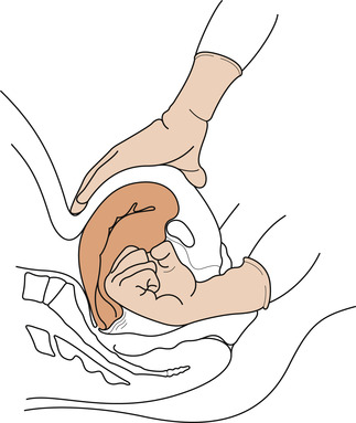 |
| Figure 45.1 Internal bimanual compression of the uterus. (From Henderson C, Macdonald S 2004, with kind permission of Elsevier.) |
Once bleeding is controlled, an intravenous infusion containing Syntocinon (oxytocin) is started to maintain uterine contraction. If blood loss is excessive or if the woman had a low haemoglobin level before delivery, a blood transfusion may be necessary. There are approximately 4000 cases of obstetric haemorrhage a year. The majority will require a blood transfusion (RCOG 2009). Therefore it is important not to underestimate the amount of blood lost. Brant (1967) found that estimates of blood loss became more inaccurate as the amount lost increased. In the case of serious PPH, the obstetrician and anaesthetist will agree on the management of fluid replacement. It is recommended that each obstetric unit should have a protocol on managing PPH, which should be followed and ‘fire-drill’ scenarios conducted to train and update practitioners on the protocol (RCOG 2009). Group O rhesus-negative blood should be available on the labour ward for use in emergencies. Where large volumes of blood are to be transfused, blood-warming coils should be used. Appropriate personnel must be included and there should be early involvement of consultant obstetrician, anaesthetist, haematologist and blood bank (RCOG 2005).
If the uterus fails to contract even though oxytocic drugs have been used, a deep intramuscular injection of the prostanoid carboprost, which is 15-methyl-PGF 2α (Rang et al 2007), can be given in a dose of 250 μg and repeated at intervals of 1.5 h. Carboprost is contraindicated in women with cardiac, renal, pulmonary and hepatic disease as well as in acute pelvic inflammation. It should be used with care in women who have asthma, hypertension, diabetes, epilepsy, hypotension or hypertension (BNF 2008). Different maternity units, according to their guidelines, may use different methods for controlling bleeding. In continuing haemorrhage, internal iliac artery ligation and uterine packing may be needed and, if all fails, a hysterectomy may be performed to save the woman’s life. A systematic review by Mousa & Alfirevic (2007) argues that there is not enough robust evidence in which to alter the present treatment of primary PPH, which is the combined use of oxytocin and ergometrine with misoprostol. Further controlled trials are required to assess the effectiveness and safety of pharmacological, interventional radiology and surgical interventions used for the treatment of PPH (Mousa & Alfirevic 2007).
Observations
Once blood loss is controlled, the total loss is estimated remembering how difficult this can be and that estimates become less accurate as blood loss increases. Fluid intake is recorded, as is the hourly urine output. Central venous pressure measurement may be required, depending on the blood loss and the severity of the woman’s condition, which may require correct fluid replacement. Maternal pulse and blood pressure are recorded every 15 min to ensure her condition remains satisfactory. The uterine fundus is palpated frequently to ensure it remains contracted and the lochia are observed. All findings can be recorded using a modified early warning system (e.g. MEWS).
If the problem involves failure of blood coagulation, a haematologist should be involved. Fresh blood is usually the best treatment as it contains both platelets and coagulation factors but fresh frozen plasma, containing factors V and VIII and fibrinogen, can be used. This is guided by the results from the blood coagulation screen (RCOG 2007).
Traumatic postpartum haemorrhage
If the blood loss is from a laceration of the genital tract, bleeding should be stopped by direct pressure if possible and then sutured. Bleeding from a cervical or lower uterine tear should be suspected if the uterus is well contracted, no superficial bleeding can be seen and the blood loss is slow and steady. Tears of the upper part of the vagina, the cervix and lower uterine segment should be sutured under spinal or epidural anaesthesia. If severe bleeding is from the uterus and cannot be stopped, a hysterectomy may be necessary to save the woman’s life.
Secondary postpartum haemorrhage
Secondary PPH is any abnormal bleeding or excessive bleeding from the birth canal occurring 24 h and 12 weeks postnatally (Alexander et al 2002). This is a complication of the puerperium and is most often seen between days 4 and 14. It is usually due to a retained piece of placenta but other causes include the presence of blood clot or a fibroid in the uterine wall. Secondary PPH is also commonly associated with infection. There may have been warning signs of heavy, red, offensive lochia and subinvolution. If infection is present, pyrexia and tachycardia may be present.
Stay updated, free articles. Join our Telegram channel

Full access? Get Clinical Tree


