Chapter Ten. Embryological systems 1—trunk, head and limbs
The trunk
Skeletal features
The vertebral column
There are three phases in the development of the vertebral column (Fig. 10.1): precartilaginous, cartilaginous and bony (Moore & Persaud 2008).
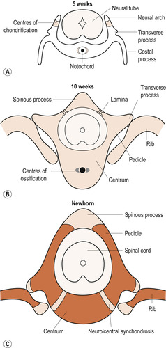 |
| Figure 10.1 (A) Blastemal vertebra with centre of chondrification (shaded dark), (B) cartilaginous vertebra with centres of ossification (shaded dark), (C) bony vertebra. (From Fitzgerald M J T, Fitzgerald M 1994, with permission.) |
Precartilaginous phase
Precursors of the vertebrae called sclerotomes are derived from the somites. Mesenchymal cells surround the notochord to form the segmented mesenchymal vertebral column. The cranial end of each mesenchymal vertebra is thinner than the caudal end because of a relative sparseness of cells. The cells at the interface between the two tissues form an intervertebral disc and the remainder of the condensed structure merges with the vertebra caudal to it to form the centrum. This is formed from parts of two sclerotomes and is an intersegmental structure. From the upper part of the centrum a pair of neural arches grows and surrounds the neural tube, giving rise to pairs of costal and transverse processes.
Cartilaginous phase
Chondrification centres appear in the centrum and neural arch late in the 5th week. In the 12 thoracic vertebrae the cartilaginous coastal processes which will develop into the ribs become detached from their parent neural arches by the formation of synovial joints. Synovial joints also appear between the costal and transverse processes. The remaining costal processes are incorporated into the vertebrae.
Bony phase
During the 8th week ossification centres appear in the centrum and neural arches and in the ribs. Ossification of the skeleton is not completed until the 25th year.
Ribs and sternum
As the lateral body folds in the 4th week, the somatic mesoderm is penetrated by the thoracic costal processes. These induce the mesoderm to add to their tips, completing the formation of the prechondrial ribs. Two sternal bars develop in the ventral part of the somatic mesoderm which meet in the midline and unite to form a prechondrial sternum. The xiphisternum often remains bifid. The ventral ends of the seven cranial costal processes fuse with the sternum and cartilage persists at the junction as the costal cartilages (Moore & Persaud 2008).
Soft tissues
During the 4th week the somites subdivide into three kinds of mesodermal primordia: myotomes, dermatomes and sclerotomes (Schoenwolf et al 2008).
Myotomes
Myotomes give rise to all the muscles that link the vertebrae and skull together and to the muscles of the abdominal and thoracic walls. Spinal nerves divide into dorsal and ventral rami. Dorsal rami supply the muscles with motor fibres and ventral rami supply the muscles and their overlying dermatome with sensory fibres. Limb muscles are not formed from myotomes.
Dermatomes
The dermatomes merge with each other to form the dermis layer of the skin. Each dermatome is accompanied by sensory nerve fibres derived from the level of the spinal cord at which the dermatome originated. That is why neurologists divide the body surface into regions called dermatomes.
The skin and mammary glands
The dermis develops from the dermatomes but the epidermis and its appendages are derived from surface ectoderm which begins as a single cuboidal layer but becomes two-layered in the 2nd month. The superficial layer is shed leaving the underlying germinal layer to form the structures of the skin. In the 3rd month the epidermis becomes stratified and its basal layer sends pegs down into the dermis to form the root sheath of the hair follicles. Lanugo, which is very soft fine hair, grows all over the body. True or vellus hair is derived from a second set of hair follicles and replaces lanugo which is shed shortly before birth.
During the 5th month the sebaceous glands bud into the dermis from the root sheath and the sweat glands grow down from the epidermis. The sebaceous glands produce a secretion which, when mixed with peridermal cells and lanugo, becomes the vernix caseosa. The mammary glands appear in the 6th week as paired strips of longitudinal ectodermal thickening on the ventral surface of the embryo called the mammary ridge. In humans only one pair of breasts forms from the thoracic part of the ridge and the rest of the ridge disappears.
The skull
The skull forms from mesenchyme around the developing brain. It consists of the neurocranium which encloses the brain and the viscerocranium making up the bones of the face. The base of the skull or chondrocranium develops out of cartilage whereas the vault bones (Ch. 24) develop from membrane (Moore & Persaud 2008). Intramembranous ossification begins from the 4th month, separate ossification centres giving rise to the parietal bones, frontal bones, occipital bone and the squamous part of the temporal bones.
The viscerocranium
All the facial bones ossify in membrane, starting with the mandible early in the 6th week. Detailed development of the face is given later.
The teeth
Tooth buds form from thickened ectoderm called the dental lamina: 10 in the upper jaw and 10 in the lower jaw. These are responsible for the deciduous (milk) teeth. Later the dental lamina forms the buds of the permanent dentition. The permanent molars do not have precursors in the deciduous dentition but develop from a backwards extension of the dental lamina. The crowns of the teeth begin when cells called odontoblasts form predentine which later calcifies to become dentine. Calcification signals cells called ameloblasts to lay down enamel on the surface of the dentine. The central cells constitute the pulp of the tooth which is richly supplied with blood and sensory nerve endings as many of us can testify!
Deep to the level of enamel production the outer and inner enamel epithelia fuse to form the epithelial root sheath. Predentine and dentine are induced to form the root of the tooth. The mesoderm of the dental sac produces a specialised form of bone called cement and the periodontal ligament which anchors the cement to the wall of the tooth socket. Eruption of the deciduous teeth occurs between 6 months and 2 years after birth.
The brain
Chapter 26 describes the complex adult brain and central nervous system. The neural tube cranial to the 4th pair of somites develops into the brain (Moore & Persaud 2008). The human nervous system begins to form 19 days after fertilisation and is the earliest system to differentiate. By 19 days three expansions are present (Fig. 10.2). These primary brain vesicles are:
• Forebrain— prosencephalon.
• Midbrain— mesencephalon.
• Hindbrain— rhombencephalon.
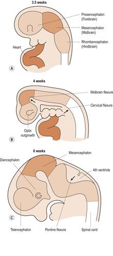 |
| Figure 10.2 Early development of the brain. (From Fitzgerald M J T, Fitzgerald M 1994, with permission.) |
From 4 weeks the major regions of the brain are distinct and neurons begin to differentiate from the epithelium of the neural tube. The thalamus and hypothalamus are differentiated by the 5th week (Fig. 10.3). By the end of the 8th week the head is equal to half the length of the embryo and controls first movement of the limbs.
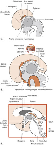 |
| Figure 10.3 Median sections of the brain (A) at 8 weeks, (B) at 12 weeks, (C) postnatal. (From Fitzgerald M J T, Fitzgerald M 1994, with permission.) |
The changing shape of the brain
The brainstem buckles and a cervical flexure appears at the junction of the brainstem and spinal cord. A midline flexure moves the mesencephalon to the summit of the brain. The rhombencephalon folds on itself, causing the walls of the neural tube to expand into the 4th ventricle. The dorsal region of the prosencephalon expands on either side to form the cerebral hemispheres or telencephalon. Within the cerebral hemispheres the neural tube dilates to form the lateral ventricles.
The remainder of the prosencephalon which straddles the midline is known as the diencephalon. The 3rd ventricle, a cavity within the diencephalon, communicates with the 4th ventricle through the aqueduct of Sylvius. An outgrowth from the diencephalon becomes the two retinas and optic nerves. The cranial end of the rhombencephalon gives rise to the pons and the cerebellum, while the caudal end becomes the medulla oblongata.
The forebrain
Neurons migrate from the ventricular zone of the telencephalon to the surface to form the cerebral cortex. The frontal, parietal, occipital and temporal lobes are present by 12–14 weeks. Two commissures of nerve fibres link the two cerebral hemispheres. The anterior commissure links the olfactory regions and the larger corpus callosum links areas of the cerebral cortex.
Other brain structures
The diencephalon gives rise to the pineal gland, the paired thalami and the hypothalamus. The basal gangliadevelop immediately below the thalamus and help control body movement.
Blood supply to the brain
The arterial blood supply develops from cranial segments of the dorsal aortae, comprising two internal carotid arteries and two vertebral arteries. The internal carotid arteries branch to form the anterior, middle and posterior cerebral arteries. Each vertebral artery gives off a branch to supply the cerebellum and medulla oblongata before uniting with its partner to form the basilar artery. This gives off two pairs of arteries to the cerebellum and upper brainstem before dividing into two terminal branches that link up with the ends of the internal carotid arteries to form the intercommunicating circle of Willis. The venous drainage is discussed in Chapter 24.
The spinal cord
Following the closure of the neural tube and the formation of the somites, neural crest cells form clusters corresponding to the somites. Corresponding levels of the neural tube develop from the primitive streak during a process of secondary neurulation. At first the neural tube is solid but becomes canalised by caudal extension of the neural canal.
Zones of the spinal cord
During the 5th week three zones can be distinguished in the side walls of the neural tube. From within outwards these are the ventricular, intermediate and marginal zones.


• The ventricular zone is where neuroepithelial cells divide. After several cell divisions daughter cells move out of the ventricular zone; the first ones become neurons and the last become the connective tissue cells called neuroglia.
Stay updated, free articles. Join our Telegram channel
Full access? Get Clinical Tree
