


Top Differential Diagnoses
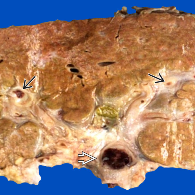
Gross photograph of this liver explant shows a large portal vein thrombus
 in the hilar region. Propagation into smaller intrahepatic portal veins
  is also grossly noticed.
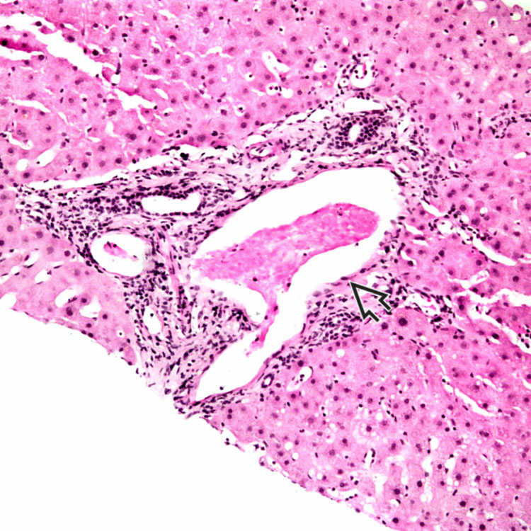
This microphotograph shows a markedly dilated portal venule
 in the setting of portal vein thrombosis. Note the presence of fibrin thrombus in the lumen.
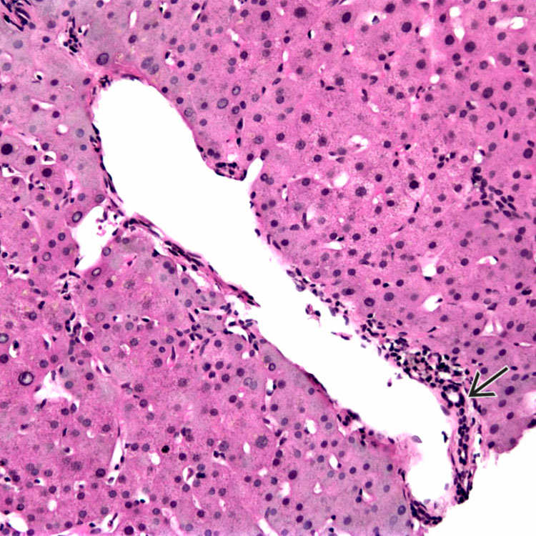
Typical changes in portal venous obstruction include dilated portal venules with “herniation” of the vessel into the surrounding parenchyma. Note the presence of a bile duct
 in compressed portal stroma.
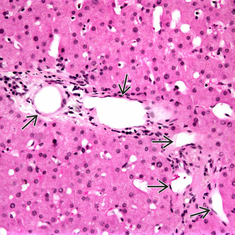
 are present in this portal tract in a case of portal venous obstruction indicating elevated portal pressures.
TERMINOLOGY
Synonyms
• Idiopathic portal hypertension


Stay updated, free articles. Join our Telegram channel
Full access? Get Clinical Tree
