Age-dependent increase in number and size of liver cysts
Higher prevalence of PLD in women
• Isolated PLD affecting < 0.01% of population
• Asymptomatic in ∼ 80% of patients
• Symptomatology due to hepatomegaly with compression of adjacent structures
Treatment options include aspiration of large dominant cyst, sclerotherapy, cyst fenestration, somatostatin analogues, partial hepatectomy, and liver transplantation
• Renal failure is main complication of ADPKD
Macroscopic
• Hepatomegaly weighing up to 13 kg
• Cysts varying from < 1 mm to > 12 cm in diameter
• Occasionally 1 lobe involved (usually left lobe)
• Clear, colorless, or straw-colored cyst fluid
Molecular
• Mutations in PKD1 gene seen in 80-85% of ADPKD cases
• Mutations in PKD2 gene seen in 15-20% of ADPKD cases
• Mutations in PRKCSH, SEC63, and LRP5 genes in isolated PLD cases
Microscopic
• Numerous variably sized cysts lined by single layer of cuboidal or flattened biliary epithelium
• Lack of communication with biliary tree
• von Meyenburg complexes commonly present
• Fibrosis and hyalinization in collapsed cysts, which may resemble corpora atretica or fibrosa of ovary
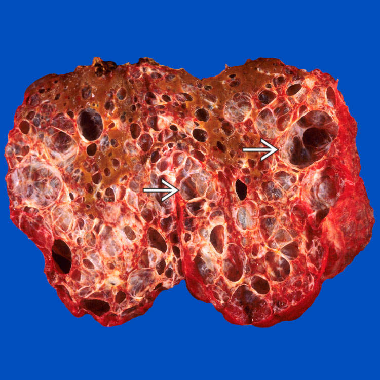
Gross Appearance This case of polycystic liver disease features massive involvement by numerous variably sized cysts, which are present throughout the liver. Note that the cyst walls are thin and smooth .
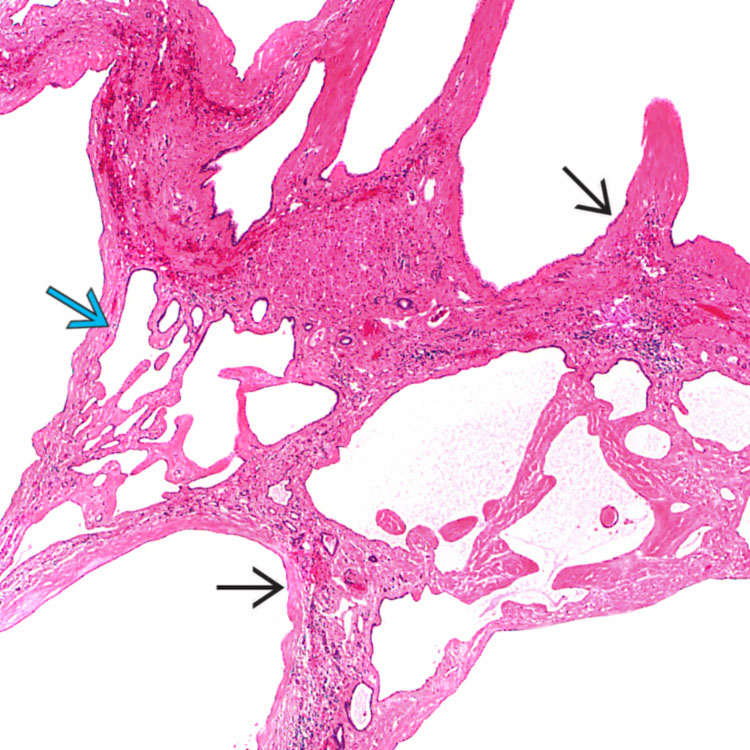
Multiple Cystic Spaces Low-power view shows numerous cystic spaces lined by a single layer of epithelial cells , supported by variable amounts of connective tissue. Only minimal liver parenchyma remains. A von Meyenburg complex is present. Slightly proteinaceous fluid is present in some of the cystic spaces.
von Meyenburg Complex A von Meyenburg complex is present adjacent to a cyst . The von Meyenburg complex is composed of dilated, angulated biliary structures. These are commonly seen in polycystic liver disease.
Collapsed Cyst A collapsed cyst consists of a corrugated and hyalinized wall . The lumen is filled with loose connective tissue, resembling a corpus atreticum or fibrosum of the ovary. Note the presence of residual liver parenchyma with a normal bile duct .


 .
.
 , supported by variable amounts of connective tissue. Only minimal liver parenchyma remains. A von Meyenburg complex
  is present. Slightly proteinaceous fluid is present in some of the cystic spaces.
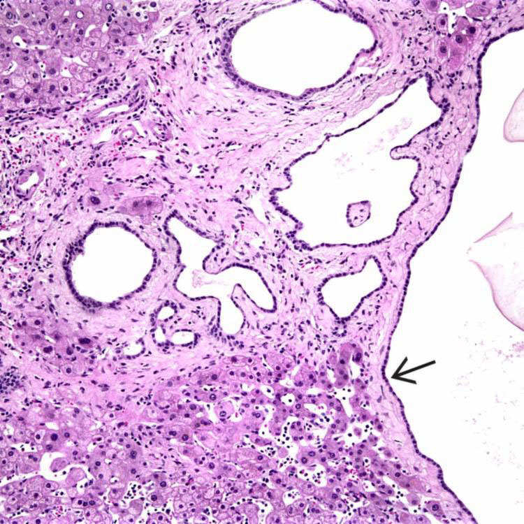
 . The von Meyenburg complex is composed of dilated, angulated biliary structures. These are commonly seen in polycystic liver disease.
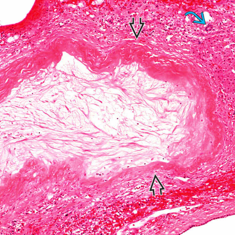
 . The lumen is filled with loose connective tissue, resembling a corpus atreticum or fibrosum of the ovary. Note the presence of residual liver parenchyma with a normal bile duct
  .
.


