QUESTION 27.2
A. Epithelioid
B. Mixed
C. Sarcomatoid
A. Epithelioid
B. Mixed
C. Sarcomatoid
A. Epithelioid
B. Mixed
C. Sarcomatoid
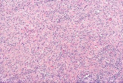
5. A 65-year-old man presents with persistent chest pain and unexplained weight loss. On initial examination, he is found to have a hemorrhagic pleural effusion, which is evacuated. A CT scan reveals irregular pleural thickening. A biopsy of the pleural lesions demonstrates the histopathologic features shown in this photomicrograph. The tumor has diffuse expression of pan-cytokeratin. Which of the following markers, if positive, would support a diagnosis of mesothelioma and rule out adenocarcinoma?
QUESTION 27.5
A. Cytokeratin 7
B. Cytokeratin 20
C. Pancytokeratin
D. Vimentin
E. None of the above
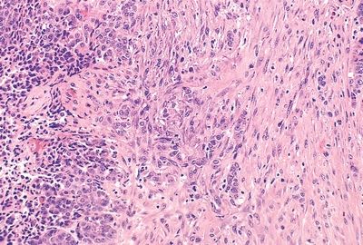
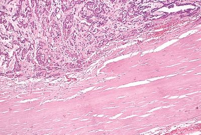
6. A pleural tumor composed of tubular and papillary structures raises the differential diagnosis between epithelioid mesothelioma and adenocarcinoma. Which of the following immunostains would be positive if the tumor were a mesothelioma?
A. Ber-EP4
B. BG-8
C. Calretinin
D. Carcinoembryonic antigen (CEA)
Stay updated, free articles. Join our Telegram channel

Full access? Get Clinical Tree


