• Treatment: Complete surgical resection with negative margins
• Local recurrences in up to 30% of cases
• Metastases in 10-20% of cases
Macroscopic
• Median: 2.5 cm
Microscopic
• Poorly marginated, usually with deep tissue extension
• Prominent cytologic and nuclear atypia and mitotic activity
• Frequent coagulative necrosis
• Lymphovascular invasion in some cases
Ancillary Tests
• Negative for keratins, S100, desmin, CD34, most others
• Immunohistochemical best utilized to exclude other diagnoses
Top Differential Diagnoses
• Atypical fibroxanthoma
• Carcinoma, melanoma
• Angiosarcoma
• Other superficial pleomorphic sarcomas
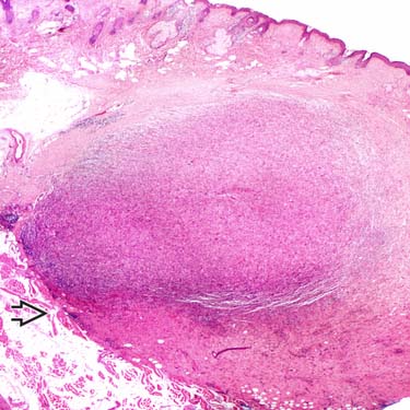
PDS Pleomorphic dermal sarcoma (PDS) originates superficially in the dermis, as depicted, but usually shows deeper extension with involvement of the subcutis, fascia, &/or underlying muscle .
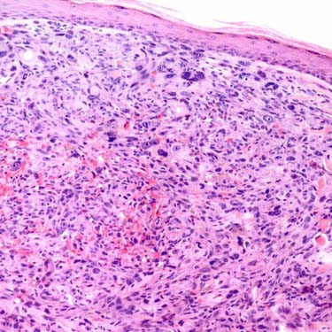
PDS With Resemblance to Atypical Fibroxanthoma PDS shows histomorphologic features similar to atypical fibroxanthoma (AFX), and these 2 entities can be indistinguishable on a superficial/partial biopsy. The presence of deep tissue extension, necrosis, or lymphovascular invasion would be most consistent with the former.
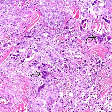
PDS With Marked Nuclear Atypia Nuclear and cytologic atypia is often prominent and diffuse in PDS, as depicted, similar to its counterpart in deep soft tissues (undifferentiated pleomorphic sarcoma). Multinucleated tumor giant cells are common, as are atypical mitotic figures.
PDS With Coagulative Necrosis Coagulative tumor necrosis is common in PDS but varies in amount and distribution. The presence of necrosis in a pleomorphic dermal neoplasm favors PDS over AFX, assuming that carcinoma, melanoma, and other tumors are first excluded.
TERMINOLOGY
Abbreviations
• Pleomorphic dermal sarcoma (PDS)
Synonyms
• Superficial malignant fibrous histiocytoma
• Undifferentiated pleomorphic sarcoma of skin
Definitions
• Pleomorphic sarcoma exhibiting morphologic similarity to atypical fibroxanthoma (AFX), but also demonstrating aggressive growth characteristics
Deep subcutaneous/fascial/muscular extension, necrosis, &/or lymphovascular invasion
Shows no identifiable line of differentiation (i.e., diagnosis of exclusion)
ETIOLOGY/PATHOGENESIS
Environmental Exposure
• History of long-term sun exposure usually exists
CLINICAL ISSUES
Epidemiology
• Incidence
Rare
– Deeply situated tumors (conventional undifferentiated sarcoma) more common
Only gold members can continue reading. Log In or Register to continue

 .
.
 are common, as are atypical mitotic figures.
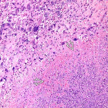
 is common in PDS but varies in amount and distribution. The presence of necrosis in a pleomorphic dermal neoplasm favors PDS over AFX, assuming that carcinoma, melanoma, and other tumors are first excluded.


