Peripheral Nervous System
LANGUAGE OF SCIENCE
 Before reading the chapter, say each of these terms out loud. This will help you avoid stumbling over them as you read.
[abdomin- belly, -al relating to, re- again, -flex bend]
[ab- away, -duc- lead, -ens process]
[acetyl- vinegar, -chole- bile, -ine made of]
(aw-toh-NOM-ik [VISS-er-al] REE-fleks)
[brachi- arm, -al relating to, plexus network] pl., plexi or plexuses
[cauda tail, equina of a horse] pl., caudae equinae
[cervic- neck, -al relating to, plexus network] pl., plexi or plexuses
[corn- horn, -al relating to, re- again, -flex bend]
[crani- skull, -al relating to]
[crani- skull, -al relating to, re- again, -flex bend]
[derma- skin, -tome cut segment or region]
[dors- the back, -al relating to, ramus branch] pl., rami
[dors- the back, -al relating to]
[gangli- knot, -on unit] pl., ganglia
[glosso- tongue, -pharyng- throat, -al relating to]
[hypo- under or below, -gloss- tongue, -al relating to]
[lumb- loin, -ar relating to, plexus network] pl., plexi or plexuses
[crani- skull, -al relating to]
[mot- movement, -or agent, crani- skull, -al relating to]
[myo- muscle, -tome cut segment or region]
[oculo- eye, mot- movement, -or agent]
[olfact- smell, -ory relating to]
[opt- vision, -ic relating to]
[phren- mind, -ic relating to]
[planta- sole, -ar relating to, re- again, –flex bend]
[plexus network] pl., plexi or plexuses
[sacr- sacred, -al relating to, plexus network] pl., plexi or plexuses
[(i)sci- hip joint, -atic relating to]
[sens- feel, -ory relating to, crani- skull, -al relating to]
[soma- body, -ic relating to, re- again, -flex bend]
[spine- backbone, -al relating to]
[spine- backbone, -al relating to, re- again, -flex bend]
[termin- boundary, -al relating to]
[tri- three, -gemina- twin or pair, -al relating to]
[trochlea- pulley, -al relating to]
[ventr- belly, -al relating to ramus branch] pl., rami
[ventr- belly, -al relating to]
[vestibulo- entrance hall, -cochle- sea shell, -ar relating to]
LANGUAGE OF MEDICINE
In Chapter 14, you learned about the structure and function of the central nervous system (CNS). In this chapter we explore the nerve pathways that lead to and from the CNS, which together make up the peripheral nervous system (PNS). The PNS is made up of the 31 pairs of spinal nerves that emerge from the spinal cord, the 12 pairs of cranial nerves that emerge from the brain, and all the smaller nerves that branch from these “main” nerves.
Recall from earlier discussions that afferent fibers carry information into the CNS. Afferent fibers, part of the sensory nervous system, help us maintain homeostasis by sensing changes in our internal or external environment—providing the feedback necessary to keep the body functioning normally. Afferent fibers belonging to the somatic sensory or special sensory nervous system feed back information regarding changes detected by receptors in the skin, skeletal muscles, and special sense organs. Afferent fibers belonging to the autonomic nervous system (ANS) feed back information regarding the effects of autonomic control of the viscera.
Recall also that efferent fibers carry information away from the CNS. Efferent fibers may belong to the somatic nervous system (SNS) that regulates skeletal muscles, allowing us to survive by defending ourselves, getting food, or performing other essential tasks. Efferent fibers may, on the other hand, belong to the ANS. Autonomic regulation controls smooth and cardiac muscle and glands in ways that help us maintain homeostasis of the internal environment.
The first sections of this chapter explore concepts regarding the structure and function of the spinal and cranial nerves. The last portion of the chapter is devoted to discussion of the peripheral elements of the SNS, with an emphasis on efferent (motor) pathways. Chapter 16 then picks up the story, emphasizing efferent pathways of the ANS. Finally, Chapter 17 explores major concepts of sensation via the afferent pathways.
This study of peripheral nerve pathways clarifies and expands the understanding of the nervous system gained in previous chapters. It also serves as a foundation for understanding important clinical applications (Box 15-1).
Box 15-1
 HEALTH matters
Peripheral Neuropathy
The term peripheral neuropathy tells you exactly what it signifies: disease of the peripheral nerves. The term is used to designate many different diseases with many different causes—but all involve damage to peripheral nerves. These disorders can also be called peripheral neuritis. If many nerves are involved, peripheral neuropathy can also be called polyneuropathy or polyneuritis. Probably the most common cause of peripheral nerve damage in the United States and Europe is diabetes mellitus (see Diabetes Mellitus online at A&P Connect), affecting some 17 million people. But additional causes include other metabolic problems such as kidney disease, injuries such as sports mishaps, poisoning as in drug neurotoxicity and alcohol abuse, infections such as in leprosy (Hansen disease; Mycobacterium leprae) or shingles (see Box 15-3, p. 472), and genetic mutations such as Leber hereditary optic neuropathy (see Chapter 37, p. 1142).
SPINAL NERVES
Thirty-one pairs of spinal nerves are connected to the spinal cord. They have no special names but are merely numbered according to the level of the vertebral column at which they emerge from the spinal cavity (Figure 15-1). Although there are only seven cervical vertebrae, there are eight cervical nerve pairs (C1 through C8), twelve thoracic nerve pairs (T1 through T12), five lumbar nerve pairs (L1 through L5), five sacral nerve pairs (S1 through S5), and one coccygeal pair of spinal nerves. The first pair of cervical nerves emerges from the cord in the space above the first cervical vertebra, and the eighth cervical nerve emerges between the last cervical vertebra and the first thoracic vertebra. Thus nerve pair C1 passes between the skull and vertebra C1, nerve pair C2 passes between vertebrae C1 and C2, nerve pair C3 passes between vertebrae C2 and C3, and so on. All the thoracic nerves pass out of the spinal cavity horizontally through the intervertebral foramina below their respective vertebrae.
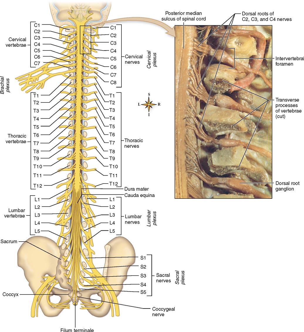
Lumbar, sacral, and coccygeal nerve roots, on the other hand, descend from their point of origin at the lower end of the spinal cord (which terminates at the level of the first lumbar vertebra) before reaching the intervertebral foramina of their respective vertebrae, through which the nerves then emerge. This gives the lower end of the cord, with its attached spinal nerve roots, the appearance of a horse’s tail. In fact, it bears the name cauda equina, which is the Latin equivalent for “horse’s tail” (see Figure 15-1).
Structure of Spinal Nerves
Each spinal nerve attaches to the spinal cord by means of two short roots, a ventral (anterior) root and a dorsal (posterior) root. The dorsal root of each spinal nerve is easily recognized by a swelling called the dorsal root ganglion, or spinal ganglion (Figure 15-2). The roots and dorsal ganglia lie within the spinal cavity, as Figure 15-2 shows.
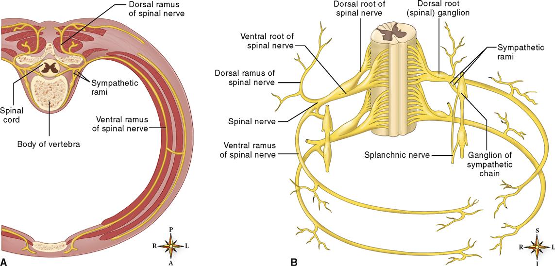
As described in Chapter 14, the ventral root includes motor neurons that carry information from the CNS and toward effectors (muscles and glands). Recall that in each somatic motor pathway, a single motor fiber stretches from the anterior gray horn of the spinal cord, through the ventral root, and on through the spinal nerve toward a skeletal muscle. Autonomic fibers, which also carry motor information toward effectors, may also pass through the ventral root to become part of a spinal nerve. The dorsal root of each spinal nerve includes sensory fibers that carry information from receptors in the peripheral nerves. The dorsal root ganglion contains the cell bodies of the sensory neurons. Because all spinal nerves contain both motor and sensory fibers, they are designated as mixed nerves.
Soon after each spinal nerve emerges from the spinal cavity, it forms several large branches, each of which is called a ramus (plural, rami). As Figure 15-2 shows, each spinal nerve splits into a distinct dorsal ramus and ventral ramus. The dorsal ramus supplies somatic motor and sensory fibers to several smaller nerves. These smaller nerves, in turn, innervate the muscles and skin of the posterior surface of the head, neck, and trunk. The structure of the ventral ramus is a little more complex. Autonomic motor fibers split away from the ventral ramus, heading toward a ganglion of the sympathetic chain. There, some of the autonomic fibers synapse with autonomic neurons that eventually continue on to autonomic effectors by way of splanchnic nerves (see Figure 15-2). However, some fibers synapse with autonomic neurons whose fibers rejoin the ventral ramus. The two thin rami formed by this splitting away, then rejoining, of autonomic fibers are together called the sympathetic rami. Motor (autonomic and somatic) and sensory fibers of the ventral rami innervate muscles and glands in the extremities (arms and legs) and in the lateral and ventral portions of the neck and trunk.
Nerve Plexuses
The ventral rami of most spinal nerves—all but nerves T2 through T12—subdivide to form complex networks called plexuses. As Figure 15-1 shows, there are four major pairs of plexuses: the cervical plexus, the brachial plexus, the lumbar plexus, and the sacral plexus. Table 15-1 summarizes important information about these major plexuses.
TABLE 15-1
Spinal Nerves and Peripheral Branches
| SPINAL NERVES | PLEXUSES FORMED FROM ANTERIOR RAMI | SPINAL NERVE BRANCHES FROM PLEXUSES | PARTS SUPPLIED | |||
| Cervical | Lesser occipital | Sensory to back of head, front of neck, and upper part of shoulder; motor to numerous neck muscles | ||||
| 1 | Greater auricular | |||||
| 2 | Cervical plexus | Cutaneous nerve of neck | ||||
| 3 | Supraclavicular nerves | |||||
| 4 | Branches to muscles | |||||
| Phrenic nerve | Diaphragm | |||||
| Cervical | Suprascapular and dorsoscapular | Superficial muscles* of scapula | ||||
| 5 | ||||||
| 6 | Thoracic nerves, medial and lateral branches | Pectoralis major and minor | ||||
| 7 | Brachial plexus | |||||
| 8 | ||||||
| Thoracic (or Dorsal) | Long thoracic nerve | Serratus anterior | ||||
| Thoracodorsal | Latissimus dorsi | |||||
| 1 | Subscapular | Subscapular and teres major muscles | ||||
| 2 | Axillary (circumflex) | Deltoid and teres minor muscles and skin over deltoid | ||||
| 3 | Musculocutaneous | Muscles of front of arm (biceps brachii, coracobrachialis, brachialis) and skin on outer side of forearm | ||||
| 4 | ||||||
| 5 | Ulnar | Flexor carpi ulnaris and part of flexor digitorum profundus; some muscles of hand; sensory to medial side of hand, little finger, and medial half of fourth finger | ||||
| 6 | No plexus formed; branches run directly to intercostal muscles and skin of thorax | |||||
| 7 | ||||||
| 8 | Median | Rest of muscles of front of forearm and hand; sensory to skin of palmar surface of thumb, index, and middle fingers | ||||
| 9 | Radial | Triceps muscle and muscles of back of forearm; sensory to skin of back of forearm and hand | ||||
| 10 | ||||||
| 11 | Medial cutaneous | Sensory to inner surface of arm and forearm | ||||
| 12 | ||||||
| Lumbar | Iliohypogastric | Sometimes fused | Sensory to anterior abdominal wall | |||
| 1 | Ilioinguinal | Sensory to anterior abdominal wall and external genitalia; motor to muscles of abdominal wall | ||||
| 2 | ||||||
| 3 | Genitofemoral | Sensory to skin of external genitalia and inguinal region | ||||
| 4 | Lateral femoral cutaneous | Sensory to outer side of thigh | ||||
| 5 | ||||||
| Sacral | Lumbosacral plexus | Femoral | Motor to quadriceps, sartorius, and iliacus muscles; sensory to front of thigh and medial side of leg (saphenous nerve) | |||
| 1 | ||||||
| 2 | Obturator | Motor to adductor muscles of thigh | ||||
| 3 | Tibial† (medial popliteal) | Motor to muscles of calf of leg; sensory to skin of calf of leg and sole of foot | ||||
| 4 | ||||||
| 5 | Common peroneal (lateral popliteal) | Motor to evertors and dorsiflexors of foot; sensory to lateral surface of leg and dorsal surface of foot | ||||
| Nerves to hamstring muscles | Motor to muscles of back of thigh | |||||
| Gluteal nerves | Motor to buttock muscles and tensor fasciae latae | |||||
| Posterior femoral cutaneous | Sensory to skin of buttocks, posterior surface of thigh, and leg | |||||
| Pudendal nerve | Motor to perineal muscles; sensory to skin of perineum | |||||
| Coccygeal | Coccygeal plexus | Anococcygeal nerves | Sensory to skin overlying coccyx | |||
| 1 | ||||||
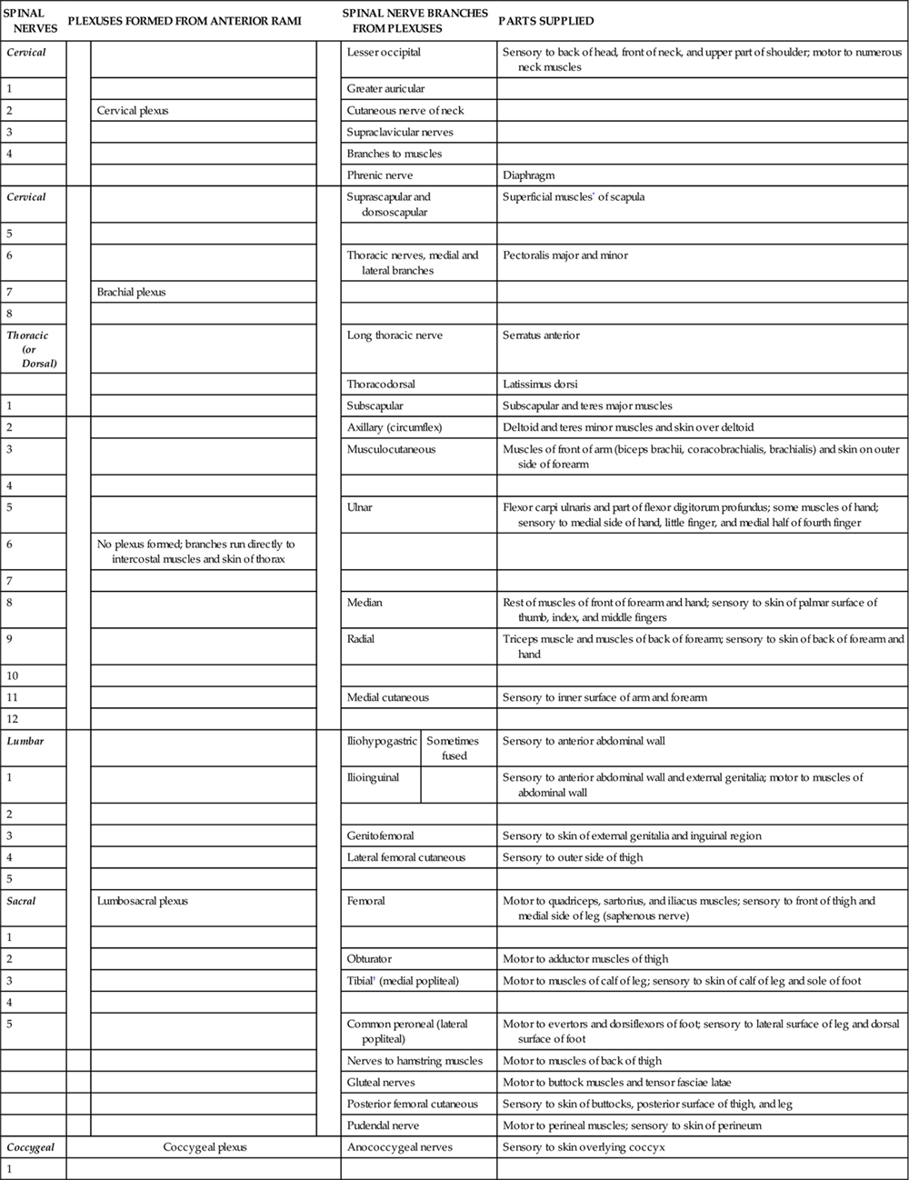
*Although nerves to muscles are considered motor, they do contain some sensory fibers that transmit proprioceptive impulses.
Stay updated, free articles. Join our Telegram channel

Full access? Get Clinical Tree


