Pelvis and Perineum
Cards 5-1 to 5-24
Bones and Joints
5-1 Bones and Ligaments of Pelvis
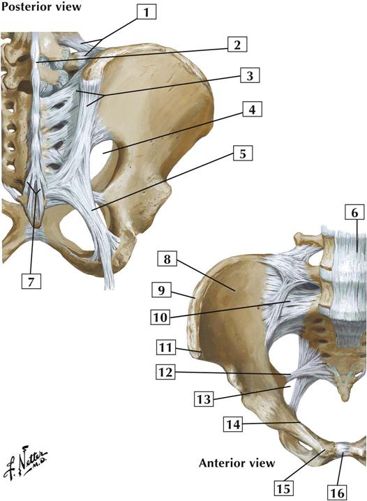
Comment:
The sacro-iliac joint is a plane synovial joint between the sacrum and ilium. It permits little movement. The sacro-iliac joint transmits the weight of the body to the hip bone when a person is standing. It is reinforced by anterior, posterior, and interosseous sacro-iliac ligaments.
The sacrococcygeal joint is a cartilaginous joint between the sacrum and coccyx. It allows some movement and contains an intervertebral disc between S5 and Co1.
The pubic symphysis is a cartilaginous (fibrocartilaginous) joint between the 2 pubic bones.
The sacrospinous ligament divides the greater sciatic foramen from the lesser sciatic foramen.
Atlas Plate 333
See also Plate 157
Muscles
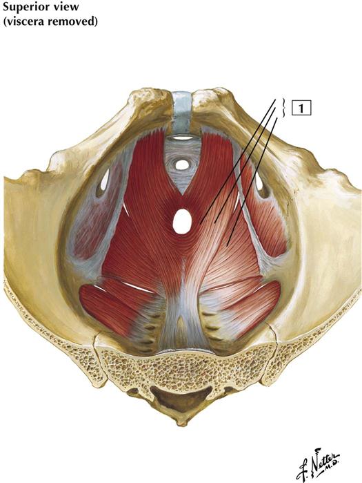
5-2 Pelvic Diaphragm: Male
Origin:
Arises from the body of the pubis, the arcus tendineus (actually a thickened portion of the obturator fascia) of the levator ani, and the ischial spine.
Insertion:
Attaches to the coccyx, anococcygeal raphe, external anal sphincter, walls of the prostate, rectum, anal canal, and central tendon of the perineum.
Action:
Supports and slightly raises the pelvic floor.
Innervation:
Ventral rami of S3 and S4 and perineal branch of the pudendal nerve.
Comment:
The levator ani has 3 parts: the puborectalis, the pubococcygeus, and the iliococcygeus muscles. With the coccygeus muscle, the levator ani forms the pelvic diaphragm.
The greater sciatic foramen exists superior to the pelvic diaphragm and provides a passageway for structures to leave the pelvic cavity and enter the gluteal region. The lesser sciatic foramen exists inferior to the pelvic diaphragm and provides a passageway for neurovascular structures to pass from the gluteal region to the perineum (importantly, the pudendal neurovascular bundle).
Atlas Plate 338
See also Plates 258, 261
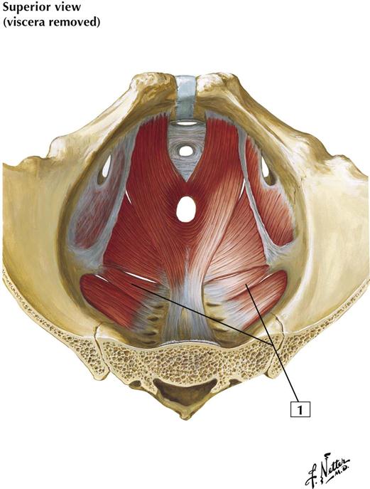
5-3 Pelvic Diaphragm: Male
Origin:
Arises from the spine of the ischium and the sacrospinous ligament.
Insertion:
Attaches to the coccyx and lower portion of the sacrum.
Action:
With the levator ani, the coccygeus supports the pelvic floor. It also draws the coccyx forward after the coccyx has been pushed back during parturition (in females) or defecation.
Innervation:
Ventral rami of S4 and S5.
Comment:
The pelvic diaphragm comprises the coccygeus and levator ani. Together, these muscles support and raise the pelvic floor.
The coccygeus muscle is the muscle used by dogs to tuck their tails between their hind legs; in humans, it is largely a mixture of skeletal muscle fibers and fibrous connective tissue.
The greater sciatic foramen exists superior to the pelvic diaphragm and provides a passageway for structures to leave the pelvic cavity and enter the gluteal region. The lesser sciatic foramen exists inferior to the pelvic diaphragm and provides a passageway for neurovascular structures to pass from the gluteal region to the perineum (importantly, the pudendal neurovascular bundle).
Atlas Plate 338
See also Plates 258, 261
5-4 Female Perineum
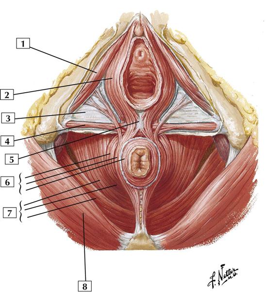
1. Ischiocavernosus muscle with deep perineal (investing, or Gallaudet’s) fascia removed
2. Bulbospongiosus muscle with deep perineal (investing, or Gallaudet’s) fascia removed
4. Superficial transverse perineal muscle with deep perineal (investing, or Gallaudet’s) fascia removed
6. Parts of external anal sphincter muscle (Deep; Superficial; Subcutaneous)
7. Levator ani muscle (Pubococcygeus; Puborectalis; Iliococcygeus)
Comment:
The muscles of the perineum are skeletal muscles. They are innervated by the pudendal nerve and its branches (ventral rami of S2-4).
The central tendon of the perineum (perineal body) is a mass of fibromuscular tissue found in the midline between the anus and vagina. It is an attachment point for many of the muscles of the perineum and is important for maintaining the integrity of this region.
Atlas Plate 356
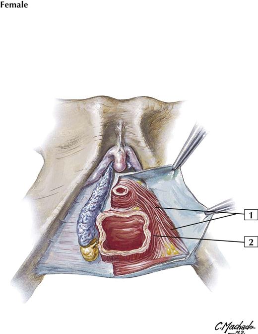
5-5 Perineum and Deep Perineum
Comment:
The anatomy of these muscles is controversial. The urethral sphincter might be more of a “urogenital sphincter,” consisting of a compressor urethrae muscle and a sphincter urethrovaginalis muscle. The sphincter action of these muscles is debatable.
These muscles are innervated primarily by the perineal branch of the pudendal nerve (S2-4).
On one side of this illustration, the ischiocavernosus and bulbospongiosus muscles have been removed to show the underlying erectile tissues of the bulb of the vestibule and the crus of the clitoris (still ensheathed in a fascial layer). Posterior to the bulb of the vestibule lies the greater vestibular (Bartholin’s) gland, which secretes mucus during sexual arousal that lubricates the vaginal opening.
Atlas Plate 356
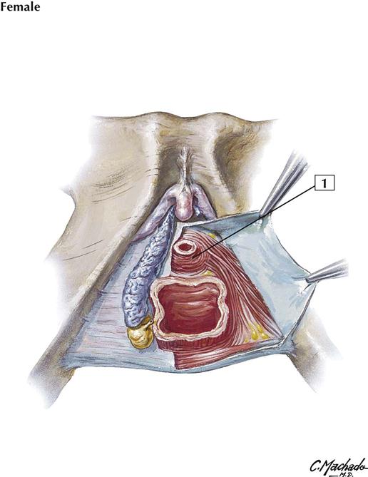
5-6 Perineum and Deep Perineum
Origin:
Arises from the inferior pubic ramus.
Insertion:
Attaches to a median raphe and the perineal body.
Action:
The muscles on both sides act together to constrict the urethra.
Innervation:
Perineal branch of the pudendal nerve (S2-4).
Comment:
In women, this muscle blends with the compressor urethrae muscle and the urethrovaginal sphincter muscles.
Although some textbooks call this muscle the “external” urethral sphincter, women do not possess an internal urethral sphincter (smooth muscle sphincter at the neck of the urinary bladder), which is a sphincter muscle found only in men.
Atlas Plate 356
5-7 Male Perineum
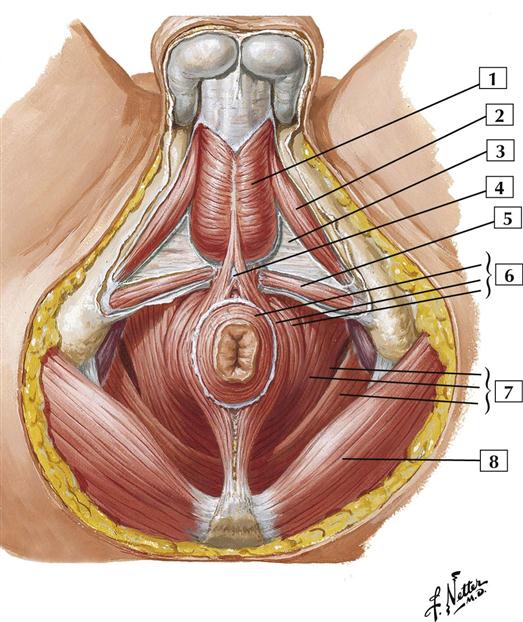
1. Bulbospongiosus muscle with deep perineal (investing, or Gallaudet’s) fascia removed
2. Ischiocavernosus muscle with deep perineal (investing, or Gallaudet’s) fascia removed
5. Superficial transverse perineal muscle with deep perineal (investing, or Gallaudet’s) fascia removed
6. Parts of external anal sphincter muscle (Subcutaneous; Superficial; Deep)
7. Levator ani muscle (Pubococcygeus; Puborectalis; Iliococcygeus)
Comment:
The muscles of the male perineum are skeletal in nature and are innervated by the pudendal nerve and its branches. Many of these muscles have attachments to the central tendon of the perineum (perineal body). The perineal body is a midline structure located just anterior to the anal canal and just behind the bulb of the penis.
This illustration shows the subdivision of the diamond-shaped perineum into an anterior urogenital triangle and a posterior anal triangle. An imaginary horizontal line connecting the 2 ischial tuberosities divides the perineum into these 2 descriptive triangles.
The ischiocavernosus and bulbospongiosus muscles cover the crus of the penis (corpus cavernosum) and the bulb of the penis (corpus spongiosum). These bodies are the erectile tissue of the penis.
Stay updated, free articles. Join our Telegram channel

Full access? Get Clinical Tree


