PEComa
Elizabeth A. Montgomery, MD
Key Facts
Terminology
PEComa is abbreviation for neoplasms with perivascular epithelioid cell differentiation
Mesenchymal neoplasms composed of distinctive perivascular epithelioid cells
Includes angiomyolipoma (AML), clear cell “sugar” tumor of lung (CCST), lymphangioleiomyomatosis (LAM), clear cell myomelanocytic tumor of falciform ligament/ligamentum teres (CCMMT)
In many respects, PEComas are simply angiomyolipomas without fat
Etiology/Pathogenesis
AML, CCST, and LAM are associated with tuberous sclerosis but not other types
Clinical Issues
Most are benign
Microscopic Pathology
PECs consist of epithelioid to spindled cells arranged around vessels extending outward radially
Clear to granular, lightly eosinophilic cytoplasm and round to oval nuclei with small nucleoli
Myoid component with densely eosinophilic cytoplasm
Adipose tissue component present in lesions termed AML
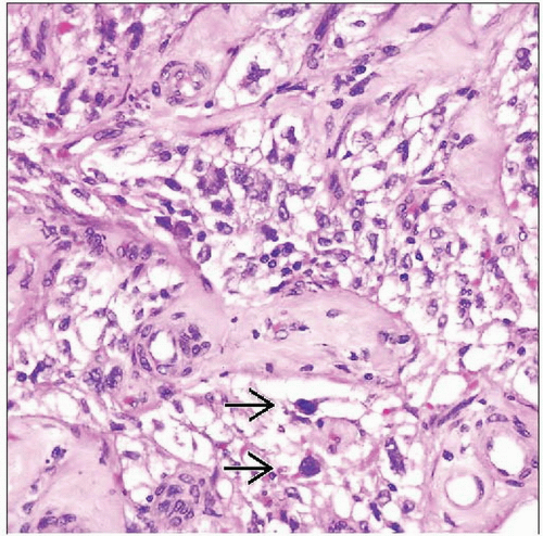 Hematoxylin & eosin shows a PEComa with epithelioid cells proliferating around vessels. There are scattered large nuclei  in this field but no mitoses. in this field but no mitoses. |
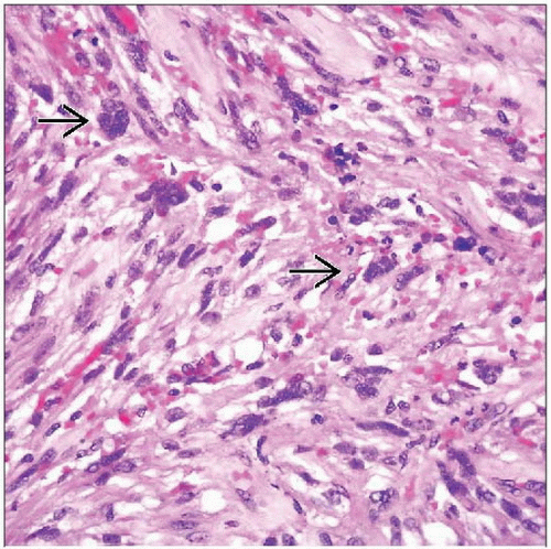 Hematoxylin & eosin shows a more myoid portion of a PEComa with more prominent cytoplasmic eosinophilia than in the previous image. This example also has scattered pleomorphic nuclei  . . |
TERMINOLOGY
Abbreviations
Perivascular epithelioid cell (PEC)
Thus neoplasms are termed “PEComa”
Synonyms
Extrapulmonary sugar tumor
Perivascular epithelioid cell tumor
Monotypic epithelioid angiomyolipoma
Definitions
Mesenchymal neoplasms composed of distinctive perivascular epithelioid cells (PEC) category includes
Angiomyolipoma (AML)
Clear cell “sugar” tumor of lung (CCST)
Lymphangioleiomyomatosis (LAM)
Clear cell myomelanocytic tumor of falciform ligament/ligamentum teres (CCMMT)
In many respects, PEComas are simply angiomyolipomas without fat
Subset displays overt histologic features of malignancy and malignant clinical behavior
ETIOLOGY/PATHOGENESIS
Association with Tuberous Sclerosis
Genetic alterations of tuberous sclerosis complex (TSC), losses of TSC1 (9q34) or TSC2 (16p13.3) genes
Autosomal dominant
Benign tumors of brain (most common), kidneys, heart, eyes, lungs, and skin
Name comes from characteristic tuber or potato-like nodules in brain, which calcify with age and become hard or sclerotic
AML, CCST, and LAM are associated with tuberous sclerosis but not other types
CLINICAL ISSUES
Epidemiology
Incidence
AML, CCST, LAM are rare
Other PEComas extremely rare
Age
CCMMT typically encountered in girls in late childhood
Most others seen in adults 50-60 years old
AML detected in younger patients in setting of tuberous sclerosis
Gender
Marked overall female predominance
Site
Reported in multiple sites
Kidney, liver, falciform ligament, deep soft tissues of extremities, skin, uterus, vulva, heart, gallbladder, gastrointestinal tract
Presentation
CCMMT presents as painful abdominal mass
Uterine examples manifest as uterine bleeding
Most other categories of PEComas present as painless masses
Brain tumors in patients with tuberous sclerosis present with seizures, developmental delay, behavioral problems
Treatment
Surgical excision
Prognosis
Most are benign
Rare documented examples of malignancy
Usually not in AML, LAM, or CCST types
Malignant examples behave as aggressive sarcomas
MICROSCOPIC PATHOLOGY
Histologic Features
Stay updated, free articles. Join our Telegram channel

Full access? Get Clinical Tree


