QUESTION 13.3
A. de Quervain (subacute) thyroiditis
B. Hashimoto thyroiditis
C. Papillary carcinoma
D. Riedel thyroiditis
E. Subacute lymphocytic (painless) thyroiditis
4. A patient with Graves disease receives antithyroid drugs and propranolol prior to subtotal thyroidectomy. Which of the following histopathologic aspects will regress following such treatment?
A. Follicular hyperplasia
B. Glandular enlargement
C. Lymphocytic infiltration
D. Presence of Hurthle cells
E. Presence of lymphoid follicles
5. Riedel thyroiditis is a pathologic process akin to:
A. Amyloidosis
B. de Quervain thyroiditis
C. Hashimoto thyroiditis
D. Progressive systemic sclerosis
E. Retroperitoneal fibrosis
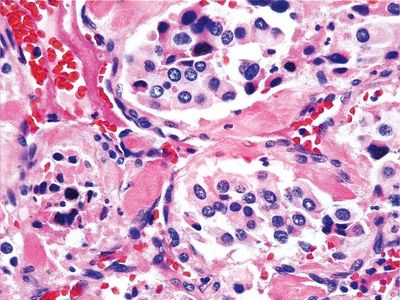
6. A thyroid nodule is excised and demonstrates the histology shown in this picture. Which of the following is the most characteristic genetic alteration of this lesion?
QUESTION 13.6
A. LOH on 10q and 3p
B. Mitochondrial gene mutations
C. p-53 mutations
D. RET oncogene rearrangements
E. Translocation t(2;3) leading to expression of PPAR-g chimeric protein
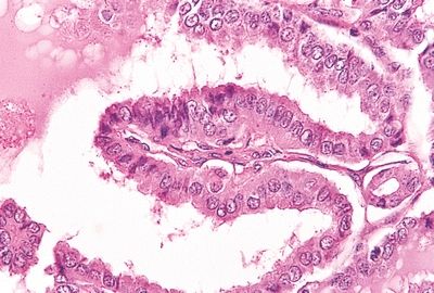
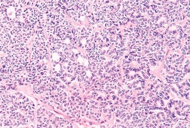
7. The variant of papillary thyroid carcinoma (PTC) with the most aggressive behavior is shown in this photomicrograph. Which one is it?
QUESTION 13.7
A. Diffuse sclerosing
B. Follicular
C. Microcarcinoma
D. Tall cell
E. Warthin-like
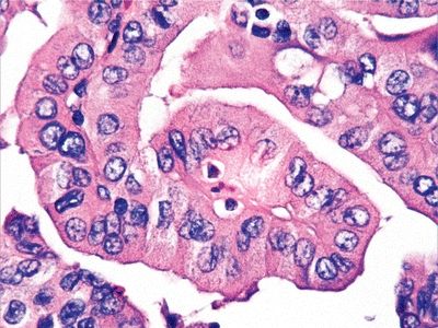
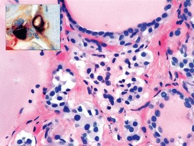
8. This picture shows a variant of PTC that has been found in 30% of patients who developed papillary carcinoma following the Chernobyl accident. Which variant is it?
QUESTION 13.8
A. Clear cell
B. Diffuse sclerosis
C. Follicular
D. Oxyphilic
E. Solid
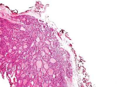
9. These photos show the external surface and cut surface of a very enlarged thyroid that was surgically removed. Histologic examination reveals nodules consisting of colloid-filled follicles of varying size. The nodules are separated by scar tissue containing hemosiderin-laden macrophages. Which of the following is the most likely diagnosis?
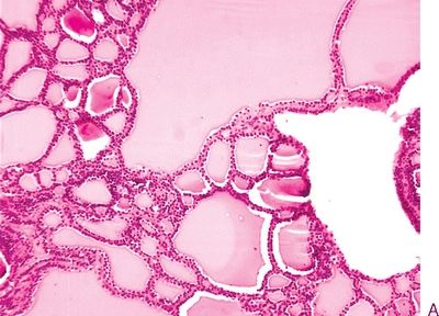
QUESTION 13.9
A. Diffuse simple goiter
B. Multinodular goiter
C. Subacute thyroiditis
D. Thyroid adenoma
E. Thyroid cyst
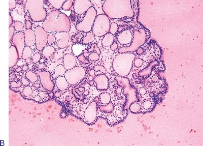
10. This photomicrograph demonstrates the histologic features of an isolated thyroid nodule. The most important step in pathologic examination of this specimen is:
QUESTION 13.10
A. Determination of Ki-67 labeling index
B. DNA ploidy analysis
C. Identification of areas of oncocytic change
D. Measurement of main diameter
E. Microscopic evaluation of capsule
A. Atypical
B. Colloid
C. Hyalinizing trabecular
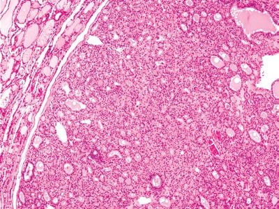
12. This photomicrograph shows an invasive thyroid tumor. Which of the following is a true statement regarding this tumor?
QUESTION 13.12
A. The brain is the most common target of hematogenous metastasis.
B. Lymphatic metastasis is the usual route of spread.
C. It is most commonly multiple and occult.
D. Prognosis is related to capsular and vascular invasion.
E. Prognosis is related to the presence of ras mutations.
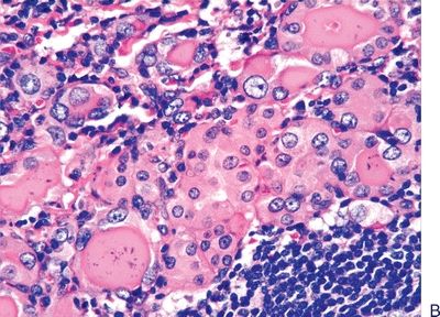
13. The thyroid tumor shown in this gross photograph is composed of large cells with well-defined cell borders, abundant granular cytoplasm, and large nucleus with prominent nucleolus. The cytoplasm of such cells is filled with:

QUESTION 13.13
A. Glycogen
B. Lipids
C. Lysosomes
D. Mitochondria
E. Thyroglobulin
14. A thyroid biopsy reveals the tumor shown in this photomicrograph. Which of the following immunohistochemical stains is likely to be positive on the eosinophilic material present in the stroma?
QUESTION 13.14
A. Calcitonin
B. Carcinoembryonic antigen (CEA)
C. Keratin
D. Synaptophysin
E. Thyroglobulin
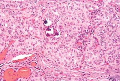
15. A 20-year-old man with a diagnosis of multiple endocrine neoplasia (MEN) type IIa undergoes prophylactic total thyroidectomy. On histologic examination, clusters of epithelial cells similar to those shown in this photomicrograph are found in the upper third of the gland. Which of the following thyroid neoplasms may arise from these cells?
QUESTION 13.15
A. Anaplastic carcinoma
B. Follicular carcinoma
C. Medullary carcinoma
D. Papillary carcinoma
E. All of the above
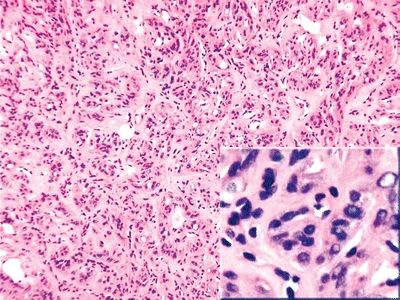
16. A 70-year-old woman undergoes thyroidectomy for removal of a large infiltrative tumor. Local lymph node metastases are found. The microscopic features of the mass are shown in this picture. Which of the following is the most likely diagnosis?
QUESTION 13.16
A. Anaplastic carcinoma
B. Follicular carcinoma
C. Medullary carcinoma
D. Papillary carcinoma
E. Poorly differentiated carcinoma
17. An elderly patient undergoes surgery for removal of a thyroid mass. The surgical specimen reveals a poorly circumscribed hemorrhagic tumor extending beyond the thyroid into adjacent tissues. The histologic appearance of the tumor is demonstrated by this picture. The tumor also contains regions with a spindle cell morphology and areas of increased vascularity. The tumor shows diffuse vimentin and focal keratin immunoreactivity. Which of the following is the most likely diagnosis?

QUESTION 13.17
A. Anaplastic carcinoma
B. Angiosarcoma
C. Giant cell tumor
D. Hemangiopericytoma
E. Malignant fibrous histiocytoma
18. Which of the following statements is correct regarding FNA of the thyroid gland?
A. Accuracy of FNA is operator independent.
B. Aspirate is nondiagnostic if it has fewer than 6 clusters of 10 to 15 follicular cells.
C. FNA has doubled the number of patients undergoing thyroid surgery.
D. Thyroid FNA is diagnostic, but never therapeutic.
E. Ultrasound guidance does not affect specimen adequacy.
19. This picture shows a fine needle aspirate from a symmetrically enlarged thyroid. Which of the following is the most likely diagnosis?
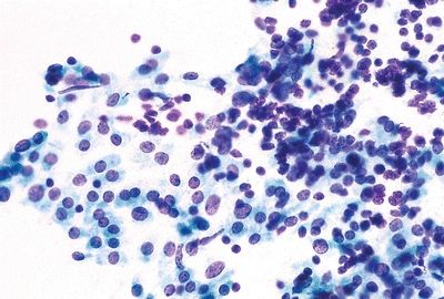
QUESTION 13.19
A. Chronic lymphocytic thyroiditis
B. Hurthle cell neoplasm
C. Lymphoma
D. Nodular goiter
E. Papillary carcinoma
20. This picture shows a fine needle aspirate from a well-circumscribed thyroid nodule. Which of the following is the most likely diagnosis?
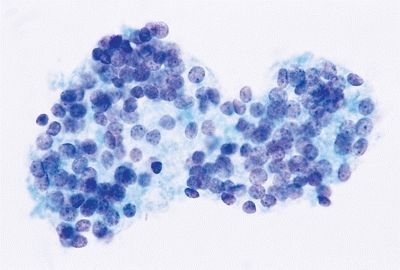
QUESTION 13.20
A. Chronic lymphocytic thyroiditis
Stay updated, free articles. Join our Telegram channel

Full access? Get Clinical Tree


