QUESTION 34.2
A. Adenomatous polyp
B. Hamartomatous polyp
C. Hyperplastic polyp
D. Juvenile polyp
E. Normal colonic mucosa
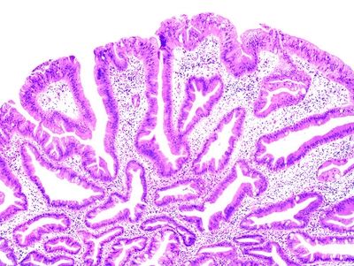
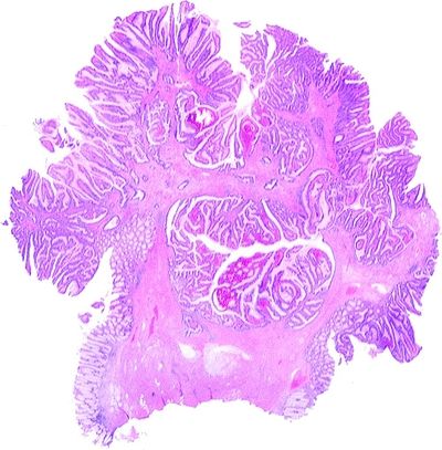
3. This photomicrograph shows a 1.6 cm pedunculated polyp excised from the sigmoid colon. What type of polyp is it?
QUESTION 34.3
A. Hyperplastic polyp
B. Sessile serrated adenoma
C. Traditional serrated adenoma
4. A polyp is resected from the proximal portion of the jejunum. Histologically, it consists of glands containing both absorptive and goblet cells supported by a stalk. Within the stalk, there are smooth muscle bundles continuous with the underlying muscularis mucosae. Which of the following additional features is most likely to be observed in this patient?
A. Innumerable adenomatous polyps of the colon
B. Malignant brain tumors
C. Osteomas of the mandible and long bones
D. Perioral and perianal pigmentation
E. Soft tissue tumors
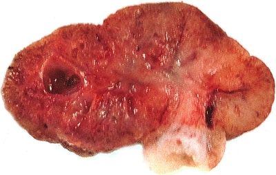
5. A 15-year-old boy undergoes excision of a solitary rectal polyp, which is shown in this picture. Which of the following types of polyp is this?
QUESTION 34.5
A. Adenomatous polyp
B. Hyperplastic polyp
C. Inflammatory polyp
D. Juvenile polyp
E. Traditional serrated adenoma
6. Proliferation of sarcoma-mimicking stroma is occasionally seen in which of the following polyps?
A. Angiomatous polyp
B. Cronkhite-Canada syndrome
C. Inflammatory polyp
D. Inflammatory fibroid polyp
E. Lymphoid polyp
7. Which of the following types of colonic polyps is associated with familial polyposis and found in up to one-third of adult individuals at autopsy?
A. Hamartomatous polyp
B. Hyperplastic polyp
C. Juvenile polyp
D. Tubular adenoma
E. Villous adenoma
A. Sessile serrated adenoma with dysplasia
B. Traditional serrated adenoma
C. Tubular adenoma with low-grade dysplasia
D. Tubular adenoma with high-grade dysplasia
E. Villous adenoma
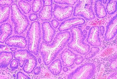
9. Which of the following characteristics is associated with the type of polyp illustrated in this photomicrograph?
QUESTION 34.9
A. Associated with early BRAF mutations
B. Centrally ulcerated mass in the sigmoid
C. Found usually in young patients
D. Low propensity for malignant degeneration
E. Sessile soft mass in the rectum
10. The definition of intramucosal carcinoma should be reserved for adenomas with:
A. Epithelial misplacement
B. Height less than two times that of normal mucosa
C. High-grade dysplasia
D. Invasion into the submucosa
E. Invasion into the lamina propria
11. Which of the following is correct about nonpolypoid adenomas?
A. Contain dysplasia at higher rate than polypoid adenomas
B. Incidence of submucosal invasion is lowest in the depressed variant
C. Majority of them are villous adenomas
D. Their height is usually between 3 and 5 mm
E. Their incidence is less than 5% of all adenomas
12. In an endoscopically removed malignant polyp, which of the following features should be evaluated and reported?
A. Grade of cancer
B. Presence or absence of lymphatic–venous invasion
C. Status of resection margin
D. All of the above
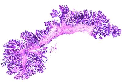
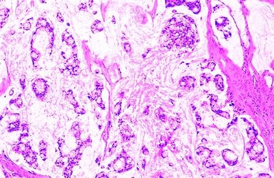
13. This photomicrograph shows a whole-mount view of an endoscopically resected polypoid adenoma. Which of the following is the most likely diagnosis?
QUESTION 34.13
A. Adenoma with pseudoinvasion
B. Hamartomatous polyp
C. Juvenile polyp
D. Malignant polyp
E. Traditional serrated adenoma
14. A 28-year-old man undergoes colonoscopic screening for family history of a polyposis syndrome. He is found to have more than 100 colorectal polyps, a germline mutation in the APC gene, and an osteoma. Which of the following lesions is known to develop in 10% of these patients?
A. Adnexal skin tumors of sebaceous differentiation
B. Desmoid tumor
C. Endometrial adenocarcinoma
D. Glioma
E. Medulloblastoma
15. Adenocarcinomas of the small bowel:
A. Are half as frequent as colonic adenocarci- nomas
B. Are most frequent in the ileum
C. Are not seen in association with Crohn disease
D. Develop most commonly in the duodenum
E. Usually arise from Brunner gland nodules
16. This photomicrograph shows a histologic type of colorectal carcinoma. Which of the following is it?
QUESTION 34.16
A. Adenocarcinoma, conventional
B. Adenosquamous carcinoma
C. Medullary carcinoma
D. Micropapillary carcinoma
E. Mucinous adenocarcinoma
F. Signet-ring cell carcinoma
G. Small cell cancer
H. Undifferentiated (medullary) carcinoma
17. A 70-year-old woman undergoes resection of a bulky, well-circumscribed mass in the proximal colon. This picture shows the histologic appearance of the tumor, which is diffusely infiltrated by lymphocytes. The tumor was found to have MSI-H and lack expression of CDX-2 or neuroendocrine markers. Which of the following is the most likely diagnosis?
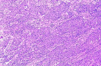
QUESTION 34.17
A. Conventional adenocarcinoma
B. Medullary carcinoma
C. Neuroendocrine carcinoma
D. Small cell cancer
E. Undifferentiated carcinoma
18. This picture shows a histologic variant of colorectal cancer, which is most commonly MSI-L or MSS and arises from traditional serrated adenoma. Which of the following is it?
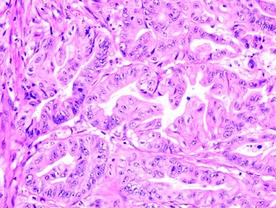
QUESTION 34.18
A. Conventional adenocarcinoma
B. Medullary carcinoma
C. Micropapillary carcinoma
D. Serrated adenocarcinoma
E. Undifferentiated carcinoma
19. Which of the following is tumor budding?
A. Feature that influences the tumor grade
B. Growth pattern specific of tumors with infiltrative borders
C. Migration of cells into the stroma at the infiltrating edge
D. Parameter without prognostic implications
20. Which of the following pathologic features is predictive of high microsatellite instability (MSI-H) in colorectal cancer (CRC)?
A. Crohn-like reaction
B. High grade
C. Medullary type
D. Mucinous and signet-ring morphology
E. Right-sided location
F. Serrated morphology
G. Tumor-infiltrating lymphocytes (TILs)
H. All of the above
21. Which of the following features of CRC is a well-established prognostic factor and is of sufficient importance to modify TNM stage?
Stay updated, free articles. Join our Telegram channel

Full access? Get Clinical Tree


