Patient Story
A 29-year-old obese woman presented with chronic headaches that were worse in the morning or while lying down. She denied nausea or other neurologic symptoms. She had no other medical problems and took no medications. On examination, she had a visual acuity of 20/20 in both eyes, bilateral papilledema (Figure 22-1), no spontaneous venous pulsations (SVPs), and no other neurologic signs. She had a brain MRI showing no mass or hydrocephalus, and elevated intracranial pressure measured by lumbar puncture. She was diagnosed with idiopathic intracranial hypertension and was followed closely for any changes in her vision. She was started on acetazolamide and assisted with a weight-loss program. Her symptoms resolved over the course of 18 months.
Figure 22-1
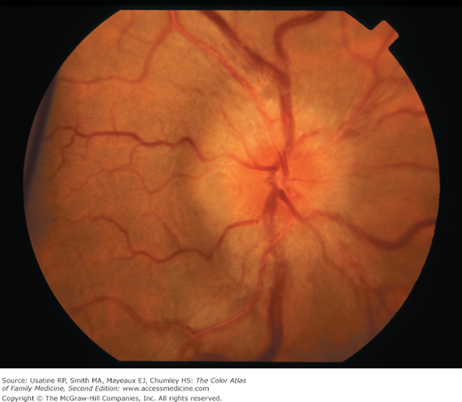
Papilledema from increased intracranial pressure. The optic disc is elevated and hyperemic with engorged retinal veins. The entire optic disc margin is blurred. Optic neuropathies can also have blurring of the entire disc margin, but often, only part of the disc is blurred. (Courtesy of Paul D. Comeau.)
Introduction
The term papilledema refers specifically to optic disc swelling related to increased intracranial pressure. When no localizing neurological signs or space-occupying lesion is present, idiopathic intracranial hypertension (IIH) is a likely cause in patients younger than age 45 years, especially obese women. Patients with IIH usually present with daily pulsatile headache with nausea and often have transient visual disturbances and/or pulsatile tinnitus. Patients often report a “whooshing” sound that they hear. Bilateral papilledema and visual field defects on a perimetry test are found in almost all patients. Elevated opening pressure on lumbar puncture is required for the diagnosis.
Epidemiology
Etiology and Pathophysiology
Diagnosis
Patients with papilledema should undergo imaging, preferably MRI, followed by lumbar puncture. IIH is a diagnosis of exclusion with the following criteria:3
- Signs and symptoms of increased intracranial pressure (headache, transient visual disturbances, papilledema).
- Normal neurologic examination, except a sixth nerve palsy may be present. This will lead to a complaint of diplopia.
- Elevated intracranial pressure is present, as measured by lumbar puncture opening pressure greater than 250 mm of water in the lateral decubitus position, with normal CSF on microscopic examination.
- No evidence of mass, hydrocephalus, or vascular lesions by MRI.
- No other identifiable cause of increased intracranial hypertension.
- More than 90% of patients with IIH are obese women of childbearing age. Look for a different diagnosis in children, men, and older patients.3
- Headaches and visual changes are the most common symptoms.
- Difficulty in thinking or concentrating is frequently reported. New studies indicate cognitive impairment, particularly in learning and memory.4
- SVP are retinal vein pulsations at the optic disc and are typically absent in IIH patients. SVP are seen in 90% of patients with normal intracranial pressure, and are absent when the CSF pressure is above 190 mm Hg. As the CSF pressure may be transiently normal in IIH, the presence of SVP does not preclude IIH, but indicates that the CSF pressure is normal at that moment.5
Papilledema is bilateral in the overwhelming majority of cases (see Figures 22-1
Stay updated, free articles. Join our Telegram channel

Full access? Get Clinical Tree


