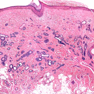
• Dermal-based adnexal tumor with variably dilated eccrine ducts
Papillary Eccrine Adenoma Low-power examination shows a dermal adnexal tumor with variably sized ductal and glandular spaces associated with a fibrous stroma.
Papillary Eccrine Adenoma at Higher Magnification On high magnification, the glands show low-cuboidal eccrine cells and areas with cribriforming and papillary projection of variable complexity . (Courtesy J. McNiff, MD.)
Papillary Eccrine Adenoma at High Magnification High magnification shows uniform, small cuboidal cells lining the glands and forming papillary projections . (Courtesy J. McNiff, MD.)
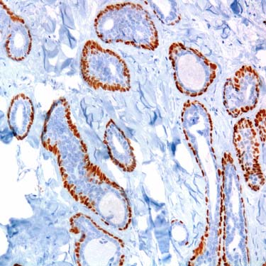
Immunohistochemistry for p63 p63 immunohistochemistry shows strong staining of the peripheral myoepithelial cell layer, consistent with a benign tumor. (Courtesy J. McNiff, MD.)
TERMINOLOGY
Abbreviations
• Papillary eccrine adenoma (PEA)
Synonyms
• Tubulopapillary hidradenoma with eccrine differentiation
Definitions
• Rare benign adnexal tumor with evidence of eccrine derivation/differentiation
In the past, many cases with apocrine features were included in this category
However, most authorities now separate PEAs from tubular apocrine adenomas
CLINICAL ISSUES
Epidemiology
• Incidence
Rare
• Sex
Women
• Ethnicity
More common in Africans/African Americans
Only gold members can continue reading. Log In or Register to continue


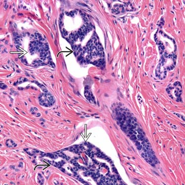
 and areas with cribriforming and papillary projection of variable complexity
  . (Courtesy J. McNiff, MD.)
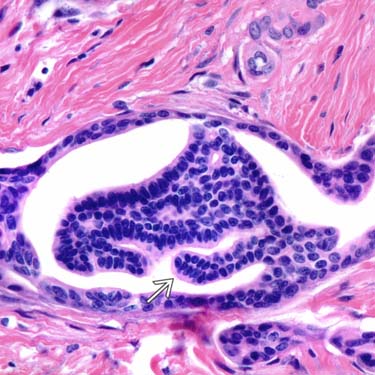
 . (Courtesy J. McNiff, MD.)