Ossifying Fibromyxoid Tumor
Cyril Fisher, MD, DSc, FRCPath
Key Facts
Terminology
Encapsulated tumor of small polygonal cells in fibromyxoid stroma with metaplastic ossification
Majority benign
Occasional atypical or malignant variants
Etiology/Pathogenesis
Schwann cell, chondroid, and myoepithelial lineages variously suggested
Clinical Issues
Most common in extremities
Mostly subcutaneous
Local recurrence in up to 27% of cases, often after long interval
Rare cases metastasize to lung
Extremities, head and neck
Microscopic Pathology
Well-defined thick fibrous capsule
Ovoid (rarely spindled) uniform cells in cords, small nests, or sheets
Partial rim of bone in 60-80%
Atypical variant
Mitoses > 2 per 50 high-power fields
High nuclear grade
Intratumoral osteoid formation, necrosis
Ancillary Tests
S100 protein typically positive
GFAP, desmin, SMA, cytokeratin sometimes positive
Ultrastructure shows external lamina and interdigitating cell processes
No consistent genetic abnormalities
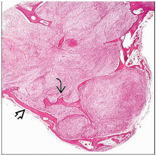 Hematoxylin & eosin shows an ossifying fibromyxoid tumor with variable cellularity and a rim of mature bone  with septal extensions with septal extensions  into the tumor mass. into the tumor mass. |
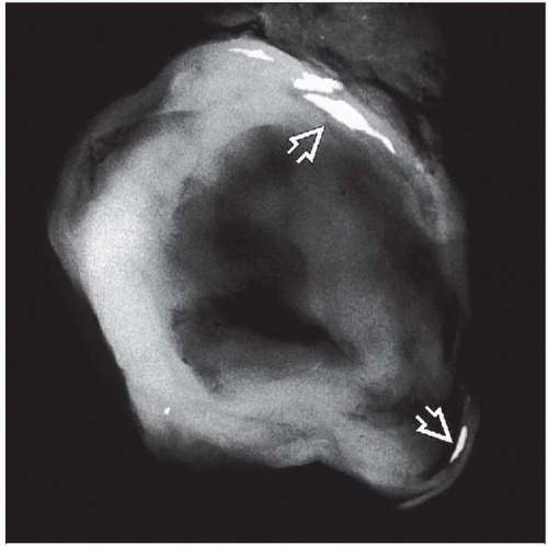 Specimen radiograph shows focal ossification  in the capsule of an ossifying fibromyxoid tumor. in the capsule of an ossifying fibromyxoid tumor. |
TERMINOLOGY
Abbreviations
Ossifying fibromyxoid tumor (OFMT)
Synonyms
Ossifying fibromyxoid tumor of soft parts
Definitions
Encapsulated tumor of small polygonal cells in fibromyxoid stroma with metaplastic ossification
Majority benign; rare atypical or malignant variants
ETIOLOGY/PATHOGENESIS
Differentiation
Various lineages suggested
Neural (Schwann cell), chondroid, myoepithelial
CLINICAL ISSUES
Epidemiology
Incidence
Rare
Age
2nd-8th decades
Mean 50 years, rare in childhood
Gender
M:F = 2:1
Site
Mostly subcutaneous
Extremities, head and neck
Also trunk, retroperitoneum, mediastinum
Presentation
Painless mass
Treatment
Surgical approaches
Local excision


