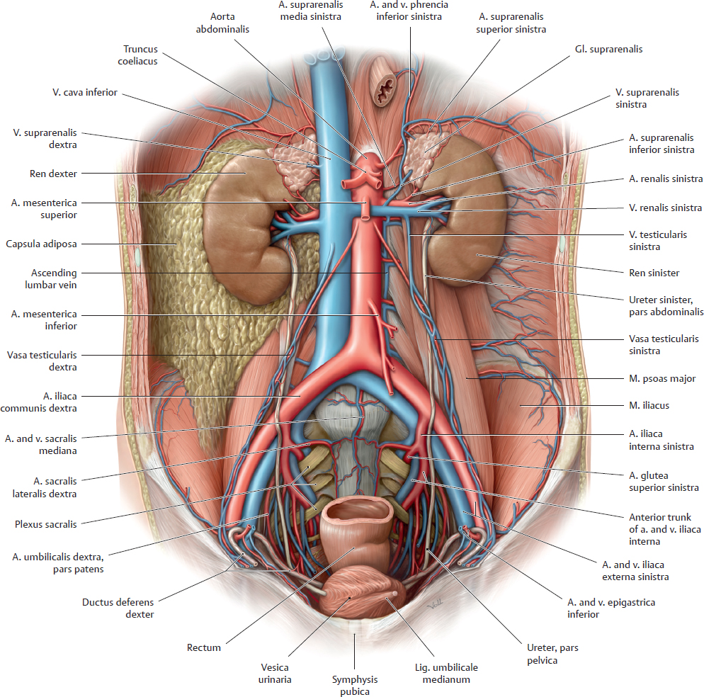
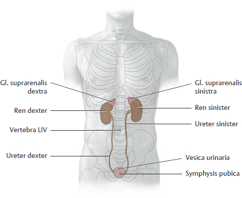
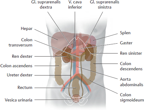
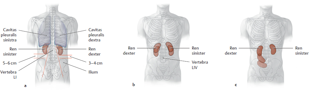
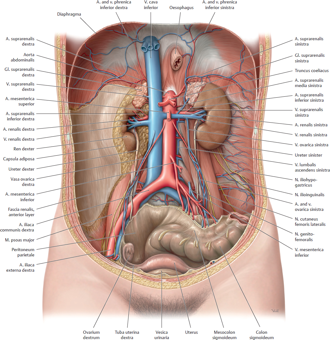
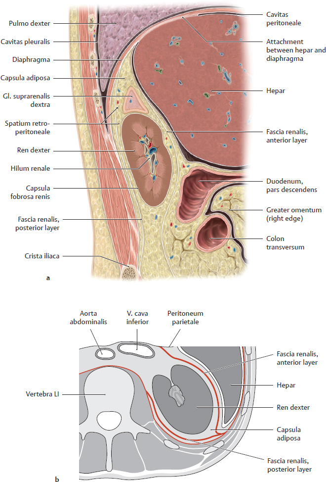
19 Organs of the Urinary System and their Neurovasculature A Projection of the renes and other urinary organs onto the skeleton Anterior view. The gll. suprarenales are also shown to aid orientation. The renes are located next to the columna vertebralis and are high enough that they overlap the eleventh and twelfth ribs. The hilum renale is situated at the L 1/L 2 level. Usually the right ren is somewhat lower than the left ren due to the space occupied by the hepar (see p. 374). The vesica urinaria is shown fully distended in the diagram. When empty, it is considerably smaller and is hidden behind the symphysis pubica. The ureteres descend in the retroperitoneum and open into the vesica urinaria from the posterior side. B Projection of the urinary organs onto the organs of the abdomen and pelvis Anterior view. Owing to its large size, the hepar displaces the right ren slightly inferiorly. The vesica urinaria is shown in a fully distended state. It is anterior to the rectum in the male and anterior to the uterus (not shown here) in the female. Because of this relationship, marked distention of the ampulla recti or enlargement of the uterus due to pregnancy exerts greater pressure on the vesica urinaria, creating an urge to urinate even when the vesica urinaria is not full. Urinary incontinence may develop due to pathological processes of longer duration, such as muscular tumors of the uterus (fibroids), or due to weakening of the vesica closure mechanism as a result of previous vaginal deliveries (descent of the muscular diaphragma pelvis). C Location of the renes, normal vs. pathological mobility a Posterior view. The cavitates pleurales overlap the renes posteriorly owing to the convexity of the diaphragma. Note that the right ren is lower than the left ren and is closer to the palpable crista iliaca. b, c Anterior view. The renes are located in the retroperitoneum just below the diaphragma. Hence they move passively with the diaphragma during respiratory excursions, moving inferiorly and slightly laterally during inspiration because of their oblique position (their poli inferiores point away from the spine, see oblique red lines in a). These passive movements may cause respiration-dependent pain in patients with renal disease. A pathological increase in renal mobility (“floating kidney,” see c) results from atrophy of the capsula adiposa that normally surrounds the renes and keeps them in a stable position. A wasting illness (e.g., metastatic tumors of varying origin) may cause such severe fatty atrophy that the renes descend to a lower level in the abdomen. As they are still tethered by the ureter and vascular stalk, this descent may kink the renal vessels or ureter and interfere with renal blood flow or urinary outflow. D The urinary organs in situ Anterior view of an opened female abdomen. The splen and gastrointestinal organs have been removed to the colon sigmoideum, and the oesophagus has been pulled slightly inferiorly. The capsula adiposa remains partially intact on the right side, removed on the left side. The renes and gll. suprarenales are incorporated into the retroperitoneum by the structural fat of this capsule. The moderately distended vesica biliaris is just visible above the symphysis pubica in front of the uterus. The peritoneum parietale has been removed to provide a clear view into the retroperitoneum. Note: The ureteres pass behind the ovarian vessels and in front of the iliac vessels as they descend in the retroperitoneum. These sites represent clinically important constrictions of the ureter where a stone from the pelvis renalis may become lodged (see B, p. 293). In most cases the renes are not oriented parallel to the coronal plane. The hilum renale, where the blood vessels and ureter enter and leave the renes, is directed anteromedially (see Ab, p. 284). Also, the renal poli superiores are closer together than the poli inferiores, so that the renes appear slightly “tilted” toward the midline. Thus the hilum renale also points slightly downward. A Position of the renes in the renal bed Right renal bed. a Sagittal section at approximately the level of the hilum renale, viewed from the right side. b Transverse section through the abdomen at approximately the L 1/L 2 level, viewed from above. The renal bed is located on each side of the spine in the retroperitoneum. It contains the renes, which are invested by a thin organ capsule (renal capsula fibrosa), and the gll. suprarenales, which are surrounded by the perirenal capsula adiposa that also encloses the kidneys. The capsula adiposa is thicker posteriorly than anteriorly. Note: Swelling of the ren (usually due to inflammation) may cause severe pain due to stretching of the capsula fibrosa. The capsula adiposa is surrounded by the fascia renalis, which separates it from its surroundings by two layers: • The anterior layer behind the peritoneum parietale (to which it is fused at some sites) • The posterior layer, which is partially attached to the fascia transversalis and muscular fasciae on the posterior trunk wall The fascia renalis, and thus the renal bed, is open inferiorly and medially to allow passage of the ureter and renal vessels. It is closed laterally and superiorly by fusion of the fascial layers. Because of this arrangement, inflammatory processes that are adjacent to the ren but within the fascia renalis tend to spread to the contralateral side or inferiorly and may spread into the pelvis. Note: The entire renal bed moves downward during inspiratory depression of the diaphragm, indirectly causing the ren and gl. suprarenalis to move as well. This differs from the hepar, which is attached to the diaphragma (area nuda) and is directly moved by diaphragma excursions. B Renal bed: Fasciae and capsulae of the renes
19.1 Overview of the Urinary Organs; The Renes in situ
19.2 Renes: Location, Shape, and Structure
Capsula fibrosa | Thin, firm connective-tissue capsule that closely invests each ren |
Perirenal capsula adiposa | Mass of fat that surrounds the renes and gll. suprarenales and completely occupies the renal bed; it is thickest lateral and posterior to the renes |
Fascia renalis | Connective-tissue fascial sac that encloses the perirenal capsula adiposa, portions of the pars abdominalis aortae and v. cava inferior close to the ren (see Ab), and the proximal ureter; subdivided into a thin anterior layer and a thick posterior layer (see Aa) |
C Structure and shape of the ren
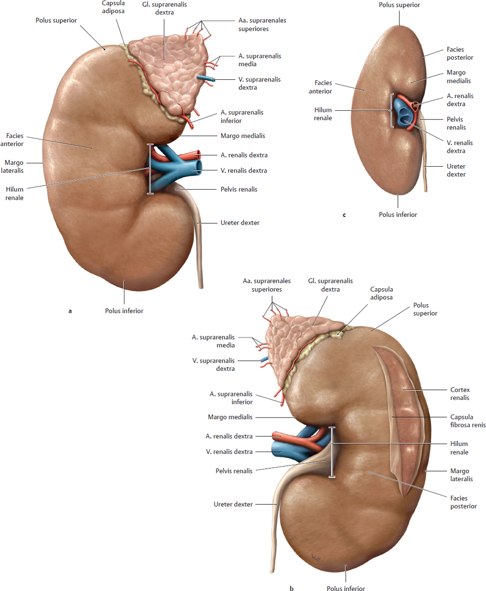
Anterior view (a), posterior view (b), and medial view (c) of the right ren. The gl. suprarenalis is left intact in a and b, and the ureter has been cut at the level of the polus inferior. The capsula fibrosa that directly invests the ren is intact in a and c and has been partially opened in b to display the underlying renal parenchyma. The sinus renalis (the deep space into which the hilum renalis opens) generally contains a certain amount of structural fat, and so the vascular structures and pelvis renalis are not exposed to view intraoperatively as they are in these drawings. The normal ren measures an average of 12 x 6 x 3 cm (L x W x T) and weighs 150–180 g. It has
• two poles (polus superior and inferior),
• two surfaces (facies anterior and posterior), and
• two borders (margo lateralis and medialis).
The margo medialis bears the hilum renale, where vascular structures and the ureter enter and leave the ren. The shallow surface grooves result from the embryonic lobulation of the ren. The hilar structures are usually arranged as follows from anterior to posterior (as shown in c): right v. renalis, right a. renalis, and right ureter.
Note: The a. renalis is usually posterior to the v. renalis because the right a. renalis passes to the right ren behind the v. cava inferior (where the vv. renales terminate), while the left v. renalis passes to the left ren i n front of the pars abdominis aortae (which gives origin to the aa. renales). The left a. renalis may also loop around the left v. renalis from above to occupy an anterior position. The ureter leaves the pelvis renalis (see p. 286) below the vessels and is usually somewhat posterior in relation to the blood vessels.
19.3 Renes: Architecture and Microstructure
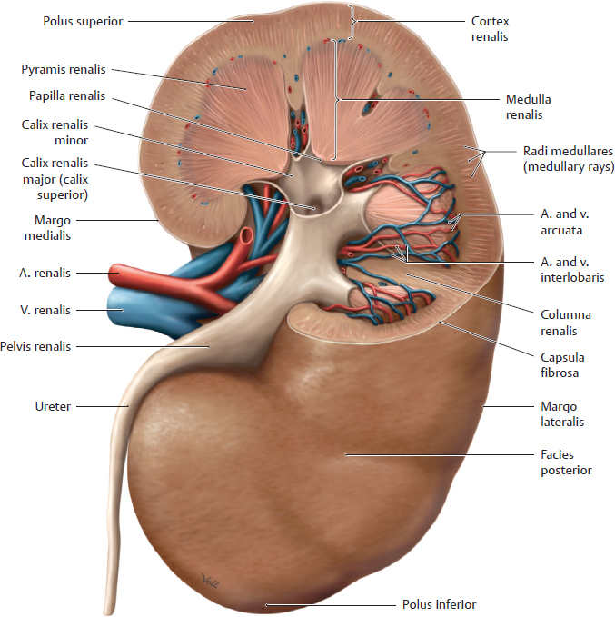
A Macroscopic structure of the ren
Posterior view of a right ren with the upper half of the ren partially removed. The renal parenchyma consists of an outer cortex renalis and inner medulla renalis:
• The cortex renalis is a relatively thin layer that lies beneath the capsula fibrosa and forms columns (columnae renales) that extend between the pyramides renales of the medulla. The cortex and columnae contain approximately 2.4 million corpuscula renalia (which contain the glomeruli, see B) as well as the proximal and distal renal tubuli (see C).
• The medulla renalis consists of approximately 10–12 pyramides renales. The bases of the pyramides are directed toward the cortex and capsula, while their apices converge toward the pelvis renalis. The medulla renalis mainly contains the ascending and descending limbs of the renal tubuli.
The pelvis renalis is described on p. 288.
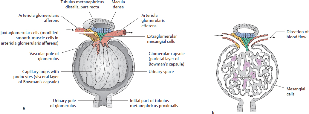
B Corpusculum renale
a With the capsula glomerularis opened; b In section.
The corpusculum renale is the interface between the blood vessels and the excretory portion of the urinary tract (see C). It consists of a central convoluted vascular loop, the glomerulus, and a bulbous envelope lined by squamous epithelial cells, the capsula glomerularis (Bowman’s capsule). Blood enters the glomerulus at the vascular pole of the corpusculum renale by flowing through the arteriola glomerularis afferens, and it leaves the glomerulus through the arteriola glomerularis efferens. The primary urine is formed within the corpusculum renale and drains through a tubular system at the urinary pole of the glomerulus. The initial portion of this tubular system that is connected to the capsula glomerularis is the tubulus metanephricus proximalis (see C)
Note: Specialized cells at the vascular pole of the corpusculum renale regulate the blood pressure that is necessary for ultrafiltration.
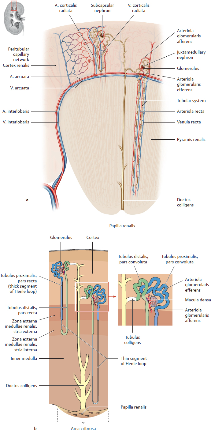
C Architecture of the renal vessels and intrarenal collecting system
a Renal vessels: Sectional view of a pyramis renalis with adjacent cortical areas. The intrarenal vascular and collecting systems are closely interrelated spatially and functionally. An ultrafiltrate from the blood (primary urine) drains into a microscopically small system of renal tubules. Blood flow to the ren (a) is supplied by aa. interlobares that pass along the sides of the pyramides renales from the hilum renale. Each a. interlobaris supplies two adjacent pyramides renales and the associated cortical zones (these branches are not shown). At the base of the pyramid, the a. interlobaris gives rise to the a. arcuata, from which the aa. interlobulares are distributed into the cortex renalis as far as the capsula fibrosa. The arteriolae glomerulares afferentes that arise from an a. interlobularis each supply one glomerulus. The arteriolae glomerulares efferentes that emerge from the glomerulus are still carrying blood at a high oxygen tension; they supply the cortex or medulla renalis.
b Intrarenal collecting system: The smallest functional unit of the kidney is the nephronum, which consists of the corpusculum renale (see B), tubuli, and ductus colligentes. There are approximately 1 million nephrona, which process approximately 1700 liters of blood daily to form approximately 170 liters of primary urine. This primary filtrate enters the tubular system at the urinary pole of the corpusculum renale and reaches the papilla renalis as the final urine, which drains into the calyceal system (approximately 1.7 liters/day). The tubular system consists of the tubuli proximales and distales (each with a pars convoluta and pars recta) and an intermediate tubule (with descending and ascending limbs). The intermediate tubule and the adjacent straight portions of the tubuli proximalis and distalis comprise the loop of Henle (ansa nephroni). While passing through the tubular system, substances contained in the filtrate (mainly water) are reabsorbed while other substances (e.g., ions) are secreted into the filtrate. This process yields the final urine, which passes through a tubulus colligens into a ductus colligens and drains through the papilla renalis into the caliceal system. It is conveyed from the calices renales and pelvis renalis to the ureter by peristalsis.
19.4 Pelvis Renalis and Urinary Transport
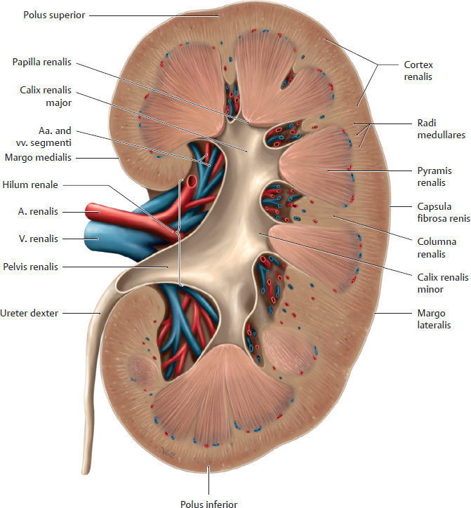
A Structure and shape of the pelvis renalis
Mid-longitudinal section through a right kidney, posterior view. The pelvis renalis lies posterior to the renal vessels and is continuous inferiorly with the ureter. It may vary in shape (see B). It is usually divided into two or three indistinctly separable calices renales majores, which further divide into calices renales minores. They encompass the tips of the papillae renales in such a way that urine drains from the papillae into the calix without entering the renal parenchyma. Smooth-muscle fibers in the calices, pelvis renalis, and ureter (for details about wall structure, see D), enable these structures to undergo peristaltic contractions (see C).
Note: Stones (see C, p. 293) that form in the calices or pelvis renalis may become so large that they more or less fill the cavity and assume its shape (caliceal stone, staghorn calculus).
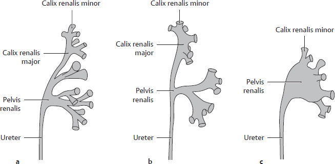
B Variations in the shape of the pelvis renalis
Anterior view of the left pelvis renalis. The pelvis renalis and ureter develop from an outgrowth of the ductus mesonephricus. This gemma ureterica grows from the bony pelvis toward the renal primordium and unites with it. Branching extensions of the pelvis renalis form the calices renales majores and minores. The calices renales majores in particular vary in number and shape: neighboring calices renales majores may fuse and “become incorporated” into the pelvis renalis. The pelvis renalis is found in basic forms along with transitional forms:
• Typus dendriticus (with extensive branching, also called linear) (a): very fine calices majores, narrow pelvis renalis;
• Transitional form (b);
• Typus ampullaris (c): indistinct calices majores with wide pelvis renalis; calices minores arise “directly” from the pelvis renalis.
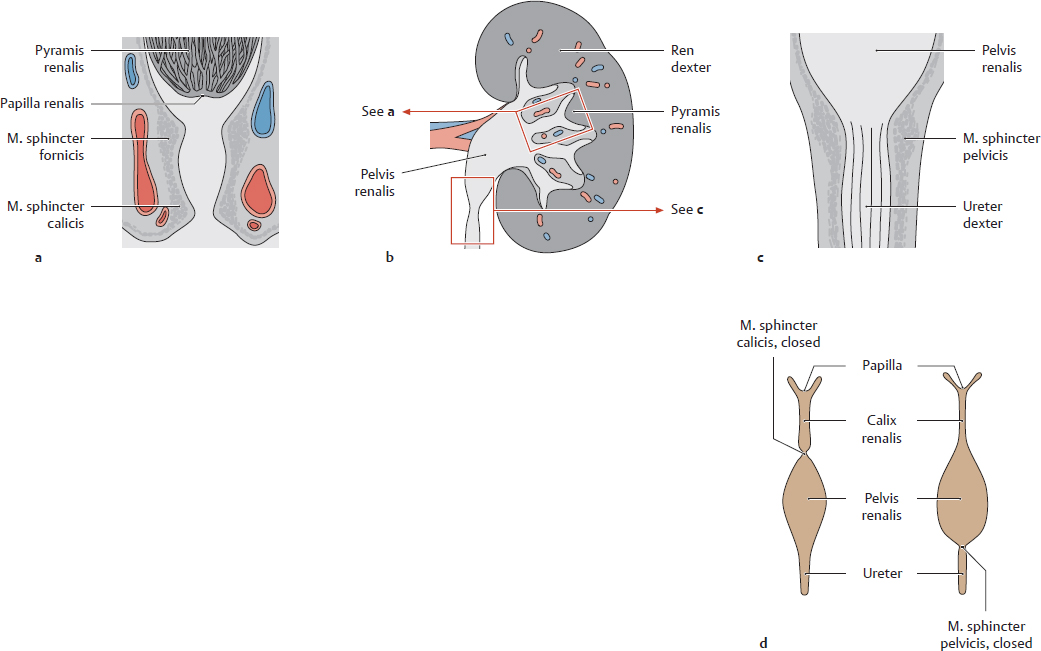
C Closure mechanisms of the calices renales and pelvis renalis; urinary transport (after Rauber and Kopsch)
Schematic representation of a ren (b) with magnified sections of a calix renalis (a) and pelvis renalis (c) and a dynamic functional diagram of the calix and pelvis renalis during urinary transport (d). Urine is transported by an active mechanism. The smooth musculature of the mm. sphincteres fornicis and calices (a) and the m. sphincter pelvicis (c) (the functional sphincter system) enables the wall of the calices and pelvis renales to contract in segments. These contractions are continuous with the peristaltic waves of the ureter, with the result that the urinary tract is never patent over its entire length but is patent in some portions and closed in others (d). This maintains a distal flow of urine from the tip of the papilla renalis into the calix renalis, through the pelvis renalis, into the ureter, and on toward the vesica urinaria while preventing the reflux of urine into the renes.
Note: If this active transport process is impaired (e.g., by renal stones or drugs that inhibit the ureteral muscles), urine may reflux into the ren and incite an inflammatory process in the pelvis renalis. The papillae, calices, and pelvis renales are often affected jointly by disease (e.g., inflammation) because of their close proximity to one another. One of the most common diseases is suppurative bacterial pyelonephritis (pyelo-, referring to the pelvis renalis, from Gr. pyelos—trough, tub, or vat).
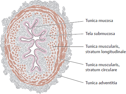
D Wall structure of the ureter
Transverse section through a ureter. A characteristic feature is the stellate lumen that appears in cross-section due to the longitudinal mucosal folds. As in the urethra and vesica urinaria, the ureteral tunica mucosa consists of a transitional epithelium of varying height (see p. 297). The smooth muscle (tunica muscularis) consists basically of a longitudinal and a circular layer. It is powerfully developed and shows a functionally spiral architecture (see E). When a renal stone enters the ureter, the smooth muscle in the ureteral wall undergoes powerful contractions in an effort to expel the stone, causing very severe pain (renal or ureteral colic). The colic may be relieved by drugs that suppress the activity of the parasympathetic nervous system, though this will also inhibit normal urinary transport to the vesica urinaria. The pelvis renalis is structurally analogous to the ureter, including the stellate shape of its lumen.
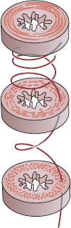
E Arrangement of the ureteral musculature (after Graumann, von Keyserlingk, and Sasse)
Schematic cross-sections at various levels of the ureter. The longitudinal and circular muscle layers of the ureter wall have a slightly oblique arrangement, forming a kind of spiral that propels urine toward the vesica urinaria by peristaltic contractions. Although the ureteres are richly innervated, the peristaltic contractions are instigated by spontaneously depolarizing smooth-muscle cells in the walls of the pelvis renalis. Peristaltic waves of contraction (with a speed of 2–3 cm/s) are propagated through direct electrical connections (gap junctions) between adjacent smooth-muscle cells. Autonomic motor innervation and local sensory reflexes serve to modulate this intrinsic activity. This mechanism may thus have some superficial similarities to the system that controls heartbeat.
19.5 Glandulae Suprarenales
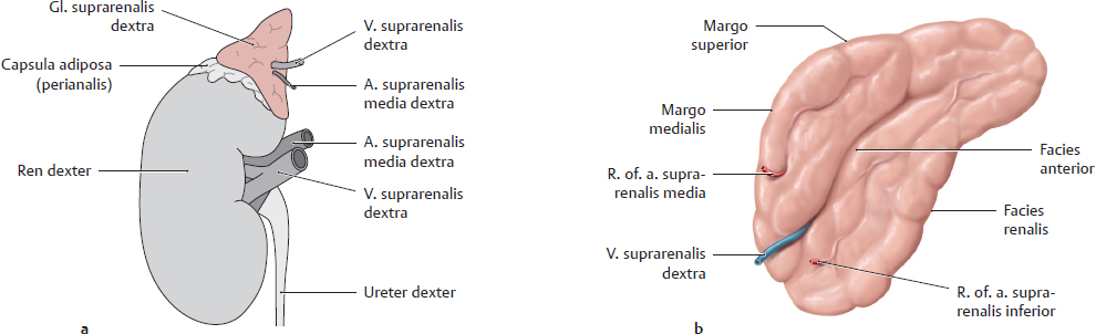
A Location and shape
a Location of the gl. suprarenalis on the right ren. b Isolated left gl. suprarenalis, anterior view.
The facies renalis of each v lies upon the polus superior of the associated ren. A thin layer of fat separates the gl. suprarenalis from the renal capsula fibrosa (making it easy to dissect the gland from the ren). The perirenal capsula adiposa, however, encompasses both the ren and the gl. suprarenalis.
Note: The entire gl. suprarenalis cannot be seen while in situ, and its true size is not appreciated until it has been detached from the ren. Portions descend on the posterior surface of the ren and are not visible in situ.
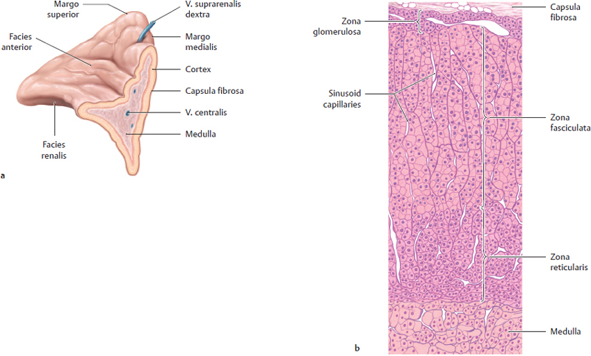
B Structure of the glandula suprarenalis
a Right gl. suprarenalis, cut open. b Histological section from a gl. suprarenalis.
The gl. suprarenalis consists of an outer cortex and an inner medulla (see a). The cortex glandulae suprarenalis is covered by a thin fibrous capsule and consists of three morphologically distinct zones (see b) in which adrenocortical hormones are produced and secreted into the bloodstream. These zones are, from outside to inside:
• Zona glomerulosa: mainly secretes mineralocorticoids (aldosterone)
• Zona fasciculata: mainly secretes glucocorticoids (hydrocortisone)
• Zona reticularis: sex hormones (estrogens and androgens)
Note: Loss or deficiency of both cortices glandularum suprarenalium leads to Addison disease, while hyperfunction of the cortex glandulae suprarenalis (or adrenocortical tumors) leads to Cushing syndrome. The medulla glandulae suprarenalis is essentially a completely different endocrine gland, of different origin, that happens to be anatomically (but also functionally) associated with the cortex glandulae suprarenalis. The cortex is derived embryonically from mesoderm lining the posterior abdominal wall. The medulla glandulae suprarenalis is, by contrast, a crista neuralis derivative, and thus has an ectodermal origin. The catecholamines epinephrine and norepinephrine are produced in the medulla glandulae suprarenalis and are released into the bloodstream. From a (neuro) functional standpoint, the medulla glandulae suprarenalis is less a gland than a ganglion sympathicum: presynaptic sympathetic neurons pass from the nn. splanchnici major and minor into the medulla glandulae suprarenalis. Because the gll. suprarenales are endocrine glands and ganglia sympathica in one, they can secrete both epinephrine and glucocorticoids (cortisone) in response to stress.
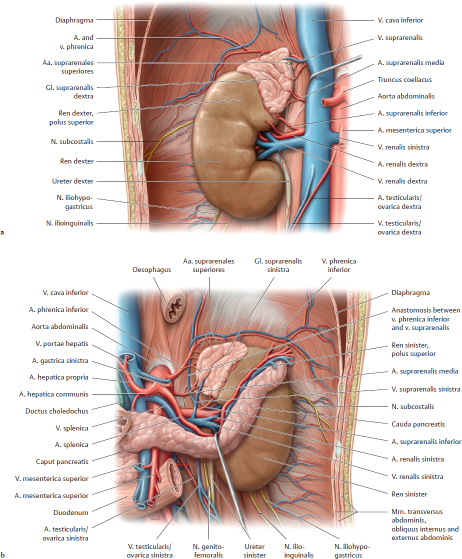
C Right and left glandulae suprarenales in situ
Anterior view of the right (a) and left (b) ren and gl. suprarenalis with the perirenal capsula adiposa removed. To demonstrate the vessels behind the gl. suprarenalis, the v. cava has been retracted medially in a and the pancreas has been retracted inferiorly in b. The principal differences between the two gll. suprarenales are as follows:
• The right gl. suprarenalis is often somewhat smaller than the left gl. suprarenalis, which frequently extends inferiorly to the hilum renale.
• The right gl. suprarenalis is pyramid-shaped while the large left gl. suprarenalis is more oblong. The right gl. suprarenalis is normally in contact with the v. cava inferior (retracted medially here), but the left gl. suprarenalis is not in contact with the aorta abdominalis.
• The v. suprarenalis dexter usually opens directly into the v. cava inferior, unlike the v. suprarenalis sinistra, which opens into the left v. renalis.
Note: The gll. suprarenales are richly vascularized because, as endocrine organs, they release their hormones directly into the bloodstream.