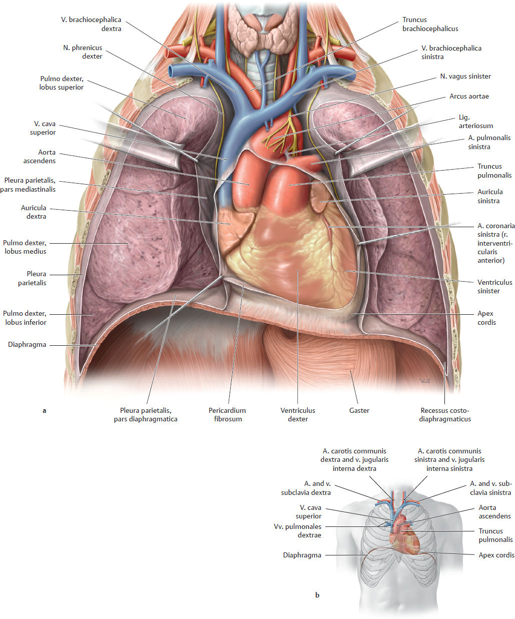
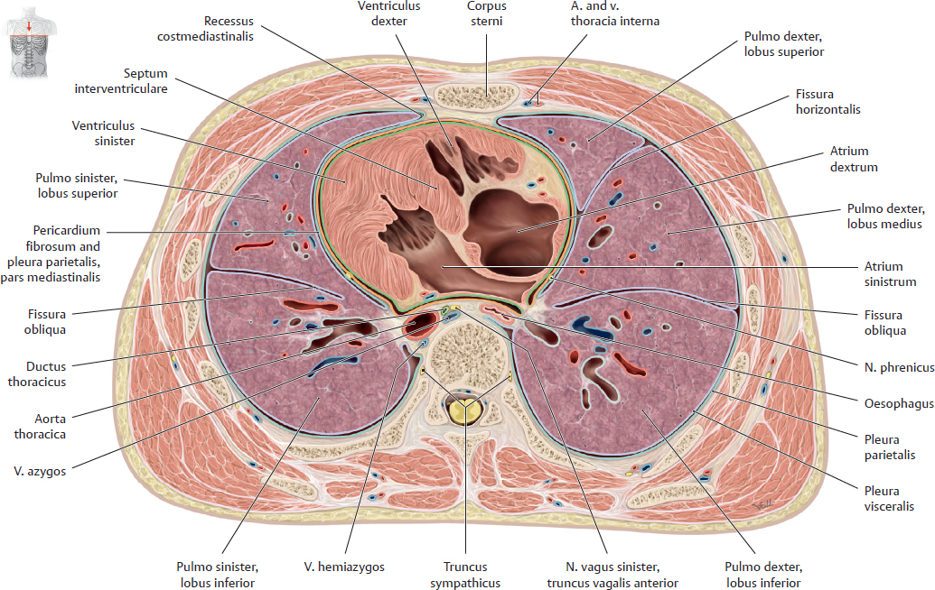
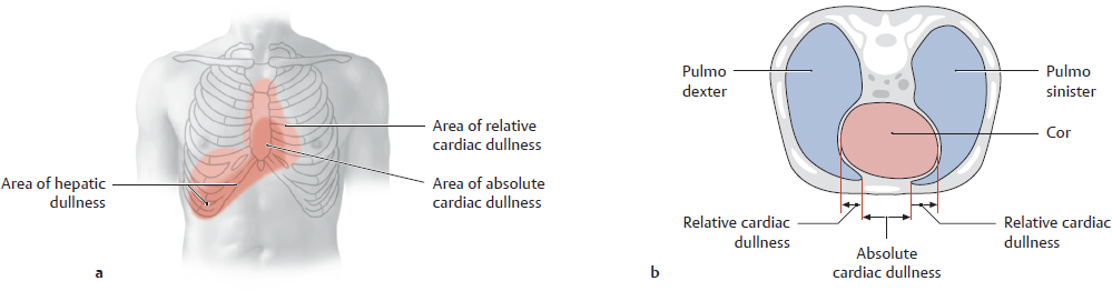
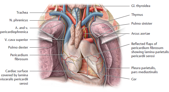
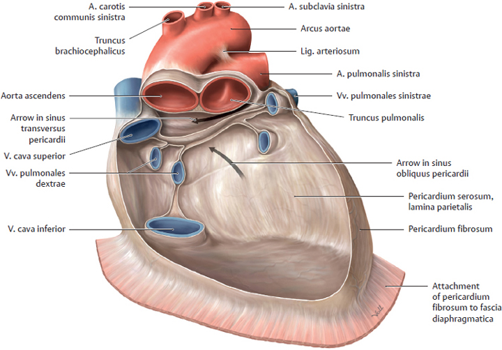
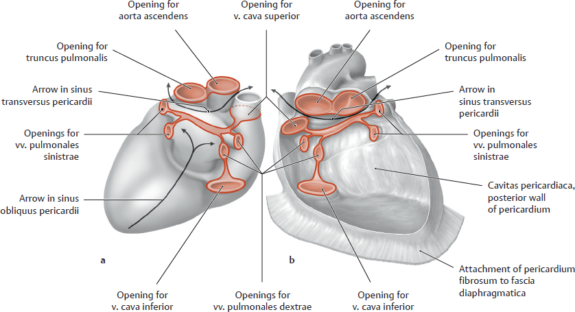
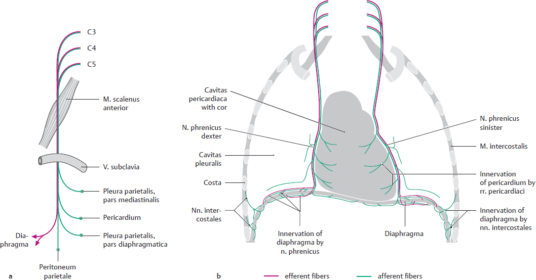
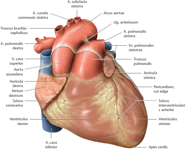
12. Organs of the Cardiovascular System and their Neurovasculature A The cor in situ, anterior view a Simplified illustration. The chest has been widely opened, and the cavitates pleurales and pericardium fibrosum have been cut open. The connective tissue has been removed from the mediastinum anterius to display the cor. Although the cavitates pleurales have been opened, the pulmones are not shown in a collapsed state. b Projection of the cor on the bony thorax. The cor lies within the pericardium, which is firmly attached to the diaphragma (see p. 90) but is mobile in relation to the pleura parietalis. A longitudinal axis drawn from the basis cordis to the apex cordis demonstrates that this “long axis” is directed forward and downward from right to left. Thus the cor, when viewed from the front, has an oblique orientation and is tilted counterclockwise within the chest. Along this axis it appears slightly “rolled” in a posterior direction. Thus the ventriculus dexter faces forward, as pictured here, while the ventriculus sinister is only partly visible. As a result, all of the great vessels cannot be seen even when the basis cordis is viewed from the front. The short vv. pulmonales are covered by the cardiac silhouette because they terminate at the atrium sinistrum, which is directed posteriorly. The auriculae dextra and dextra (atrial appendages) are clearly visible. The apex cordis points downward and to the left. Most of it is still covered by pericardium in this dissection. Its movement, called the apical beat, is palpable as a fine motion in the fifth spatium intercostale on the left midclavicular line (see p. 101). The thin tunica serosa of the lamina visceralis pericardii serosi (see p. 90) gives the surface of the heart a shiny appearance. Under this membrane are clusters of fatty tissue in which the coronary vessels are embedded. B The cor in situ, superior view Transverse section through the thorax at the level of T8 vertebra. Viewing the transverse section demonstrates the asymmetrical position of the cor in the mediastinum medium and its slight degree of physiologic counterclockwise rotation: the ventriculus sinister faces downward and to the left, while the ventriculus dexter faces forward and to the right. The ventriculus dexter thus lies almost directly behind the posterior wall of the sternum (with only the narrow mediastinum anterius intervening, see p. 71). The atrium sinistrum is in very close relationship to the oesophagus. The recessus costomediastinalis is interposed between the cor and sternum on the right and left sides. A relatively small space remains between the cor and columna vertebralis for the passage of neurovascular structures and organs: pars thoracica aortae, oesophagus, ductus thoracicus, vv. azygos and hemiazygos, and portions of the autonomic nervous system. Each pulmo bears an indentation from the cor called the impressio cardiaca. This impression is larger in the pulmo sinister than in the pulmo dexter because of the cor’s asymmetric position. The potential spaces between the pleural layers and the pericardium serosum are considerably smaller than pictured here. C Cardiac dullness on percussion of the chest Anterior view (a) and transverse section viewed from above (b). In contrast to the sonorous sound that is produced by the percussion of air-filled pulmo (see p. 128), the fluid-filled cor produces a flat sound on percussion known as cardiac dullness. The dullness may be absolute (at sites where there is no pulmo tissue to moderate cardiac dullness) or relative (at sites where pulmo tissue overlies the cor and adds resonance to the percussion sound). Accordingly, the area of absolute cardiac dullness is located between the chest wall and cor while the area of relative cardiac dullness is located over the recessus costomediastinales dexter and sinister, which contain small expansions of pulmo tissue (see B). Note: Cardiac dullness gives way to hepatic dullness in the epigastrium and right hypochondriac region due to the anatomical extent of the hepar (see a). The boundaries of the cor can be roughly estimated from the area of cardiac dullness because the sound characteristics at the cardiac borders contrast with the more resonant pulmo sounds. A Location of the pericardium in the thorax, anterior view The chest has been opened to display the pericardium, which is the dominant structure in the mediastinum inferius. It is attached inferiorly to the fascia diaphragmatica by connective tissue. Anteriorly, it is separated from the posterior surface of the sternum only by connective tissue of the mediastinum anterius (removed here, see p. 71). The pericardium is bounded laterally by the cavitates pleurales, from which it is separated by pars mediastinalis pleurae. B Cavitas pericardiaca and structure of the pericardium Anterior view of the empty pericardial sac. The pericardium consists of two layers, one within the other, that enclose and protect the heart: • Lamina parietalis. The lamina parietalis pericardii forms a sac with an outer surface, the pericardium fibrosum, composed of tough and indistensible connective tissue which is partially attached to the diaphragma. Its inner surface, facing the heart, is lined with a serous membrane. • Lamina visceralis (epicardium). This is a thin serous membrane which covers, and is firmly adherent to, the cor itself and the proximal parts of the great vessels. The two tunicae serosae of the laminae parietalis and visceralis are closely apposed, but move freely over one another, allowing a gliding motion during the heartbeat. These two tunicae serosae are referred to together as the pericardium serosum. In the locations where the lamina parietalis is folded back onto the lamina visceralis covering the vessels, two sinuses are formed (see arrows): • The sinus transversus pericardii located between the arteries and veins • The sinus obliquus pericardii located between the vv. pulmonales sinistrae and dextrae Note: Because the pericardium cannot expand significantly, bleeding into the cavitas pericardii (e.g., from a ruptured myocardial aneurysm) will place increasing pressure on the cor as the blood accumulates within the sac. This condition, called cardiac tamponade, seriously compromises the ability of the ventriculi to fill and pump blood, creating a threat of cardiac arrest. Similar problems may arise from inflammation of the pericardium (pericarditis). C Openings in the pericardium a Posterior view of the cor with the epicardium. b Anterior view of the “empty” cavitas pericardiaca. An empty pericardium typically has eight openings by which vessels enter and leave the cor: • One opening for the aorta ascendens • One opening for the truncus pulmonalis • Two openings for the two venae cavae • Up to four openings for the four vv. pulmonales D Innervation of the pericardium a Somatosensory and somatomotor components of the n. phrenicus b Sensory and motor distribution of the n. phrenicus Like the serous membranes of the diaphragma (pars diaphragmatica pleurae and peritoneum parietale), the pericardium (pericardium fibrosum and lamina parietalis of pericardium serosum) is innervated by the nn. phrenici, which arise from segmenta cervicales C3–5 medullae spinalis. A Cor, facies sternocostalis Anterior view. The cor is a muscular hollow organ shaped approximately like a flattened cone. It consists topographically of a base, apex, and three surfaces: • The basis cordis, which is occupied by entering and emerging vessels, is directed superiorly, posteriorly, and to the right. • The apex cordis is directed inferiorly, anteriorly, and to the left. • The surfaces are described as facies anterior (sternocostalis), facies posterior, and facies inferior (diaphragmatica) (see B). The facies sternocostalis of the cor is formed chiefly by the ventriculus dexter, whose boundary with the ventriculus sinister is marked by the sulcus interventricularis anterior. The ventriculus sinister (occupying the inferior and posterior cordis) forms the left border and apex cordis. The sulcus interventricularis anterior contains the r. interventricularis anterior of the a. coronaria sinistra (see p. 112) and the v. interventricularis anterior (v. cardiaca magna). Both vessels are embedded in fat and almost completely occupy the groove, so that the facies anterior of the cor appears nearly smooth. The atria sinistrum and dextrum are separated from the ventriculi by the sulcus coronarius, which also transmits coronary vessels (the intrinsic vessels of the cor, see pp. 112–115) The right auricula atrii lies at the root of the aorta ascendens, the auricula sinistra at the root of the truncus pulmonalis. The origin of the a. pulmonalis dextra from the truncus pulmonalis is hidden by the pars ascendens aortae. For clarity, all three illustrations in this series (A, C, D) show sites where the lamina visceralis of the pericardium is reflected to form the lamina parietalis. The pericardium extends onto the roots of the great arteries. B Facies of the cor
12.1 Location of the Heart (Cor) in the Thorax
12.2 Pericardium: Location, Structure, and Innervation
12.3 Cor: Shape and Structure
Surface | Orientation | Cardiac chambers that form the surface (with vessels) |
Facies anterior (sternocostalis) | Directed anteriorly toward the posterior surface of the sternum and the ribs | • Atrium dextrum with auricula dextra • Ventriculus dexter • Small part of ventriculus sinister with apex cordis • Auricula sinistra • Pars ascendens aortae, v. cava superior, truncus pulmonalis |
Facies posterior | Directed posteriorly toward the mediastinum posterius | • Atrium sinistrum with termination of four vv. pulmonales • Ventriculus sinister • Part of atrium dextrum with termination of vv. cavae superior and inferior |
Facies inferior (diaphragmatica) (clinically: the posterior wall) | Directed inferiorly toward the diaphragma | • Ventriculus sinister with apex cordis • Ventriculus dexter • Part of atrium dextrum with termination of v. cava inferior |
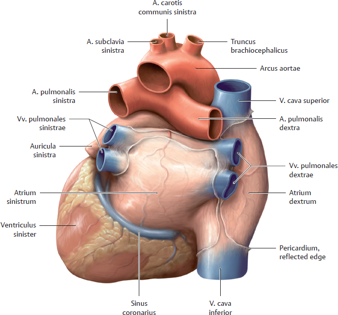
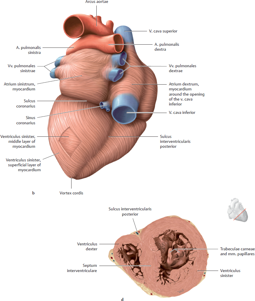
C Cor, facies posterior
Posterior view. This dissection shows how the arcus aortae crosses over the truncus pulmonalis at the point where the trunk divides into the aa. pulmonales sinistra and dextra. At that site the aorta gives off the three major arteries to the upper limbs, neck, and head: the truncus brachiocephalicus, left a. carotis communis, and left a. subclavia. This view also clearly shows the terminations of the vv. pulmonales (usually four in number) in the atrium sinistrum and the terminations of the two vv. cavae in the atrium dextrum. Note also the sinus coronarius in the posterior part of the sulcus coronarius, which runs between the ventriculus sinister and atrium sinistrum. This sinus is the collecting vessel for venous blood returned from the heart by the vv. cardiacae.
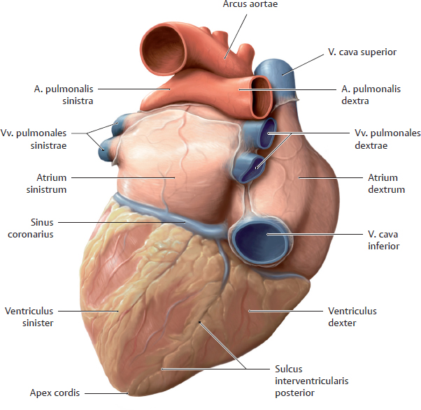
D Cor, facies diaphragmatica
Posteroinferior view. The cor is tilted forward to give a better view of its facies diaphragmatica, which is formed by both ventriculi and the atrium dextrum with the termination of the v. cava inferior. If the cor were viewed from below, from the perspective of the diaphragma (not shown here), it would be obvious that both vv. cavae are in alignment: Looking into the v. cava inferior, one can see through the terminal part of the v. cava superior.
E Structure of the cardiac wall
Layer | Location | Composition |
Endocardium | Innermost layer, lines the cavities of the cor and lines the cuspides and pockets of the cardiac valves | Single layer of epithelial cells with a subendothelial layer composed of collagen and elastic fibers; both layers are continuous with the intima of the vessels |
Myocardium | Middle layer and thickest part of the cardiac wall; motor for the pumping action of the cor (see pp. 94 and 95) | Complex arrangement of muscle fibers |
Epicardium | Outermost layer of the cardiac wall; part of the pericardium (see p. 90), forming its lamina visceralis | Serous membrane (single layer of epithelial cells with underlying layer of connective tissue) |
12.4 Structure of the Cardiac Musculature (Myocardium)
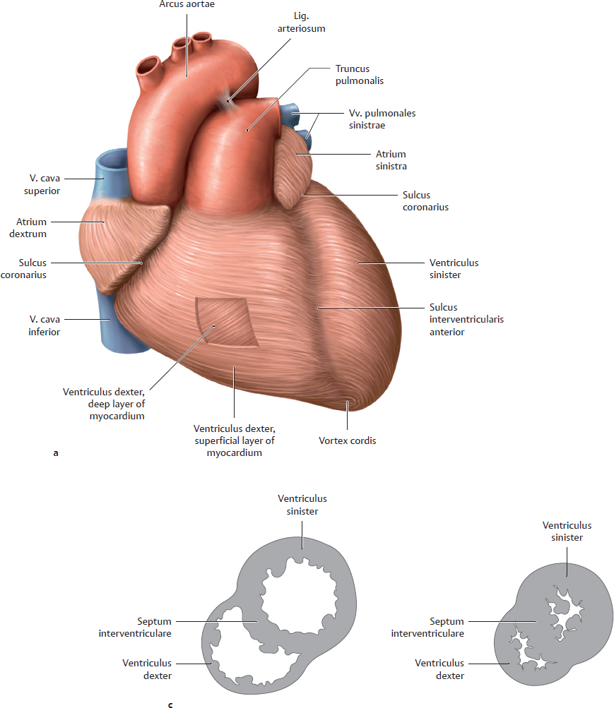
A Myocardial architecture
a, b External musculature of the cor, simplified anteroinferior view. The muscular walls of the ventriculi dexter and sinister have been windowed to display the deeper fibers.
Note: The epicardium has been removed in a and b along with the subepicardial fat. The coronary vessels are not shown in order to display more clearly the cardiac surface grooves (sulci interventriculares anterior and posterior).
The musculature of the atria is arranged in two layers, superficial and deep. The superficial layer (shown here) extends over the atria and is common to both, whereas each atrium has its own deep layer. Looped and annular muscle fibers extend down to the atrioventricular boundary and also encircle the ostia venarum. The ventricular musculature has a complex arrangement, consisting basically of a superficial (subepicardial), middle, and deep (subendocardial) layer. The superficial layer joins apically with the deeper layers to form a whorled arrangement of muscle fibers around the apex cordis (vortex cordis). The ventriculus dexter, which is a low-pressure system (see c), is less muscular than the ventriculus sinister and almost completely lacks a middle layer. The subendocardial layer forms the trabeculae carneae and mm. papillares (see d and p. 101). The histological unit of the myocardium is the cardiac myocyte, a specialized form of cardiac muscle cell. Unlike their electronically isolated counterparts in skeletal muscle, cardiac myocytes form a syncytium in which membrane depolarization and contraction spread in a wave.
c, d Myocardial cross-sections perpendicular to the long axis of the cor, viewed from above. c Schematic representation: The ventriculi in an expanded state (diastole, left figure), and in a contracted state (systole, right figure). d Transverse section through a specimen during diastole.
All the sections clearly demonstrate the difference in thickness between the left and right ventricular myocardia: The ventriculus sinister is part of the high-pressure system, and therefore its myocardium must generate a significantly higher pressure (120–140 mmHg during ventricular contraction) than the ventriculus dexter (approximately 25–30 mmHg). The difference in thickness is most pronounced during ventricular contraction (see c). Section d shows how the coronary vessels and subepicardial fat fill the sulci in the cor.
12.5 Cardiac Chambers
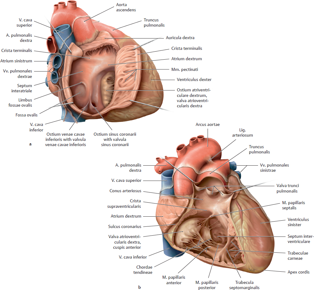
A Chambers of the right cor
a Right lateral view of the atrium. b Anterior view of the ventriculus. The ventricular and atrial walls have been opened widely, and the heart wall has been cut open to display the internal chambers.
The atrium dextrum (see a) consists of a posterior and anterior segment. The posterior segment with the smooth-walled sinus venarum (not visible here) bears the ostia venarum cavarum superioris and inferioris venae cavae. A small valve at the ostium venae cavae inferioris (valvula venae cavae inferioris) directs blood in the prenatal circulation through the foramen ovale in the septum interatriale. Because the foramen ovale is sealed shut postnatally, becoming the fossa ovalis (surrounded by a rounded margin, the limbus fossae ovalis), this valve atrophies after birth. The ostium sinus coronarii also bears a small crescent-shaped valve (valvula sinus coronarii). The anterior segment, which comprises the actual atrium with the auricula, is separated from the posterior segment by a ridge, the crista terminalis. Small muscular trabeculae, the mm. pectinati, arise from the crista terminalis, giving this segment an irregular wall texture. The wall of the atrium dextrum is thin (low-pressure system).
The ventriculus dexter is divided into two segments by two muscular ridges, the crista supraventricularis and trabecula septomarginalis: the inflow tract posteroinferiorly (with the cor positioned in situ) and the outflow tract anterosuperiorly (see also p. 111). The muscular ridges of the trabeculae carneae project into the lumen of the ventricular inflow tract. Specialized extensions of the trabeculae, the mm. papillares, are attached to the cuspides of the valva atrioventricularis dextra by collagenous cords, the chordae tendineae (see p. 101). The outflow tract is cone-shaped and consists mainly of the conus arteriosus, which has a smooth wall. The right ventricular outflow tract expels blood into the truncus pulmonalis, whose ostium is guarded by the valva trunci pulmonalis. The wall of the ventriculus dexter is relatively thin (low-pressure system).
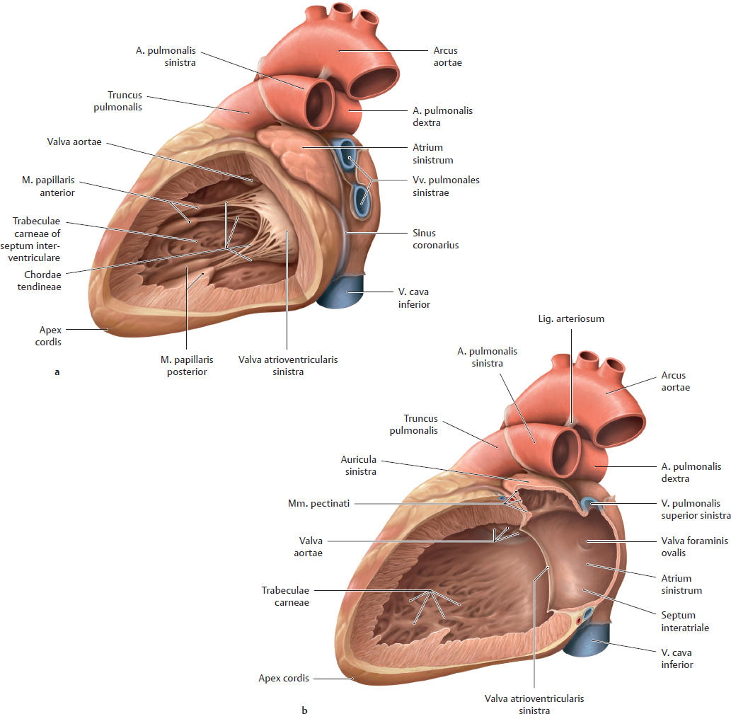
B Chambers of the left cor
Left lateral view. a Ventriculus, b ventriculus and atrium. The ventricular and atrial walls have been opened.
The atrium sinistrum is smaller than the atrium dextrum (see Aa). Its muscular wall is thin (low-pressure system) and is smooth in areas derived embryologically from the ostia venarum pulmonalium. The rest of the atrium is lined by mm. pectinati. The vv. pulmonales, usually four in number, terminate in the atrium sinistrum. Occasionally a narrow tissue fold (valvula foraminis ovalis) is found on the septum interatriale, formed by a protrusion of the fossa ovalis into the atrium sinistrum. It marks the site of fusion between the embryonic septum primum and septum secundum.
The ventriculus sinister has an inflow tract and outflow tract. The inflow tract begins at the left ostium atrioventriculare, which is guarded by the valva atrioventricularis sinistra (see p. 99). As in the ventriculus dexter, the wall of the left ventricular inflow tract is studded with trabeculae carneae, and mm. papillares are attached by chordae tendineae to the valva atrioventricularis sinistra. The outflow tract of the ventriculus sinister has smooth inner walls and lies close to the septum interventriculare. It leads to the aorta and is capped by the valva aortae at the root of the aorta ascendens (see p. 99). The septum interventriculare consists largely of muscle tissue (pars muscularis), and only a small portion near the aorta consists entirely of connective tissue (pars membranacea). The placement of the septum interventriculare between the cardiac chambers is marked externally by the sulci interventriculares anterior and posterior on the cardiac surface. The muscular wall of the ventriculus sinister is thick (high-pressure system), having approximately three times the thickness of the right ventricular wall (see Ab). The chambers of the left (and right) cor are lined by endocardium.
12.6 Overview of the Cardiac Valvae (Valve Plane and Cardiac Skeleton)
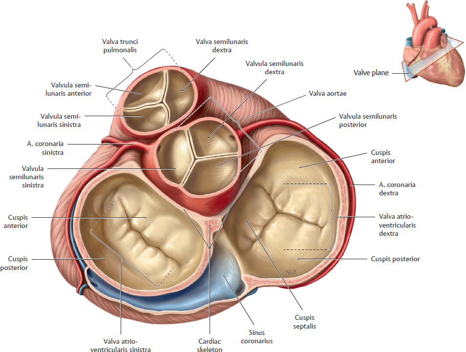
A Overview of the cardiac valvae
Plane of the cardiac valvae viewed from above. The atria have been removed, and the great arteries have been transected at their roots. The cardiac valvae are classified into two types—atrioventricular and semilunar. All heart valves lie in a plane, the valve plane. The cardiac valvae function as one-way valves. They ensure the unidirectional flow of blood between the atria and ventriculi (valvae atrioventriculares sinistra and dextra), and out of the heart (valva aortae and valva trunci pulmonalis). Valvae atrioventriculares. Located between the atria and ventriculi, the valvae atrioventriculares sinistra and dextra are composed of thin, avascular connective tissue covered by endocardium. They are classified mechanically as sail valves (see C) because the chordae tendineae (see C) constrain the movement of each cuspis like the tethering ropes on a sail. The function of these valvae is to prevent the reflux of blood from the ventriculi into the atria.
• The valva atrioventricularis sinistra (valva mitralis) has two cuspides (bicuspid valve): a cuspis anterior (anteromedial) and a cuspis posterior (posterolateral). The cuspis anterior is continuous with the wall of the aorta. The alternate term valva mitralis is derived from the two major cuspides, which are similar in shape to a bishop’s miter. Subdivisions in the lateral margins of the otherwise smooth valve have led some anatomists to describe small accessory cusps called the cuspides commissurales (usually two). These are not true cuspides, however, and are not connected to the anulus fibrosus sinister of the cardiac skeleton (see B). The cuspides are tethered by mm. papillares (see C).
• The valva atrioventricularis dextra has three cuspides (valva tricuspidalis): cuspides anterior, posterior, and septalis. One or two small accessory cusps may also be found; they do not extend to the anulus fibrosus dexter.
Semilunar valves. These valves have three crescent-shaped valvulae of approximately equal size placed at the ostia of the truncus pulmonalis (valva trunci pulmonalis) and aorta (valva aortae). Like the valvae atrioventriculares, they are composed of thin connective tissue covered by endocardium. The semilunar valves are classified mechanically as pocket valves because their valvulae pouch into the ventriculus like bulging pockets. The wall of the aorta and truncus pulmonalis show slight dilations just above the valve (the sinus trunci pulmonalis and sinus aortae). The sinus aortae expand the cross-section of the aorta, forming the bulbus aortae. The aa. coronariae dextra and sinistra branch off the base of the aorta just past the valva aortae (see pp. 112–115 for details).
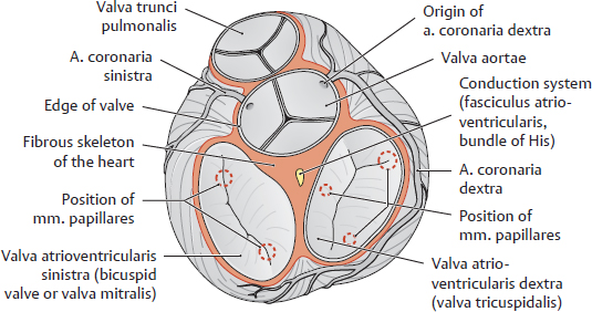
B Skeleton of the cor
The skeleton of the cor is a layer of connective tissue (often with considerable fat) that completely separates the myocardium of the ventriculi from that of the atria. The components of the cardiac skeleton in a narrow sense are as follows:
• The anuli fibrosi dexter and sinister and intervening trigona fibrosa
• The anulus fibrosus of the valva aortae, which is connected to both anuli fibrosi
• The pars membranacea of the septum interventriculare (not shown here).
In a broad sense, the anulus fibrosus of the valva trunci pulmonalis also contributes to the cardiac skeleton. It is connected by a collagenous band (tendo infundibuli) to the anulus fibrosus of the valva aortae. The valvae atrioventriculares are anchored to the anuli fibrosi, while the semilunar valves are each attached by connective tissue to their valvular anuli fibrosi. Thus, the cardiac skeleton in the broad sense provides a mechanical framework for all the cardiac valvae. Besides mechanically stabilizing the heart, the fibrous skeleton also functions as an electrical insulator between the atria and ventriculi. The electrical impulses that stimulate cardiac contractions (see p. 108 f) can pass from the atrium to the ventriculus only through the fasciculus atrioventricularis (bundle of His), and there is only one opening in the fibrous skeleton (in the trigonum fibrosum dextrum) which transmits that bundle).
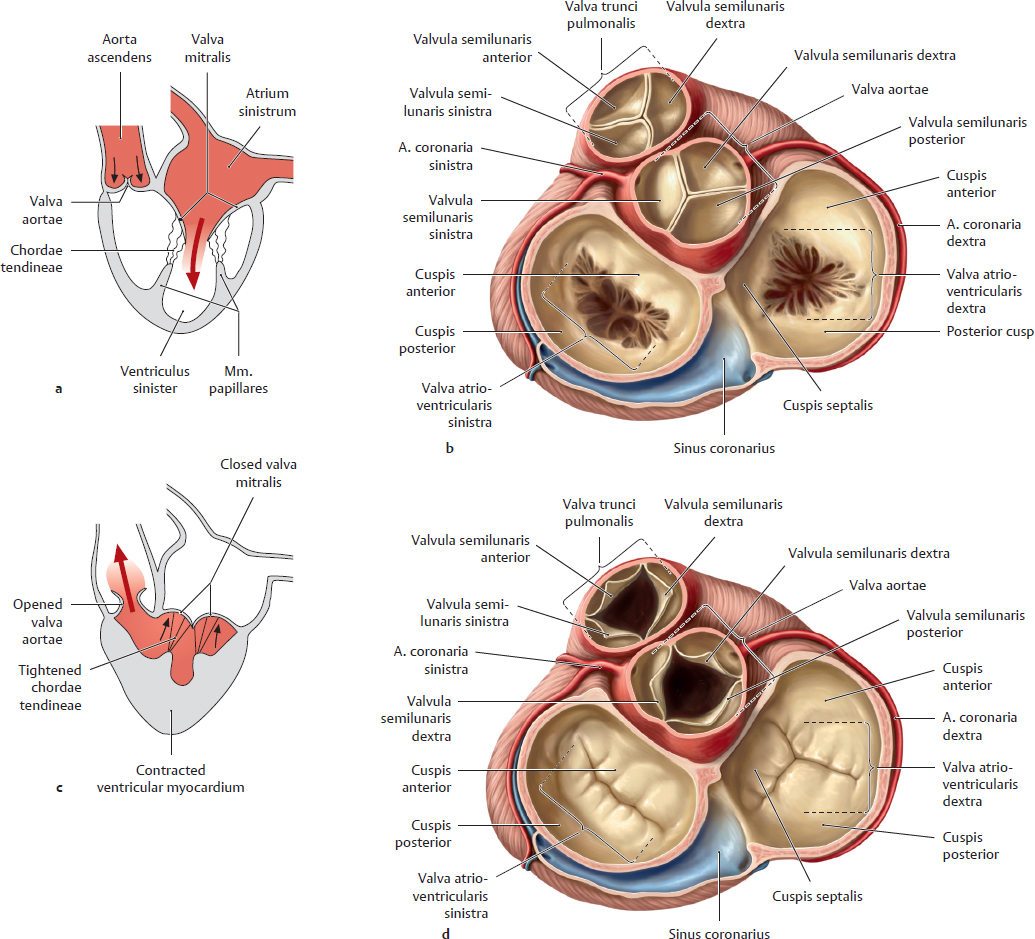
C Function of the valvae cordis during the cardiac cycle
a and b Ventricular diastole; c and d Ventricular systole. a and c Direction of blood flow in the left cor; b and d Valve plane viewed from above.
12.7 Cardiac Valves and Auscultation Sites
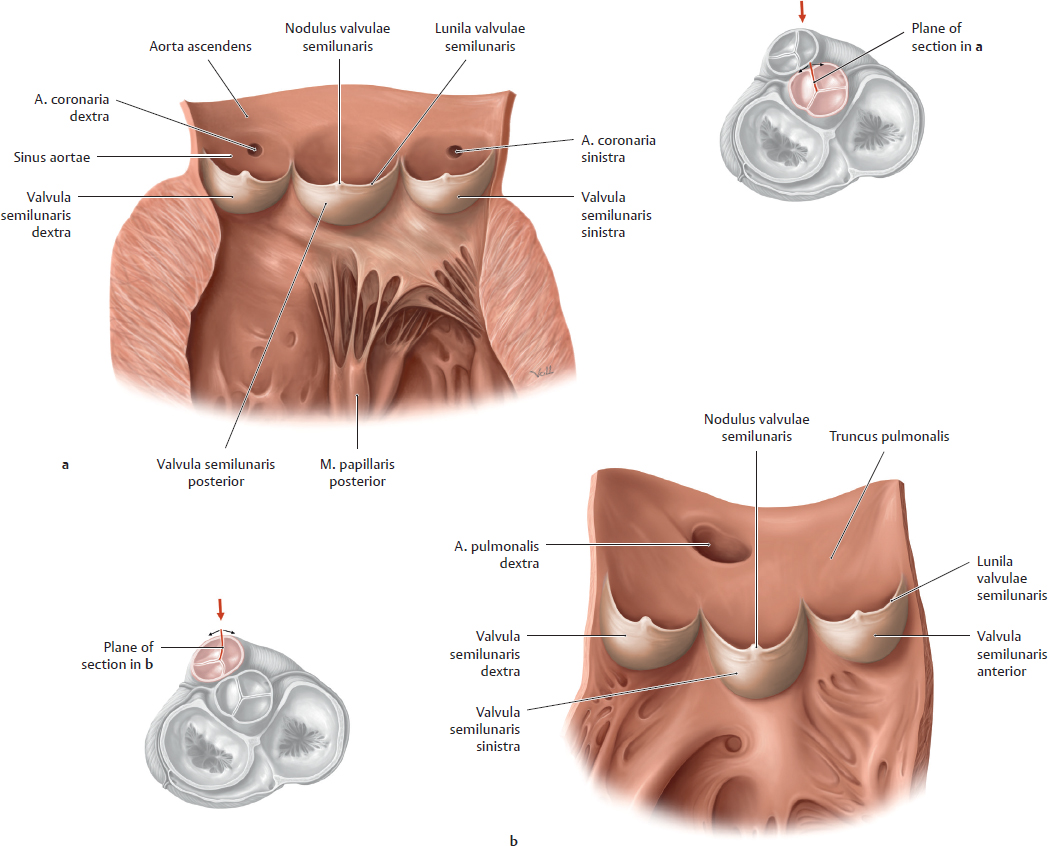
A Semilunar valves of the outflow tracts (valva aortae and valva trunci pulmonalis)
The valva aortae (a) and valva pulmonalis (b) have been displayed by cutting open the pars ascendens aortae and truncus pulmonalis and opening them up like a book. The valva aortae and valva trunci pulmonalis close the ventricular outflow tracts during diastole:
• The valva aortae closes the left ventricular outflow tract.
• The valva trunci pulmonalis closes the right ventricular outflow tract.
These valves almost completely prevent the regurgitation of blood expelled by the ventriculi. The origins of the arteriae coronariae sinistra and dextra can be clearly identified in the sinus aortae past the valvulae semilunares (a), and the origin of the a. pulmonalis dextra can be identified in the truncus pulmonalis (b). The free margin of each valvula semilunaris is thickened centrally to form a nodulus valvulae semilunaris, and on each side of the nodulus is a fine rim called the lunula valvae semilunaris. The nodulus and lunula ensure that the margins of the valvulae appose tightly and completely during valva closure. Both the valvae atrioventriculares (see p. 98) and the semilunar valvae may undergo pathological changes, usually due to inflammation (endocarditis). Inflammation may result in secondary vascularization of the initially avascular valvae, causing them to undergo fibrotic changes that stiffen the valvae and compromise their function. There are two main abnormalities of valvular mechanics, which may coexist in the same valva:
• Valvular stenosis: Opening of the valva is impaired, causing a reduction of blood flow across the valva. Usually this creates a pressure overload on the chamber proximal to the obstruction.
• Valvular insufficiency: Closure of the valva is impaired, allowing blood to regurgitate into the chamber proximal to the valva. Such a pathological reflux creates a volume overload on the affected cardiac segments. When the load exceeds a certain magnitude, surgical replacement of the valva may be necessary to prevent further damage to the cor.
• Stenosis and insufficiency may coexist: A valva may become stuck in an intermediate position, unable to open or close completely.
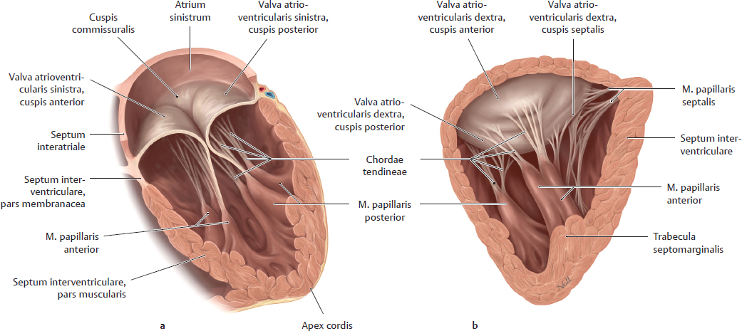
B Valvae atrioventriculares and musculi papillares
Anterior view of the valvae atrioventriculares sinistra (a) and dextra (b). The drawings represent a very early phase of ventricular contraction in which the valvae atrioventriculares have just closed. The mm. papillares are clearly displayed. There are three mm. papillares for the three cuspides of the valva atrioventricularis dextra (mm. papillares anterior, posterior, and septalis) and two mm. papillares for the two cuspides of the valva atrioventricularis sinistra (mm. papillares anterior and posterior). The mm. papillares (specialized extensions of the trabeculae carneae) are attached to the free margins of the valve cuspides by chordae tendineae. When the mm. papillares contract (valva closure), the chordae tendineae are shortened to restrict the motion of the valve cuspides. This keeps the cuspides from opening into the atria during ventricular contraction (systole), thereby preventing the regurgitation of blood back into the atria.
Note: Like other myocardial regions, the myocardium of the mm. papillares may suffer necrosis due to a myocardial infarction, leaving the corresponding cuspis prone to prolapse into the atrium. Conversely, pathological shortening of the chordae may also prevent the valva from closing completely. This also would allow blood to regurgitate into the atrium during ventricular systole, producing an audible heart murmur (see p. 110).
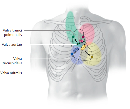
C Auscultation of the valvae cordis
The diagram shows the anatomical projection of the valvae onto the thorax, thus illustrating both the sites for auscultating the valvae cordis on physical examination and the areas to which abnormal heart murmurs of the respective valvae are transmitted. The auscultation sites are more or less located at the center of the areas for the transmitted murmurs. There are both physiological (see p. 110) and pathological heart sounds. Table D summarizes the anatomical location and auscultation sites of the valvae.
In the healthy cor, blood does not generate a perceptible sound as it flows across the valvae cordis. But if the valvae are functionally impaired as a result of disease (stenosis, insufficiency), the blood flow at the valvae cordis becomes turbulent. This type of flow produces audible sounds that are transmitted via the bloodstream. In most cases these sounds (murmurs) are not heard best over the anatomical projections of the valvae on the chest wall (due to sound muffling by the thick cardiac wall), but are heard more clearly at sites located downstream from the valvae (see D).
D Anatomical projections and auscultation sites of the cardiac valves
Valve | Anatomical projection | Auscultation site |
Valva atrioventricularis sinistra (valva mitralis) | Fourth / fifth cartilago costalis on the left side | Left fifth spatium intercostale on the linea medioclavicularis |
Valva atrioventricularis dextra (valva tricuspidalis) | Sternum at the level of the fifth cartilago costalis | Right fifth spatium intercostale close to the sternum* |
Valva aortae | Left sternal border at the level of the third costa | Right second spatium intercostale close to the sternum |
Valva trunci pulmonalis | Left sternal border at the level of the third cartilago costalis | Left second spatium intercostale close to the sternum |
12.8 Radiographic Appearance of the Cor
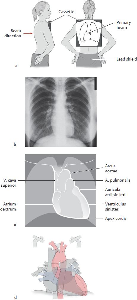
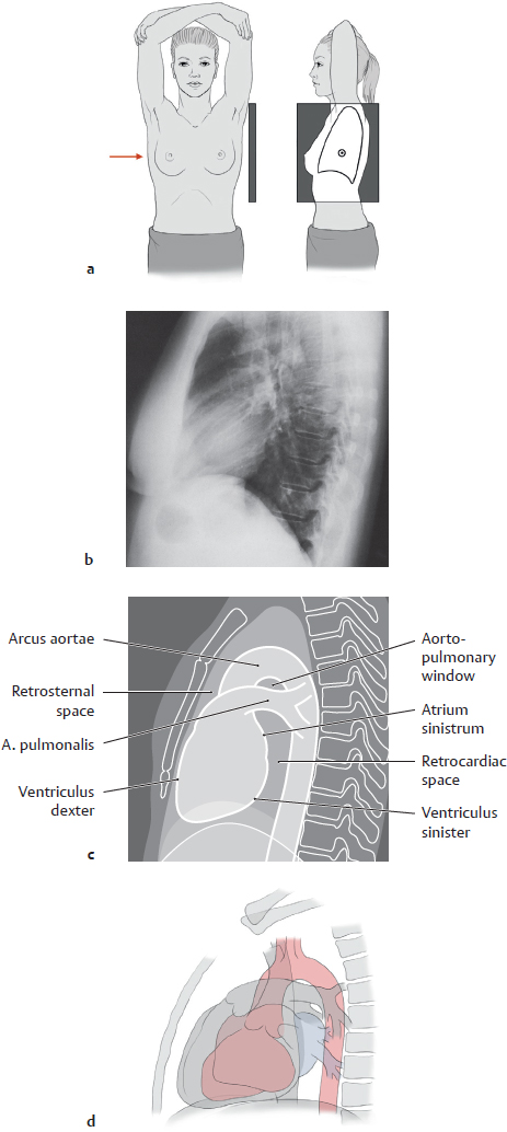
A Postero-anterior (PA) chest radiograph
a The patient stands with the anterior chest wall on the cassette (the beam “passes” through the patient in a posterior-to-anterior direction with the central beam targeted at the level of the T6 vertebra). The radiographs are taken with the patient keeping the mouth open, breathing in and holding the breath. The back of the hands are placed on the hips with the elbows turned forward;
b Posterior-anterior radiograph (viewed from an anterior to posterior direction);
c Heart shadow (cardiac silhouette) with structures that form the cardiac borders;
d Topography of the cardiac shadow: right cor with inflow and outflow tract (gray); ventriculus sinister with outflow tract (red), atrium sinistrum with inflow tract (blue).