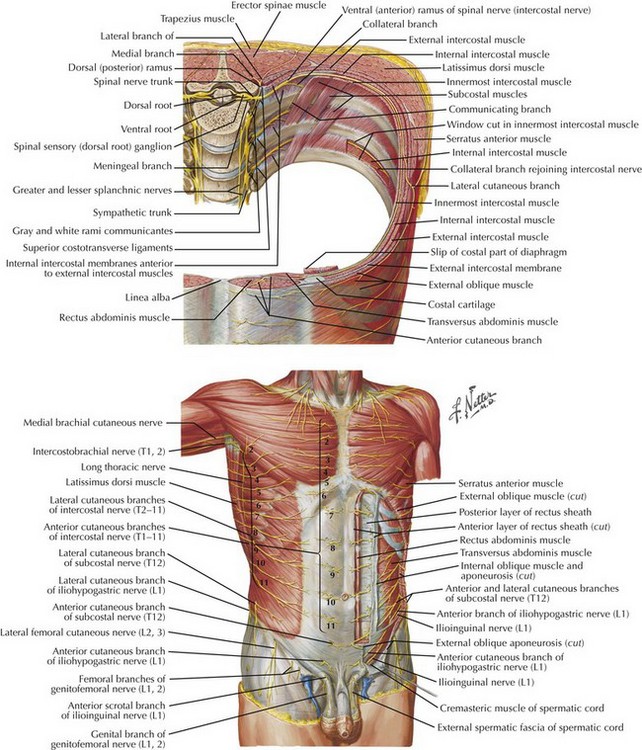
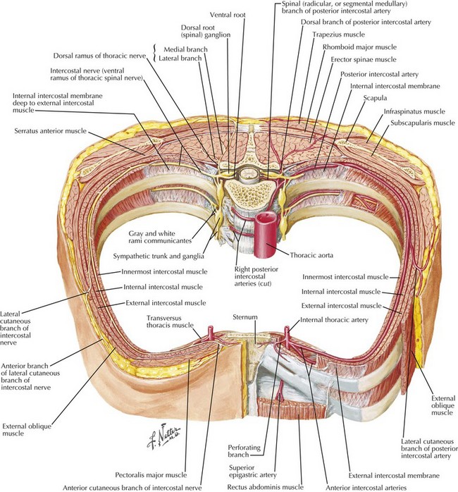
Open Ventral Hernia Repair

Chapter 31


Open Ventral Hernia Repair


Introduction


Ventral (abdominal) hernia repair is one of the most common procedures performed by general surgeons, with more than 200,000 done each year in the United States. Despite the prevalence of this procedure, there is little consensus as to the indications for repair, optimum technique, or appropriate position of the prosthetic mesh. Given the wide variety of patient and hernia factors, no single approach will likely suffice to repair all abdominal wall defects. Options include a laparoscopic or an open repair. The laparoscopic approach is typically reserved for obese patients with small to medium-sized defects, to avoid extensive subcutaneous dissection and potential mesh infections. The open repair can be performed with several methods.


Most surgeons agree that all incisional hernias should be repaired with prosthetic (synthetic or biologic) mesh, because recurrence rates are reduced by half. Prosthetic mesh can be placed as an inlay (sewn to the fascial edge), an onlay (sewn above the fascia), or a sublay (underneath the fascia). Sublay mesh can be placed in the intraperitoneal, preperitoneal, or retrorectus position. The inlay approach has been largely abandoned because of high recurrence rates, and the onlay approach is discouraged because the prosthetic mesh is placed in the subcutaneous position at highest risk for mesh sepsis. Most herniologists agree that the prosthetic mesh should be placed as a sublay. This chapter focuses on the sublay repair, with particular attention to the retrorectus placement of the mesh.


Surgical Principles


General principles of incisional hernia repair include gaining access to the reoperative abdomen, complete adhesiolysis, preparation of the abdominal wall for prosthetic mesh placement, mesh deployment, and reconstruction of a dynamic abdominal wall with reapproximation of the linea alba. Safe access to the reoperative abdomen typically involves extending incisions in a cephalad direction to enter the undissected peritoneum. Complete adhesiolysis of the anterior abdominal wall is important to free the abdominal wall musculature and allow it to advance to the midline during eventual reconstruction of the midline.


Reestablishing the linea alba is an important concept in abdominal wall reconstruction. If the linea alba is seen as the tendinous insertion of the rectus abdominis muscle and oblique muscles, it is critical to achieve appropriate physiologic loading of the abdominal wall. In particular, reconstructing a completely tension-free repair in fact renders the oblique muscles nonfunctional, with constant lateral displacement of the abdominal wall leading to mesh displacement at the mesh-tissue interface. This chapter emphasizes surgical methods to reapproximate the rectus complex.


Preoperative Imaging


Preoperative imaging of the abdominal wall is very helpful. The author typically performs abdominal pelvic computed tomography (CT) scans in all patients undergoing complex abdominal wall reconstruction. CT images can help delineate the location, size, and complexity of the hernia, particularly in obese patients. Attention to the relationship of the hernia to bone structures (e.g., xiphoid, costal margin, iliac crest, pelvis) is important for surgical planning. Recognizing the appropriate plane is paramount to gain sufficient overlap of the mesh and achieve the most durable repair. Likewise, the integrity of the rectus abdominis muscle is important. If the rectus muscle is narrow, the mesh cannot be placed in the retrorectus position and will need to be located in the preperitoneal position, as described later. If the rectus muscle is destroyed, absent, or atrophic, alternative methods are necessary.


Some surgeons have advocated abdominal wall ultrasound to detect hernias, but the author has found this to be very user dependent, with minimal experience.


Anatomy of Abdominal Wall


Figures 31-1 and 31-2 show the anatomy of the vascular supply and innervation of the abdominal wall. Understanding the relationships of these nerves and vessels and their location in the abdominal wall is critical to preserve them during dissection, to maintain an innervated functional abdominal wall.


Stay updated, free articles. Join our Telegram channel

Aug 12, 2016 | Posted by in ANATOMY | Comments Off on Open Ventral Hernia Repair

Full access? Get Clinical Tree

Get Clinical Tree app for offline access