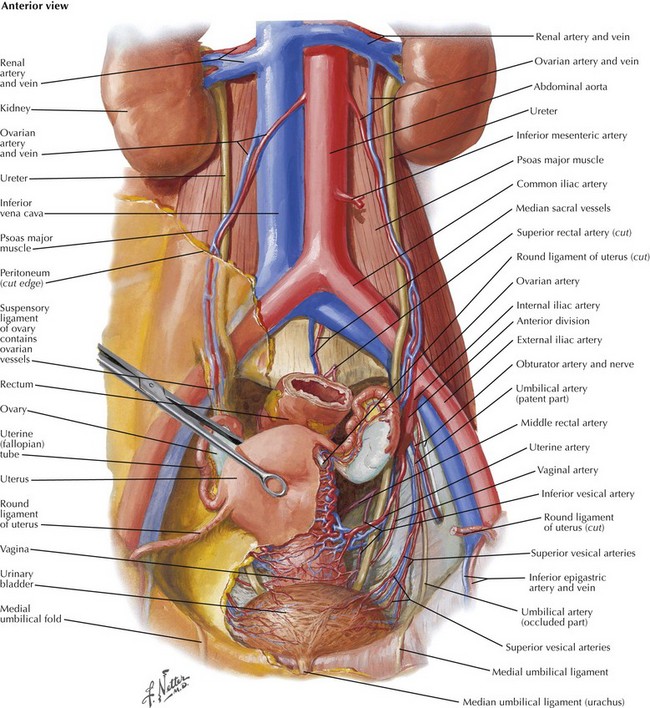
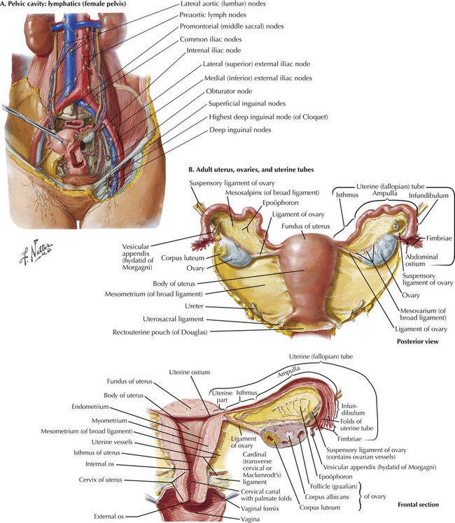
Oophorectomy for Benign and Malignant Conditions

Chapter 52


Oophorectomy for Benign and Malignant Conditions


Introduction


The ovary is a complex organ from both a histologic and a functional standpoint. As a result, numerous tumors, both benign and malignant, can arise in the adnexa. The surgical approach is often determined by the pathology as well as the desire to preserve gonadal function and fertility.


Although the vast majority of tumors arising in the ovary will be benign, especially in younger women, proper surgical management of ovarian or tubal malignancy is much more complex. Epithelial ovarian and tubal malignancies tend to metastasize early and spread along peritoneal surfaces throughout the abdomen. These surgeries are designed to render the patient with minimal residual disease and often require pelvic peritonectomy with en bloc rectosigmoid resection to clear the pelvis. Proper surgery often includes equally radical upper abdominal resection. Multiple studies have shown that complete cytoreduction of metastatic disease impacts both overall survival rates and progression-free survival rates in women with epithelial ovarian malignancy. Overall survival rates of 66 to 120 months is achievable even in women with advanced disease.


Preoperative Imaging


Preoperative imaging of an adnexal mass helps not only to characterize the tumor, but also to assess for ascites, hydronephrosis, lymphadenopathy, and omental implants that may impact the preoperative counseling and surgical approach. Ultrasonography is the most frequently used modality to assess a pelvic mass. It is readily accessible, noninvasive, and provides excellent characteristics of the lesion itself. The ultrasonographer should comment on lesion size, cystic/solid components, complexity, and Doppler flow along with evidence of hydronephrosis and ascites. Magnetic resonance imaging (MRI) can provide significantly more information about an ovarian tumor, but in reality MRI is rarely helpful in triaging an adnexal mass. Most lesions believed to be complex should be removed in all age groups. Computed tomography (CT) scans are essential to evaluate the retroperitoneum and upper abdomen in women with an ovarian mass that may be malignant.


Surgical Approach


The most prudent approach to a patient with an adnexal mass is made on the basis of the patient’s age, desire for future fertility/hormonal preservation, and imaging characteristics. Almost all pelvic masses in children, premenopausal girls, and postmenopausal women should be evaluated. Triage is made on the basis of imaging characteristics, symptoms, and concern for malignancy. Low-risk lesions, especially in premenopausal girls, can often be followed for spontaneous resolution, especially if these are primarily cystic in nature. Solid masses or complex masses in any age group are more likely to be malignant and usually require surgical evaluation. Tumor markers (e.g., CA125) should be obtained preoperatively, although these may be informative in only 90% of cases. The surgical approach, whether laparoscopic, robotic, or conventional laparotomy, depends on the nature of the lesion and the likelihood of identifying a malignancy.


Anatomy and Dissection of the Adnexa


The adnexa refers to both the ovary and fallopian tube. An intimate understanding of the vascular supply to the adnexa and the relationship to the underlying ureter and uterus is required before commencing surgery (Fig. 52-1). The general principles are similar regardless of surgical approach (open, laparoscopic, or robotic).


Gonadal Vessels and Infundibulopelvic Ligament


The best approach to removing a pelvic mass is first to open the retroperitoneum and identify the gonadal vessels and ureter. The gonadal blood supply, or infundibulopelvic ligament (IP), originates from the aorta and runs parallel to the ureter, crossing into the pelvis over the bifurcation of the common iliac vessels (Fig. 52-2, A).


Stay updated, free articles. Join our Telegram channel

Aug 12, 2016 | Posted by in ANATOMY | Comments Off on Oophorectomy for Benign and Malignant Conditions

Full access? Get Clinical Tree

Get Clinical Tree app for offline access