Chapter 6 Nose and Mouth
“How do you know that I’ve told a lie?” Pinocchio asked the Fairy.
“Lies, my dear boy, are quickly discovered; because they are of two kinds. There are lies with short legs, and lies with long noses. Yours is clearly of the long-nosed variety.”
A. The Nose
(1) Generalities
If you can excuse the pun, it is wise to be nosy about the nose. In fact, examination of this important facial appendix may reveal unsuspected abnormalities that lead to a diagnosis of either systemic diseases or capital sins (such as cocaine abuse).
(2) The External Nose
2 What is rhinophyma?
From the Greek rhino (nose) and phyma (tumor), this is a variety of acne rosacea characterized by a bulbous and rather prominent nose. It is also referred to as copper-, hammer-, or potato-nose since the skin is thickened, erythematous, nontender, and often covered with multiple telangiectasias (rum or gin blossoms). In fact, other colorful lay terms for this condition include brandy nose, rum nose, and toper’s nose—all paying tribute to its alleged association with the bottle, which is actually rather loose. Pathology shows follicular dilation and sebaceous hyperplasia, with fibrosis and hypervascularity. Of note, rhynophima may degenerate into basal cell carcinoma.
3 What are the causes of rhinophyma?
The ones conventionally blamed are climatic exposure and alcohol, with the best example of gin blossoms being those of the great comedian W.C. Fields, a man whose nose was directly proportional to his affinity for liquor. Rhinophyma is also rather common among politicians, with an early phase of it noted in the 42nd U.S. president, Bill Clinton. If this prompts you to wonder whether Pinocchio had rhinophyma, too, the answer is no. Pinocchio had variable rhinomegaly, a peculiarly reversible condition related to his telling lies.
4 Is there any scientific basis for the “Pinocchio effect”?
Yes. Recent evidence indicates that liars may indeed experience nasal changes, resulting in a telltale itch that eventually prompts them to unconsciously touch their nose. To corroborate this phenomenon, Alan Hirsch, director of the Chicago-based Smell and Taste Research Foundation, gathered 23 giveaway signs (both verbal and nonverbal) of “mendacious speech” and recently used them to analyze Bill Clinton’s famous 1998 grand jury testimony on the Lewinsky affair. Overall, Clinton did 20 of the 23 signs, but whenever being particularly untruthful (or “legally accurate”), he consistently touched his nose, which brings us back to Collodi’s Pinocchio and science. The nose contains abundant vascular erectile tissue (an unfortunate property it shares with the penis), which involuntarily dilates whenever a person lies, thus making the node redder, bigger, and itchy. Hence, the repetitive nose-touching of liars. Hirsch honored Collodi’s insight by naming this phenomenon the “Pinocchio effect.” His observation has been confirmed in other famous cases. For example, even O.J. Simpson touched his nose rather frequently during trial when describing Nicole Brown Simpson’s murder. Hence, simple observation might allow policemen, attorneys, psychiatrists, and everyone else to tell whether someone is lying. Or, as Yogi Berra used to say, “You can observe a lot by watching.”
5 What is a saddle nose? Does it really exist outside of board questions?
A saddle nose is the congenital (or acquired) erosive indentation of the nasal bone, turning the distal tip of the nose upward and outward. A true saddle nose (caused by destruction of the bony portion of the nose) is typical of congenital lues, but actually more frequent in Wegener’s granulomatosis. Given the increasing availability of good therapy for both these conditions, a saddle nose has now become less common. A pseudo-saddle nose is instead a feature of relapsing polychondritis, although the destruction in this case involves cartilage and not bone.
6 What are nasal fractures?
They are the most common trauma-related disorders of the nose. They present with severe pain and anterior epistaxis, often from both nares. Periorbital ecchymoses invariably develop 24 hours after trauma, along with significant sequelae, such as septal hematoma or septal deviation. Because these fractures are open, antibiotics are necessary to prevent osteomyelitis.
7 What is a septal hematoma? How does it differ from septal deviation?
A hematoma is a purple and painful nodule in the nasal septum, easily spottable through the nostril. Conversely, traumatic displacement of the nasal septum (i.e., septal deviation) may be difficult to see until later, since edema of the early posttraumatic stages makes physical exam difficult to perform. Both of these complications require referral to an ear-nose-throat (ENT) specialist for drainage or reduction. If undrained, the hematoma may cause the septal cartilage to become ischemic and necrotic, with a resulting permanent nasal deformity.
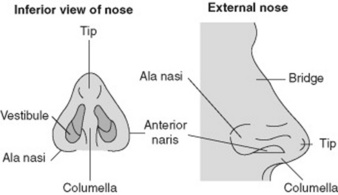
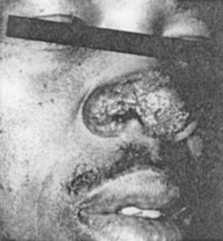
8 What is lupus pernio?
It is a chronic, nonblanching, diffuse, and purple skin discoloration of the external nose, in the absence of true nasal enlargement (Fig. 6-2). Hence, it differs from rhinophyma. A sign of active sarcoid, it may occur with uveitis, erythema nodosum, and pulmonary involvement. It may also coexist with lesions of the ears, cheeks, hands, and fingers. The term lupus refers to any disfiguring skin condition that, like a wolf (lupus in Latin) “devours” the patient’s facial features. It is thus used with modifying terms to designate various disfiguring skin diseases, such as lupus verrucosus, lupus erythematosus, lupus tuberculosis, lupus vulgaris, and, of course, lupus pernio. Pernio is Latin for frostbite and refers to the peculiar violet-bluish hue of the condition (see Chapter 3, The Skin, question 265).
9 What did Rudolph of the reindeer story really have?
Probably rhinophyma, but that begs the question: Did Santa spill the grog while feeding his reindeer?
(3) The Internal Nose
10 What are the normal structures of the internal nose?
The normal internal structures are shown in Fig. 6-3:
1. The vestibules. As indicated by the term, these are paired internal widenings, immediately beyond each naris. They are delimited:

2. Deeply beyond the vestibules are the turbinates, or conchae. These are curving bony structures that project into the internal nose. There are three turbinates (and three corresponding meatuses) in each nasal cavity: superior, middle, and inferior. Their main function is to increase the nasal surface for humidification, temperature control, and filtering of inhaled air. To do so, they are covered by a well-vascularized and erectile mucosa.
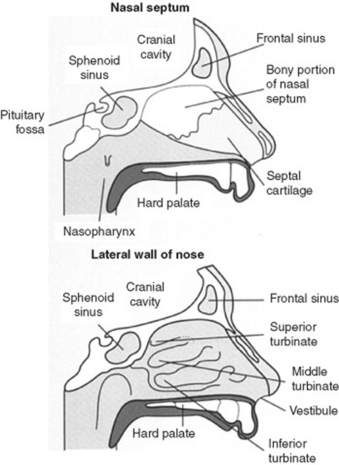
(From Epstein O, Perkin CD, de Buono DP, Cookson J: Clinical Examination, 2nd ed. St. Louis, Mosby, 1998.)
11 What are paranasal sinuses?
They are the frontal, maxillary, ethmoid, and sphenoid sinuses: four hollow and air-filled paired cavities that open through small ostia into recesses of the nasal cavities called meatuses. These are covered by bony shelves lined by erectile soft tissue (the turbinates)—three on each side.
12 And what about the adenoids?
They are aggregates of lymphatic tissue located on the roof of the nasopharynx, just below the sphenoid sinus. They typically regress with puberty and so in adults are usually absent. Yet in infants and children, they may enlarge considerably and cause nasal obstruction, nasal voice, snoring, restless sleep, and mouth breathing. Since mouth breathing in times of facial formation may result in bony changes, affected children often develop an adenoid facies, with a high, arched palate; prominent upper teeth; pinched-in nose; shortened upper lip; a staring expression of the eyes; and a slightly elongated and dumb-looking face. Adenoidal hypertrophy also may obstruct the eustachian tubes and thus result in recurrent middle ear effusions and otitis media.
13 What is the significance of flaring of the nostrils?
Flaring of the nostrils (i.e., of the alae of the nose) is a sign of increased work of breathing, typical of impending respiratory failure. It is often associated with other findings of distress, such as respiratory alternans and abdominal paradox (see Chapter 13, Chest Inspection, Palpation, and Percussion, questions 52–58). It also can occur in peritonitis as a result of impaired and painful excursion of the diaphragm.
14 What are the best tools for inspecting nares and internal nose?
Otoscope: This can be mounted with a nasal speculum (instead of an ear speculum) and thus used to shine a light directly into each vestibule.
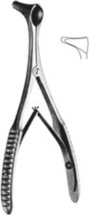
Handheld Vienna nasal speculum: This is a speculum that opens upon closure of the handles. It is commonly used with a head mirror (Fig. 6-4).
In the absence of these tools, you may simply use fingers and penlight. Either way, always instruct the patient to first blow the nose (gently but forcibly) to expel any mucus.
15 Is inspection of nasal secretions useful?
Yes. In fact, there is quite some value in snot, the colloquial term for excessive flow of nasal mucosa. Although the butt of vulgar jokes, snot is actually noble in origin, going all the way back to the Old English gesnot. It also retains diagnostic value, since different diseases tend to produce different snots:
A clear discharge is suggestive of viral or atopic rhinitis.
A yellow discharge may instead indicate an early suppurative process.
A green discharge is quite consistent with purulent sinusitis.
Obviously, a bloody discharge suggests anterior epistaxis.
A dark and almost black discharge (especially in comatose diabetics) argues for mucormycosis.
16 What is an abscess of the nasal vestibule?
It is a superficial abscess of hair follicles (furunculosis), usually caused by Staphylococcus aureus and not uncommonly located in the nasal vestibule or septum. Quite painful, it presents as an erythematous and fluctuant nodule in the septal mucosa. Usually treated with antibiotics and warm compresses, it may require incision and drainage to avoid cavernous sinus thrombosis.
17 What are the causes of swelling/bumps in the nasal septum?
All require ENT referral, since an untreated hematoma (or abscess) may result in septal perforation.
18 What are the most common causes of airflow obstruction in one or both nares?
In addition to the conditions previously listed, other causes include nasal mucosa edema, septal deviation, and foreign bodies—all diagnosable by exam. Foreign bodies are especially common in children, given their unique penchant for inserting rocks, twigs, and crayons into nares and various body orifices.
19 What are nasal polyps?
They are pedunculated, fleshy, and friable structures that hang from the lateral or septal mucosa. Polyps originate from localized swellings of either sinus or nasal mucosa, initially small, but eventually growing with each recurrence of submucosal edema until they may even protrude from the vestibule and cause nasal obstruction. They are often multiple, clearly visible, easy to move back and forth, and not tender (which differentiates them from other internal nasal structures). They are common in chronic (atopic) rhinitis and aspirin sensitivity (often in association with asthma) and can be easily removed through endoscopy.
20 What is a papilloma of the nasal vestibule?
It is a wart of the inner nose. In contrast to the more common polyp, papillomas are jagged in appearance and more likely to bleed. Like polyps, they can interfere with the sense of smell and cause obstruction. They are easily visible on exam and must all be removed since they can undergo neoplastic degeneration. Like warts elsewhere, they often recur.
21 What is a nasopharyngeal carcinoma (lymphoepithelioma)?
It is a sequela of Epstein-Barr virus (EBV) infection, rare in Europe but frequent in Southeast Asia, where it represents the third most common cancer. Tumor growth is often asymptomatic, and presentation is late, typically with cervical lymphadenopathy. Hence, the prognosis is poor, even though screening for anti-EBV antibodies may help detect early and treatable disease.
22 What does a nasal septal perforation look like?
Like a hole in the septum. This can be demonstrated on inspection or by shining a light into one nostril and seeing it transilluminate both sides.
23 What are the common causes of a septal perforation?
Traumatic: Facial injury or self-induced lesions (nose picking/piercing)
Iatrogenic: Prior septal surgery, nasogastric tube placement, or nasal intubation
Inflammatory/malignant: Often the sequela of untreated septal hematomas or abscesses
Cocaine snorting: One of the most frequent causes today, often presenting with large and expanding perforations. Cocaine contains adulterants that may irritate the mucosa, plus has strong alpha-agonist effects (causing vasoconstriction and ischemia of the nasal cartilage). Nasal obstruction often accompanies perforation.
24 What are the less-common causes of perforation?
Chemical irritants (chromic or sulfuric acid fumes, glass dust, mercurials, and phosphorous)
Infections (tuberculosis, syphilis, and, more rarely, leprosy)
Collagen vascular diseases (Wegener’s, midline granuloma, systemic lupus erythematosus, rheumatoid arthritis, mixed connective tissue disease, and progressive system sclerosis)
25 What are the nasal manifestations of a basilar skull fracture?
In addition to the direct facial trauma, patients often show evidence of nasal fracture, such as periorbital ecchymoses, swelling/tenderness of the nasal bridge, and anterior epistaxis. There also may be cerebrospinal fluid rhinorrhea (leakage of cerebrospinal fluid [CSF] through the fracture site). This is a risk factor for the development of meningitis and thus necessitates prompt surgical attention.
26 How do you recognize CSF rhinorrhea?
By placing a drop of nasal secretions over a paper tissue. In CSF rhinorrhea, this will serve as a “poor man’s” paper chromatography, showing a clear halo around “nasal” secretions that represent instead spinal fluid leakage. Alternatively, the glucose content of secretions may be measured at the bedside by using a Chemstrip. In patients with CSF rhinorrhea, this will reveal a high glucose concentration, close to spinal fluid levels of 40–80 mg/dL.
27 What is a cold? What are its nasal manifestations?
The common cold, one of humankind’s most frequent afflictions, has a plethora of nasal manifestations: swollen mucosa (to the point of obstruction), stuffy feeling, and serous or purulent nasal discharge. Since it represents a viral infection of the upper respiratory tract, there may be concomitant serous otitis media, nonexudative pharyngitis, and shotty nodes.
28 What results in swelling of the nasal mucosa?
Viral: Nasal and oropharyngeal infections by either rhinoviruses or adenoviruses
Atopic: Pollen or dander exposure causing nasal congestion, allergic (serous) conjunctivitis, and sneezing
Vasomotor: Response to a specific inhalant, characterized by boggy edema of the mucosa and marked tearing. Inhalants may either be noxious to all (such as tear gas) or only to some. For example, perfumes may elicit an idiosyncratic response in a few predisposed individuals, causing pronounced swelling of the nasal mucosa.
29 Can you diagnose the cause of GI bleeding by peeking into the patient’s nose?
Yes, bleeding in the gut may indeed be linked to the nose. The most common reason is simple epistaxis, wherein patients swallow nasal blood, resulting in guaiac-positive stools and even melena. More rare are the multiple nasal telangiectasias of Rendu-Osler-Weber syndrome, an autosomal dominant disorder characterized by multiple (and often bleeding) vascular lesions of the gut, mouth, face, extremities, and chest. The tongue and lips also may have telangiectasias.
30 Who was Rendu?
Henry J.L. Rendu (1844–1902) was the grandson of a distinguished Parisian painter and the son of an agricultural inspector. An art lover, he was so fascinated by his father’s profession as to pursue a medical career only after a stint in agriculture, geology, and botany. Still, he never lost his passion for plants and eagerly maintained it as a lifelong hobby. At age 43, he finally joined the staff of the Necker Hospital in Paris (the same one where Laënnec had been chief of chest medicine), rapidly gaining fame as a charismatic lecturer and gifted clinician.
31 Who was Weber?
Frederick P. Weber (1863–1962) was a British physician, already encountered in the ear chapter because of Sturge-Weber disease. Educated in Cambridge, Vienna, and Paris, Weber cultivated throughout his life an interest in medical philosophy, the arts, and numismatics, which he then expressed in many books and articles. His father was Herman D. Weber (1823–1918), himself a famous and long-lived physician (he is the same Weber who described the midbrain syndrome that still carries his name). Weber père was a charming man with many interests, who taught himself English so that he could read Shakespeare and befriend, among others, Addison, Carlyle, and several Waterloo veterans (including the famous Sir Peregrine Maitland of Wellington’s cry: “Now it’s your time, Maitland, now it’s your time!”). Increasingly fascinated by England, Weber senior eventually moved there in 1854, married an Englishwoman, and became member of the Royal College of Physicians. Both father and son were avid climbers and advocates of physical exercise as key to long and productive lives (which served them well, since both lived well into their 90s). The older Weber climbed several mountains in the Italian Alps (including one for his 80th birthday) and walked 40–50 miles/week.
32 Who was Osler?
Sir William Osler (1849–1919) is such a legend that a few lines in this chapter will do him a disservice. Born in the backwoods of Canada as son of a missionary, Osler was so spiritual that early in life he even considered joining the clergy. A charming and compassionate man with a prankish twist, he was a charismatic teacher, a superb bedside diagnostician, and a good person who never lost his respect for patients as fellow humans in need of help. After teaching in Canada and the United States, he moved to England, where he became regius professor of Medicine at Oxford. The last part of his life was unfortunately quite sad, tormented by the memory of his only son, who had died in Flanders toward the end of World War I.
33 What is anosmia?
It is the congenital or acquired absence of smell (from the Greek an, lack of, and osme, smell).
Acquired anosmia may result from a long list of disease processes, affecting either the central nervous system or nose. Among them are multiple sclerosis, Parkinson’s disease, diabetes mellitus, pernicious anemia, liver cirrhosis, chronic renal insufficiency, Cushing’s syndrome, cystic fibrosis, sarcoidosis, allergic rhinitis, nasal polyposis, and zinc deficiency. Still, sequelae of a viral infection are often the most common reasons for acquired reversible anosmia.
Congenital anosmia is almost always caused by Kallmann’s syndrome. Described by the German psychiatrist Franz J. Kallmann (1897–1965), this consists of familial hypogonadotropic hypogonadism with or without anosmia (usually characterized by congenital absence of olfactory lobes). Kallmann’s is inherited through sex-linked recessive or autosomal transmission, with expression mostly in males. It can be treated with gonadotropins.
34 Do smell and taste interact?
Yes. Patients whose olfaction is weakened or absent (hyposmia or anosmia) can only taste the five fundamental sensations of sweet, sour, bitter, salty, and umami (i.e., the taste of glutamate). This is because flavor results from oral combination of the five basic tastes plus the foodstuff’s odor and various chemical characteristics, such as texture, temperature, and other sensations. Hence, flavor is typically absent when olfaction is also absent. Overall, smell disorders are more common than isolated taste disorders (such as hypogeusia and ageusia). Perversion of taste can also occur, with misreadings and distortions, such as foul tastes from otherwise pleasant substances.
35 Is perception of alcohol odor an indication that the sense of smell is intact?
No. Alcohol is an irritant. Hence, it stimulates the trigeminal rather than the olfactory endings of the nasal mucosa. That is why even patients without olfactory lobes (such as those with Kallmann’s syndrome) can “feel” an alcohol sponge. Same is true for other irritants, such as ammonia and pepper. To test the sense of smell, you should use nonirritating substances with strong odors. Coffee and spices (such as cinnamon, cloves, nutmeg) are excellent choices.
B. The Oral Cavity
(1) Generalities
If it is true that you should not look a gift horse in the mouth, it is also true that patients are not horses. Hence, always do a good inspection of the oropharynx, since mouth and tongue lesions may provide important clues to systemic disorders. Even the lowly teeth, which represent important risk factors for serious anaerobic infections of the respiratory tract, can hint at an otherwise unsuspected systemic or psychiatric disorder. In fact, lead poisoning, congenital syphilis, and bulimia can all be recognized by an astute dental examination. Unfortunately, far too many patients have gone undiagnosed because of a poor or incomplete mouth exam.
(2) Posterior Pharynx and Tonsils
36 What are the two main structures of the posterior pharynx?
The hard and the soft palate (Fig. 6-5). These are supported by the anterior and posterior pillars.
37 What is a cleft palate?
A congenital deformity that is usually corrected in infancy but may persist into adulthood. It is quite common (1 case in 1000 live births, with or without cleft lip), with greater prevalence in Native Americans (3.6 cases per 1000 live births), and lower in African Americans (0.3 cases per 1000 live births). Of all cases, 20% are an isolated cleft lip; 50% are a cleft lip and palate, and 30% are a cleft palate alone. Cleft lip and palate together are more common in males, whereas isolated cleft palate is more common in females. Bifid uvula occurs in 1 of 80 patients, often in isolation (see question 38).
38 What is the uvula? What disease processes may affect it?
The uvula (little grape in Latin) is the midline structure that projects from the roof of the posterior pharynx, and whose main function is probably to prevent fluids from entering the nasopharynx upon swallowing. Interesting presentations include:
Absent uvula: Usually due to surgical removal as part of uvulopalatopharyngoplasty (UPPP) for obstructive sleep apnea. May result in inhalation of swallowed fluids.
Bifid uvula: Fascinating but entirely benign normal variant, wherein the uvula is congenitally forked. Not a sign of lying, it may instead be associated with an occult cleft palate. Look for it.
Bobbing uvula: Patients with chronic and severe aortic insufficiency may have a rhythmic pulsatile movement of the uvula (Mueller’s sign). This is the equivalent of the de Musset sign (see Chapter 1, Facies, General Appearance, and Body Habitus, questions 51 and 54) and is quite rare nowadays, thanks to timely valvular treatment.
Neoplastic uvula (squamous cell degeneration): As for many other oropharyngeal structures.
39 What is uvulomegaly? What are its causes?
Uvulomegaly is a swollen and elongated uvula. Causes are mostly two: one quite common (pharyngitis), and the other exceptionally rare (gamma-heavy chain disease). Uvulomegaly of pharyngitis is often associated with oral manifestations, whereas that of heavy chain disease is associated instead with pancytopenia and “B” symptoms of lymphoproliferative disorders. In both conditions, the uvula may become large enough to cause coughing, snoring, and even gagging.
40 What is a localized reddening of both anterior pharyngeal pillars? What are its causes?
It is a condition that is different from diffuse inflammation of the oropharynx. It was first described by Cunha in association with chronic fatigue syndrome (CFS) and dubbed the crimson crescents. As many as 80% of CFS patients may indeed present with a peculiar purplish discoloration of the two anterior pharyngeal pillars. These crescents are always bilateral, of a vivid crimson color, and quite briskly demarcated from the rest of the pharynx. There is no pain, no sore throat, and no other evidence of pharyngitis. Crescents last for months and gradually fade as disease goes into remission. During exacerbations, however, they may redden again. Although present in 3–5% of nonchronic fatigue patients with nonspecific sore throats, crimson crescents are typically absent in pharyngitis due to group A streptococcus, mononucleosis, cytomegalovirus, or the common viral upper respiratory tract infection.
41 What are the causes of a diffuse reddening of the oropharynx?
The most common are viral and bacterial infections:
42 How common is sore throat?
Very common. In fact, it is the third most frequent presenting complaint in office-based practice, accounting for 4.3% of all visits. Identifying patients with group A beta-hemolytic streptococcal pharyngitis (strep throat) is often difficult.
43 What are the causes of an exudate (i.e., pus) on the posterior pharynx?
Many agents can cause exudative pharyngitis (Table 6-1), the most important being upper respiratory tract viruses, group A beta-hemolytic streptococci, and the EBV.
Table 6-1 Causes of exudative posterior pharyngitis
Pathogen | Probability (%) |
---|---|
Viral | 50–80 |
Streptococcal | 5–36 |
Epstein-Barr virus | 1–10 |
Chlamydia pneumoniae | 2–5 |
Mycoplasma pneumoniae | 2–5 |
Neisseria gonorrhoeae | 1–2 |
Haemophilus influenzae type b | 1–2 |
Candidiasis | <1 |
Diphtheria | <1 |
(Data from Ebell M, et al: Does this patient have strep throat? JAMA 284:2912–2918, 2000.)
44 What are the clinical features of viral upper respiratory tract infections?
Pharyngeal vesicles and ulcers. These are so typical as to argue against group A streptococci being the cause of the patient’s sore throat (see herpangina, questions 87 and 89).
45 What are the clinical features of group A beta-hemolytic streptococcal infection?
Marked and diffuse pharyngeal swelling and redness, with coating of lymphoid tissue by a punctuated or confluent gray exudate. This eventually causes whitish tonsillar spots (follicles), thus giving the disease its name of follicular tonsillitis. Even though the beefy red color of the pharynx typically ends at the soft palate with just a few scattered petechiae, there are often erythema and swelling of the surrounding structures (faucial pillars, uvula, and base of the tongue). Cough and rhinorrhea are usually minimal, but high fever, chills, and mild nausea can be present. Enlarged (and tender) jugulodigastric and anterior cervical lymph nodes are typically palpable, especially at the angle of the jaw. This is particularly true in the early phase of the disease. Throat pain is persistent, often radiating to the ear upon swallowing. In fact, mouth opening is painful and at times difficult. Tongue coating and halitosis can occur too. Local suppurative complications include peritonsillar abscess (<1% of patients treated with antibiotics), retropharyngeal abscess, suppurative cervical lymphadenitis, otitis media, sinusitis, and mastoiditis. More serious complications (such as meningitis, pneumonia, bacteremia, rheumatic fever, and scarlatina) are instead less common.

Stay updated, free articles. Join our Telegram channel

Full access? Get Clinical Tree

