QUESTION 1.5
A. Acute spongiotic dermatitis
B. Lichen simplex chronicus
C. Pityriasis rosea
D. Psoriasis
E. T-cell lymphoma
6. A skin biopsy shows changes of subacute spongiotic dermatitis. The clinical diagnosis is pityriasis rosea. Which of the following features would favor a diagnosis of syphilis over that of pityriasis rosea?
A. Focal papillary hemorrhages
B. Parakeratosis
C. Perivascular lymphocytosis
D. Plasma cell infiltration
E. Spongiosis
7. A skin biopsy reveals spongiotic changes associated with acanthosis and parakeratosis. Neutrophils are found in the stratum corneum. The granular layer is of normal thickness. Which of the following diagnoses is most likely?
A. Dermatophytosis
B. Irritant contact dermatitis
C. Pityriasis rosea
D. Pustular psoriasis
E. Stasis dermatitis
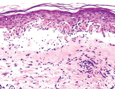
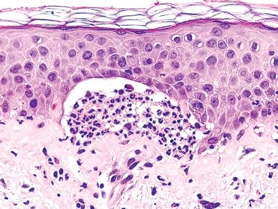
8. This photomicrograph depicts some of the most characteristic changes of a common skin condition. Which of the following histopathologic features becomes prominent in the pustular variant of this condition?
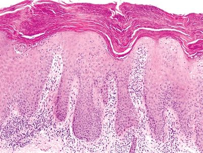
QUESTION 1.8
A. Confluent parakeratosis
B. Focal hemorrhages in the papillary dermis
C. Lymphocytic infiltration of the lower epidermis
D. Munro microabscesses
E. Suprapapillary thinning of epidermis
9. A skin biopsy of an erythematous lesion shows lymphocytic infiltration along the dermal–epidermal junction in a band-like distribution. There are scattered colloid bodies in the basal layer, numerous melanophagocytes in the papillary dermis, and disruption of the basement membrane. Which of the following disorders should be considered in the differential diagnosis?
A. Arthropod bite reaction, gyrate erythema, urticaria
B. Contact dermatitis, atopic dermatitis, pityriasis rosea
C. Lichen planus, lichen nitidus, lichenoid drug eruption
D. Lupus erythematosus, dermatomyositis, graft versus host disease
E. Pemphigus vulgaris, pemphigus foliaceus, keratosis follicularis
10. The histopathologic picture of a skin biopsy is shown in this picture. There are numerous colloid bodies scattered immediately above and below the basement membrane. Which of the following is the most likely clinical presentation?
QUESTION 1.10
A. Erythematous plaques covered by silvery scale on extensor surfaces
B. Malar erythema in a butterfly distribution
C. Painful subcutaneous nodule on the anterior surface of the leg
D. Pruritic plaques with accentuation and thickening of skin markings
E. Pruritic purple papules on the flexor aspects of arms and legs
11. A 10-year-old girl is brought to a pediatrician for evaluation of a skin eruption characterized by tiny, skin-colored, nonitchy papules on the trunk, abdomen, and arms. A skin biopsy is performed. The photomicrograph depicts the histopathologic changes found on a biopsy of one of the papules. Which of the following is the most likely diagnosis?
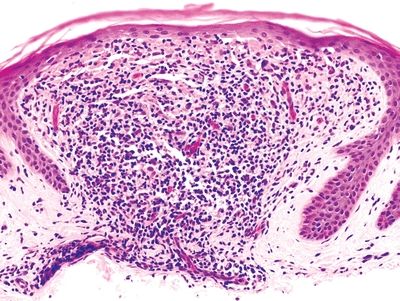
QUESTION 1.11
A. Discoid lupus erythematosus
B. Lichenoid drug eruption
C. Lichen planus
D. Lichen nitidus
E. Pityriasis lichenoid and varioliformis acuta (PLEVA)
12. A 17-year-old female presents with bilateral malar rash in a butterfly distribution and signs of acute pleuritis. A skin biopsy shows liquefactive degeneration of the basal cell layer, variable epidermal atrophy, perivascular and periadnexal lymphocytic infiltration, and a thickened basement membrane. Which of the following patterns of immunofluorescence would be expected?
A. Deposits of IgA at the tips of dermal papillae
B. Granular deposits of IgG and C3 along the dermal–epidermal junction
C. Intercellular IgG deposition within the epidermis
D. Linear deposition of IgG and C3 along the dermal–epidermal junction
E. Smudgy IgG deposits around vessels of papillary dermis
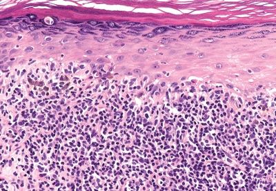
13. A skin biopsy taken 4 weeks after bone marrow transplantation shows marked vacuolar degeneration of basal keratinocytes, sparse lymphocytic infiltration along the dermal–epidermal junction, and apoptosis of individual keratinocytes, as shown in the picture. In its chronic stage, this condition mimics which of the following disorders?
QUESTION 1.13
A. Actinic keratosis
B. Discoid lupus erythematosus
C. Late radiation dermatitis
D. Mycosis fungoides, plaque stage
E. Scleroderma
14. A 25-year-old man presents with target lesions, characterized by papules with central bullae and peripheral erythema. Histologic examination shows a lichenoid dermal infiltrate and vacuolar degeneration of the basal cell layer epidermis, with small subepidermal bullae. Within the epidermis, there are keratinocytes with bright eosinophilic cytoplasm and pyknotic nuclei. Which of the following is the most common cause of this condition?
A. Herpes simplex virus
B. Histoplasma capsulatum
C. Mycoplasma pneumoniae
D. Nonsteroidal anti-inflammatory drugs
E. Sulfonamides
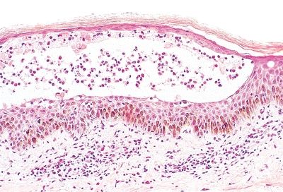
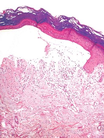
15. A 25-year-old man presents to the emergency room with a widespread eruption of macules, papules, and flaccid bullae involving at least 30% of skin and mucosal surfaces. The patient reports taking sulfonamides for an ear infection. The histopathologic skin changes characteristic of this condition are shown in this photomicrograph. Which of the following is the most likely diagnosis?
QUESTION 1.15
A. Lichenoid drug reaction
B. Staphylococcal scalded skin syndrome
C. Stevens-Johnson syndrome
D. Subcorneal pustular dermatosis
E. Toxic epidermal necrolysis
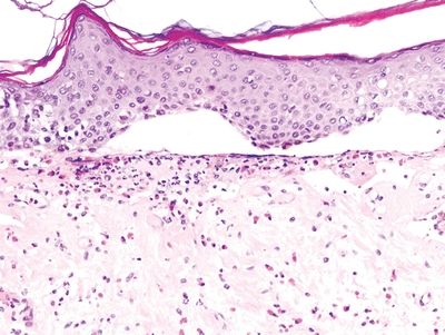
16. An elderly patient presents with extensive areas of erosions and crusts on the trunk. Histopathologic examination of the lesions reveals acantholysis of the granular layer with formation of bullae containing scanty inflammatory cells, as shown in this picture. Which of the following is the most likely diagnosis?
QUESTION 1.16
A. Bullous impetigo
B. Erythema toxicum neonatorum
C. Pemphigus foliaceus
D. Pemphigus vulgaris
E. Staphylococcal scalded skin syndrome
17. Which of the following bullous diseases is pathogenetically related to autoantibodies against antigens of epidermal intercellular junctions?
A. Bullous pemphigoid
B. Dermatitis herpetiformis
C. Epidermolysis bullosa
D. Erythema multiforme
E. Pemphigus vulgaris
18. A 45-year-old man suffers from a skin disease characterized by chronic eruption of brown hyperkeratotic lesions on the neck and trunk. The lesions have a follicular distribution. Several family members on his father’s side suffered from a similar disorder. Biopsies show the histopathologic changes depicted in this picture. Which of the following is the most likely diagnosis?
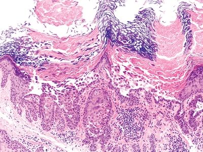
QUESTION 1.18
A. Benign familial pemphigus
B. Keratosis follicularis
C. Pemphigus vulgaris
D. Transient acantholytic dermatosis
E. Warty dyskeratoma
19. Which of the following skin conditions is most likely to be associated with subepidermal bullae accompanied by inflammatory infiltration?
A. Bullous pemphigoid
B. Epidermolysis bullosa
C. Pemphigus foliaceus
D. Pemphigus vulgaris
E. Porphyria cutanea tarda
A. Bullous pemphigoid
B. Herpesvirus infection
C. Keratosis follicularis
D. Pemphigus vulgaris
E. Toxic epidermal necrosis
21. A patient with gluten intolerance complains of an itchy vesicular eruption on the back. A skin biopsy shows the histologic changes in this photomicrograph, whereas direct immunofluorescence demonstrates IgA deposition at the tips of dermal papillae. Which of the following is the most likely diagnosis?
QUESTION 1.21
A. Bullous pemphigoid
B. Dermatitis herpetiformis
C. Epidermolysis bullosa, junctional type
D. Erythema multiforme
E. Porphyria cutanea tarda
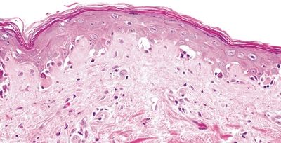
22. A 26-year-old man presents to a dermatologist with recent onset of blisters on his face and hands. On physical examination, hypertrichosis is noted on temporal and malar areas. The family history is negative for similar skin disorders. A skin biopsy reveals the histopathologic changes shown in this photomicrograph. Which of the following is the most likely cause of this skin condition?
QUESTION 1.22
A. Autoantibodies to basement membrane proteins
B. Autoimmune reaction against gliadin
C. Mutation of gene encoding an intracellular calcium pump
D. Mutations of genes encoding structural epidermal or dermal proteins
E. Reduced activity of uroporphyrinogen decarboxylase enzyme
23. Consider the following conditions: Vitiligo, anetoderma, ichthyosis, acanthosis nigricans, tinea versicolor, postinflammatory pigmentary alteration. Which of the following histologic abnormalities would be seen in biopsies of these skin conditions?
A. Interface dermatitis
B. Nil lesion
C. Prominent acanthosis
D. Spongiosis
E. Subepidermal bulla
24. A 20-year-old woman presents to her physician with intensely itchy erythematous papules around the ankles. The lesions appeared a few days after she moved to a house with roommates who have cats and dogs. A biopsy of the skin lesions shows the changes depicted in the picture. Which of the following is the most likely diagnosis?
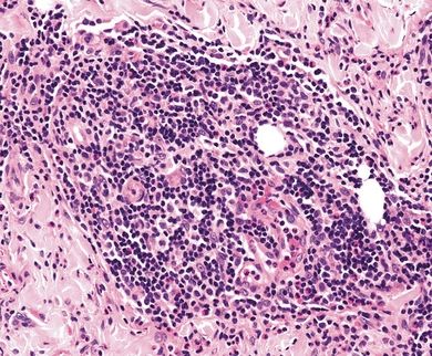
QUESTION 1.24
A. Arthropod bite reaction
B. Leukocytoclastic vasculitis
C. Necrobiosis lipoidica
D. Pigmented purpura
E. Polymorphous light reaction
F. Pyoderma gangrenosum
G. Urticaria pigmentosa
H. Sarcoidosis
I. Urticaria
25. A patient with a history of a recent tick bite presents with a febrile illness characterized by skin lesions and lymphadenitis. The skin lesions are described as erythema chronicum migrans. Which of the following is the underlying etiology?
A. Borrelia burgdorferi
B. Cancer
C. Group A streptococcus
D. Rickettsia rickettsii
E. Treponema pallidum
26. A skin biopsy reveals the dermal abnormality demonstrated in this photomicrograph. Which of the following clinical findings is most likely associated with this histopathologic change?
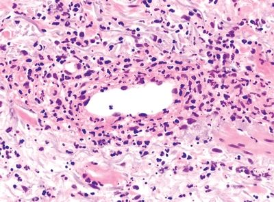
QUESTION 1.26
A. Gyrate erythema
B. Hives
Stay updated, free articles. Join our Telegram channel

Full access? Get Clinical Tree


