QUESTION 2.2
A. Acanthosis nigricans
B. Keratoacanthoma
C. Seborrheic keratosis
D. Subcorneal bullae
E. Xanthomas
3. Pseudoepitheliomatous hyperplasia is a form of verruciform acanthosis that extends deep into the dermis and may mimic squamous cell carcinoma. This pattern of epithelial growth may be associated with which of the following conditions?
A. Fungal infections
B. Granular cell tumors
C. Halogenodermas
D. Prurigo nodularis
E. All of the above
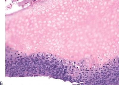
4. A 55-year-old woman undergoes excision of a 0.8-cm dome-shaped reddish nodule on her right leg. The microscopic features of the lesion are shown in this photomicrograph.
Which of the following is the diagnosis?
QUESTION 2.4
A. Actinic keratosis
B. Benign lichenoid keratosis
C. Clear cell acanthoma
D. Large cell acanthoma
E. Seborrheic keratosis
5. Which of the following epidermal cysts contains ciliated epithelium?
A. Branchial cleft cyst
B. Epidermal cyst
C. HPV-associated cyst
D. Steatocystoma
E. Trichilemmal cyst
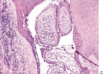
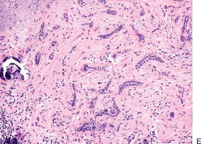
The following are photomicrographs of five different benign adnexal tumors. Match each picture with the most likely clinical presentation in the next five questions.
6. A 25-year-old woman with multiple flesh-colored, round, 1- to 3-mm papules around the lower eyelids.
8. A 15-year-old boy with a hairless plaque-like lesion in the scalp that recently has become larger and nodular.
10. A 5-year-old child with an asymptomatic, stony hard nodule in the skin of the neck. The “tent sign” is observed upon stretching the skin.
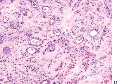
11. A 35-year-old woman presents with a long-standing history of numerous coalescing nodular lesions on the scalp. An excisional biopsy shows a dermal lesion composed of nests that fit together like a jigsaw puzzle, embedded in a basement membrane–like substance. Which of the following is the most likely diagnosis?
A. Chondroid syringoma
B. Cylindroma
C. Eccrine poroma
D. Sebaceous adenoma
E. Trichilemmoma
12. A painful nodule is excised from the lower leg of a young patient. Histologically, it shows extreme cellular density and a two-cell population, epithelial and myoepithelial, along with a rich lymphocytic infiltrate within perivascular spaces and between tumor cells. Overall, its morphologic features are reminiscent of thymoma. Which of the following is the most likely diagnosis?
A. Cylindroma
B. Glomus tumor
C. Metastatic thymoma
D. Spiradenoma
E. Synovial sarcoma
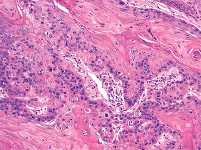
13. Which of the following adnexal tumors is morphologically similar to mixed tumors arising in salivary glands?
A. Chondroid syringoma
B. Cylindroma
C. Eccrine poroma
D. Syringoma
E. Trichoepithelioma
14. Sweat gland carcinomas are rare. These tumors are most likely to simulate histologically which of the following neoplasms?
A. Melanoma
B. Metastatic breast or renal cell carcinomas
C. Metastatic salivary gland tumors
D. Metastatic small cell carcinoma of the lungs
E. Metastatic thymoma
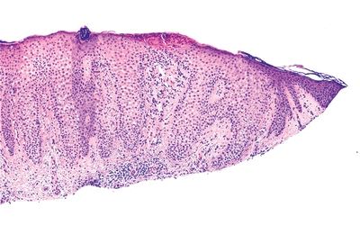
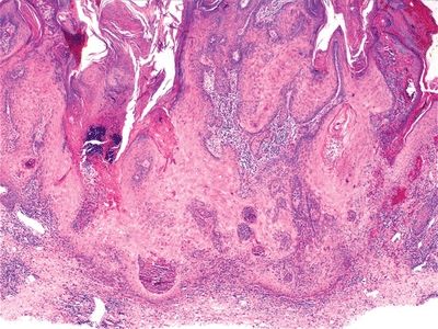
15. A 65-year-old man undergoes excisional biopsy of a pigmented, rough-surfaced patch on the forehead. The histologic appearance of the lesion is shown in this picture. The percentage of metastases that develop from carcinomas arising in such lesion is approximately:
QUESTION 2.15
A. Less than 5%
B. 20%
C. 40%
D. 60%
E. 80%
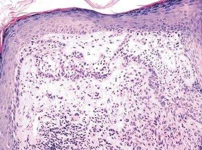
16. The clinical condition known as Bowen disease of the skin is characterized histologically by:
A. Acantholytic suprabasal bullae
B. Epidermal atypia with koilocytosis
C. Low-grade squamous intraepithelial lesion
D. Melanocytic atypia
E. Squamous cell carcinoma in situ
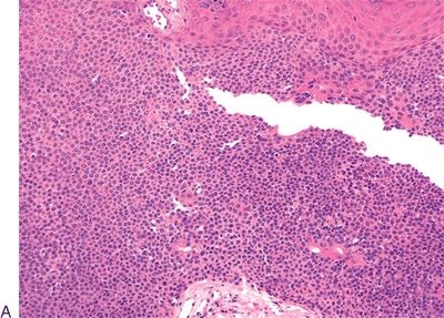
17. This low-power photomicrograph shows a cutaneous neoplasm histologically similar to squamous cell carcinoma. Which of the following is the most important morphologic aspect in differentiating this neoplasm from squamous cell carcinoma?
QUESTION 2.17
A. Architecture
B. Cytologic atypia
C. Mitotic activity
D. Nuclear features
E. Perineural invasion
18. This picture shows a common skin malignancy. Which of the following is the most important predictor of this tumor’s behavior?
QUESTION 2.18
A. Degree of differentiation
B. Histologic subtype
C. History of immune suppression
D. Origin from actinic keratosis
E. Tumor thickness and depth of invasion
19. Which of the following variants of squamous cell carcinoma may mimic angiosarcoma?
A. Carcinosarcoma
B. Pseudovascular adenoid
C. Signet ring cell
D. Spindle cell
E. Verrucous
20. Which of the following microscopic types of basal cell carcinoma is associated with the greatest propensity for metastatic spread?
A. Adenoid
B. Basosebaceous
C. Basosquamous
D. Fibroepithelioma of Pinkus
E. Infiltrative
F. Micronodular
G. Morpheaform
H. Nodulocystic
I. Pigmented
J. Superficial
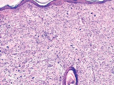
21. A 50-year-old man presents with a dome-shaped nodule growing on his right middle finger. The nodule is removed, and histologic examination reveals hyperkeratotic and acanthotic epidermis overlying dense collagenous bundles oriented perpendicular to the skin surface, as shown in this picture. Which of the following is the most likely diagnosis?
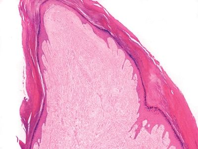
QUESTION 2.21
A. Accessory digit
B. Acquired digital angiofibroma
C. Collagenous nevus
D. Dermatofibroma
E. Morpheaform basal cell carcinoma
22. This photomicrograph shows a common dermal tumor. The immunohistochemical staining most helpful in differentiating this lesion from dermatofibrosarcoma protuberans (DFSP) is:
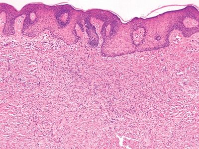
QUESTION 2.22
A. Actin
B. CD34
C. Factor XIIIa
D. HMB-45
E. Vimentin
23. A biopsy of a polypoid skin lesion on the back of a 45-year-old man reveals a tumor characterized by a prominent “cartwheel” pattern as shown in this picture. On immunohistochemistry, tumor cells are positive for CD34 and negative for S100 protein. Which of the following is the most likely diagnosis?
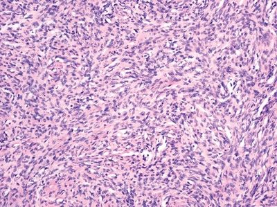
QUESTION 2.23
A. Atypical fibroxanthoma
B. Dermatofibrosarcoma protuberans (DFSP)
C. Leiomyoma
D. Neurofibroma
E. Schwannoma
24. A 70-year-old man presents with a slowly growing, exophytic, reddish 1-cm nodule on the face. Excisional biopsy reveals a poorly circumscribed, highly cellular tumor within the dermis, extending to, but not continuous with, the epidermis, as shown in this photomicrograph. It is composed of a mixture of fibroblast-like spindle cells and histiocyte-like cells. Many multinucleated giant cells are present. Mitoses are plentiful (including atypical ones), and cellular pleomorphism is marked. Neoplastic cells are immunoreactive for CD10 and negative for S100, keratin, actin, and desmin. Which of the following is a correct statement about this tumor?
QUESTION 2.24
A. Has same prognosis as malignant fibrous histiocytoma (MFH).
B. Is more frequent in young individuals.
C. Metastases are rare.
D. Is most frequent in acral locations.
E. Reliable specific immunohistochemical marker is available.
25. A 45-year-old woman undergoes removal of a soft nodule on the neck. This picture shows the microscopic features. Mitotic figures are rare. Tumor cells are immunoreactive for S100 and negative for HMB-45 and p53. The Ki-67 labeling index is very low. Immunohistochemistry for neurofilaments shows scattered axons throughout the tumor. Which of the following is the diagnosis?
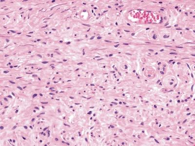
QUESTION 2.25
A. Desmoplastic melanoma
B. Malignant peripheral nerve sheath tumor (MPNST)
C. Neurofibroma
D. Neurotized nevus
E. Schwannoma
26. A painful dermal tumor is removed and shows the microscopic features in this picture. The tumor is strongly immunoreactive for S100, and immunostain for neurofilaments fails to reveal any axons. Which of the following is the most likely diagnosis?
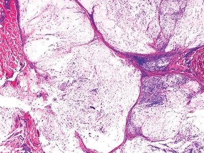
QUESTION 2.26
A. Dermatofibroma
B. Glomus tumor
C. Neurofibroma
D. Schwannoma
E. Traumatic neuroma
27. An ulcerated violaceous nodule is excised from the face of a 55-year-old man. As shown in this photomicrograph, the lesion is an intradermal tumor composed of small round cells with high nucleocytoplasmic ratio and brisk mitotic activity. The chromatin pattern is finely granular. Which of the following immunohistochemical or ultrastructural features would favor a diagnosis of Merkel cell carcinoma?
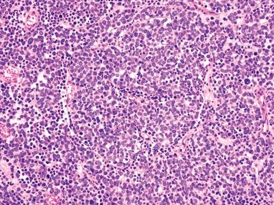
QUESTION 2.27
A. Diffuse cytoplasmic immunoreactivity for high molecular weight keratin
B. Identification of Birbeck granules on electron microscopy
C. Immunoreactivity for CD99
Stay updated, free articles. Join our Telegram channel

Full access? Get Clinical Tree


