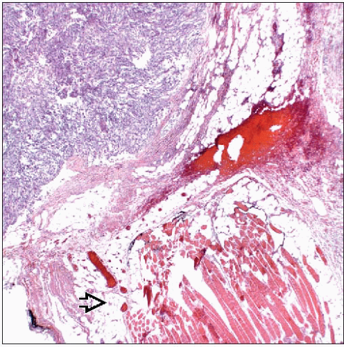
Nodular Fasciitis
Key Facts
Terminology
Benign self-limited proliferation of fibroblasts and myofibroblasts
Clinical Issues
Presents as rapidly growing painful or tender mass
Without treatment, tumors will spontaneously regress within a few months
Location may be in subcutaneous tissue, muscle fascia, or within breast parenchyma
Image Findings
There are no specific imaging findings that would identify a lesion as NF
Many lesions have irregular margins and can closely resemble invasive carcinoma
Microscopic Pathology
Mitoses are typically frequent
Large myoid-type cells or multinucleated cells may be present
Nodular fasciitis is composed of short spindle-shaped cells
Cells are loosely organized and do not have specific pattern
Described as “tissue culture” appearance
Top Differential Diagnoses
Fibromatosis
Spindle cell carcinoma
Myofibroblastoma
Proliferative fasciitis and proliferative myositis
Sarcoma
TERMINOLOGY
Abbreviations
Nodular fasciitis (NF)
Definitions
Benign self-limited proliferation of fibroblasts and myofibroblasts
CLINICAL ISSUES
Presentation
Presents as rapidly growing painful or tender mass
Occurrence within breast is very rare
Location may be in subcutaneous tissue, muscle fascia, or within breast parenchyma
Treatment
No treatment is necessary
In most cases, lesion will be excised to exclude other neoplasms
Prognosis
Tumors spontaneously regress within a few months
IMAGE FINDINGS
Mammographic Findings
Lesions present as mammographic densities with irregular margins
Ultrasonographic Findings
Many lesions have irregular margins
MICROSCOPIC PATHOLOGY
Histologic Features
Nodular fasciitis is composed of short spindle-shaped cells
Cells loosely arranged and are not organized in specific patterns
Tumor has a “tissue culture” appearance
Short fascicles or small whorls may be present
Nuclei are small and uniform
Prominent nucleoli may be present
Mitoses are typically frequent
Cells form solid masses and do not infiltrate around ducts and lobules
Thick bands of collagen are typical
Chronic inflammation may be present; cells are distributed throughout and not restricted to periphery
Stay updated, free articles. Join our Telegram channel

Full access? Get Clinical Tree


