Patient Story
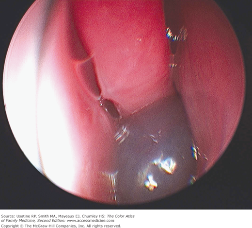
A 35-year-old man complains of unilateral nasal obstruction for the past several months of gradual onset. On examination of the nose, a nasal polyp is found (Figure 30-1).
Introduction
Epidemiology
- Prevalence of 1% to 4% of adults; 0.1% of children of all races and classes.
- The male-to-female ratio in adults is approximately 2:1.
- Peak age of onset is 20 to 40 years old; rare in children younger than 10 years old.
- Associated with the following conditions:
- Nonallergic and allergic rhinitis and rhinosinusitis.
- Asthma—In 20% to 50% of patients with polyps.
- Cystic fibrosis.
- Aspirin intolerance—In 8% to 26% of patients with polyps.
- Alcohol intolerance—In 50% of patients with polyps.
- Nonallergic and allergic rhinitis and rhinosinusitis.
Etiology and Pathophysiology
- The precise cause of nasal polyp formation is unknown.
- Infectious agents causing desquamation of the mucous membrane may play a triggering role.
- Activated epithelial cells appear to be the major source of mediators that induce an influx of inflammatory cells, including eosinophils prominently; these in turn lead to proliferation and activation of fibroblasts.2 Cytokines and growth factors play a role in maintaining the mucosal inflammation associated with polyps.
- Food allergies are strongly associated with nasal polyps.
Diagnosis
- The appearance is usually smooth and rounded (Figure 30-1).
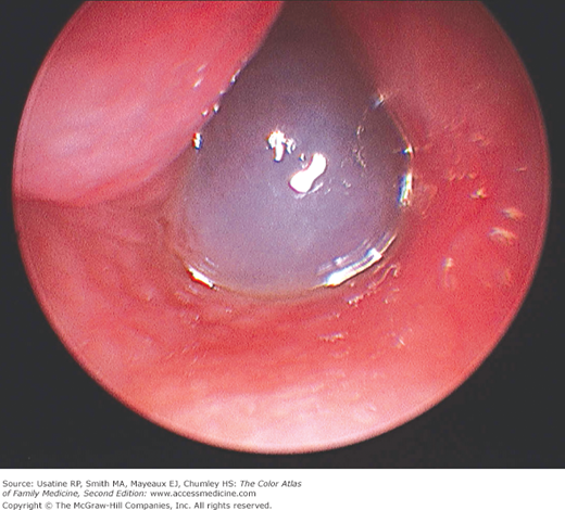
- Moist and translucent (Figure 30-2).
- Variable size.
- Color ranging from nearly none to deep erythema.
- Consider allergy testing.
- In children with multiple polyps, order sweat test to rule out cystic fibrosis.
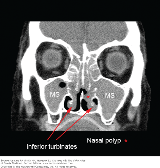
- CT of the nose and paranasal sinuses may be indicated to evaluate extent of lesion(s) (Figure 30-3).